What does the thoracic duct empty into?
The thoracic duct continues superiorly to empty into the junction of the left subclavian and internal jugular veins. The right lymphatic duct drains the right side of the thorax, right upper extremity, and right side of the neck and head.
What is the role of the thoracic duct?
- it is responsible for the removal of interstitial fluid from tissues.
- it absorbs and transports fatty acids and fats as chyle from the digestive system.
- it transports white blood cells to and from the lymph nodes into the bones.
Where does the right lymphatic duct drain lymph from?
The right lymphatic duct, also called the right thoracic duct, is about 1.25 cm long. It drains lymphatic fluid from the right thoracic cavity (this is the section of the trunk on the upper right side), the right arm, and from the right side of the neck and the head. In some people, it also drains lymph from the left lung’s lower lobe.
Where is the air condition drain hose located?
They are usually under the passenger side of the vehicle. If the drain was plugged, you should have wet floor boards on the passenger side. Look by the firewall, right side of engine. 44 people found this helpful. Mark helpful. Report. FordNut answered 5 years ago. 4 is drain hose. 20 people found this helpful.
What drains into thoracic duct quizlet?
Lymph from the entire body drains eventually into the thoracic duct. There are two lymph ducts in the body—the right lymphatic duct and the thoracic duct.
Which of the following lymphatic vessels drain into the thoracic duct?
The right lymphatic duct drains lymph from the right side of the body above the diaphragm. The Cisterna Chyli is where the thoracic duct begins.
Which region of the body is drained by the thoracic duct quizlet?
The thoracic duct drains lymph from the right side of the head, neck, right arm, and the right side of the thoracic cavity.
Does Cisterna Chyli drain into thoracic duct?
The cisterna chyli tapers at its superior aspect and becomes the thoracic duct. Most frequently the cisterna chyli is replaced by a confluence of lymph trunks in the abdominal region. The thoracic duct subsequently enters the thorax through the aortic hiatus just to the right of the aorta.
What is the thoracic duct?
Thoracic Duct. The thoracic duct is the largest lymphatic vessel within the human body, and plays a key role in the lymphatic system. It is also called the left lymphatic duct or the alimentary duct. A large portion of the body’s lymph is collected by this duct and then drained into the bloodstream near the brachiocephalic vein between ...
Where does the duct originate?
It originates from the second lumbar vertebra level and goes to the neck’s root. The duct arises from the combination of the left and right lumbar trunks and the intestinal trunk in the abdomen.
What is the left lymphatic duct?
It is also called the left lymphatic duct or the alimentary duct. A large portion of the body’s lymph is collected by this duct and then drained into the bloodstream near the brachiocephalic vein between the internal jugular and the left subclavian veins.
How much lymphatic fluid does the aortic duct transport?
It travels through the aortic aperture diaphragm and rises along the posterior mediastinum. It transports up to four liters of lymphatic fluid each day. This process is primarily caused by the breathing action and is assisted by the smooth muscle of the duct. Last medically reviewed on January 21, 2018.
Where does the thoracic duct drain?
It drains into the systemic (blood) circulation at the junction of the left subclavian and internal jugular veins, at the commencement of the brachiocephalic vein. When the duct ruptures, ...
What is the thoracic duct?
Anatomical terminology. In human anatomy, the thoracic duct is the larger of the two lymph ducts of the lymphatic system. It is also known as the left lymphatic duct, alimentary duct, chyliferous duct, and Van Hoorne's canal. The other duct is the right lymphatic duct. The thoracic duct carries chyle, a liquid containing both lymph ...
What is the lymph transport in the thoracic duct?
The lymph transport, in the thoracic duct, is mainly caused by the action of breathing, aided by the duct's smooth muscle and by internal valves which prevent the lymph from flowing back down again. There are also two valves at the junction of the duct with the left subclavian vein, to prevent the flow of venous blood into the duct.
Which lymphatic duct is drained by the right lymphatic duct?
The other duct is the right lymphatic duct. The thoracic duct carries chyle, a liquid containing both lymph and emulsified fats, rather than pure lymph. It also collects most of the lymph in the body other than from the right thorax, arm, head, and neck (which are drained by the right lymphatic duct ). The thoracic duct usually starts ...
Where is the Virchow's node located?
The first sign of a malignancy, especially an intra-abdominal one, may be an enlarged Virchow's node, a lymph node in the left supraclavicular area, in the vicinity where the thoracic duct empties into the left brachiocephalic vein, right between where the left subclavian vein and left internal jugular join (i. e., the left Pirogoff angle). When the thoracic duct is blocked or damaged a large amount of lymph can quickly accumulate in the pleural cavity, this situation is called chylothorax .
Where does the thoracic duct develop?
The thoracic duct develops from lymphatic trunks on either side of the aorta that anastomoses to form a channel from the jugular lymph sacs to the cisterna chyli. Trunks continue to anastomose and enlarge, forming embryonic right and left thoracic ducts. The adult thoracic duct is derived from both of these embryonic thoracic ducts.
What is the function of the thoracic duct?
The function of the thoracic duct is to transport lymph back into the circulatory system. Interstitial fluid is collected by lymph capillaries from the interstitial space. Lymph then moves through lymphatic vessels to lymph nodes. Lymphatic vessels merge to create the lymphatic ducts which drain into the venous system.
How many vessels can a thoracic duct terminate?
The thoracic duct can also terminate as a single vessel (up to 87.5%), bilateral ducts (up to 25%), or several terminal branches (up to 7%). The thoracic duct displays physiologic adaptation to certain disease processes by increasing in diameter.
What are the two lymphatic ducts?
Introduction. Lymphatic ducts empty lymph fluid into the venous system. The two lymphatic ducts of the body are the right lymphatic duct and the thoracic duct. The thoracic duct is the larger of the two and responsible for lymph drainage from the entire body except for the right sides of the head and neck, ...
How long is the thoracic duct?
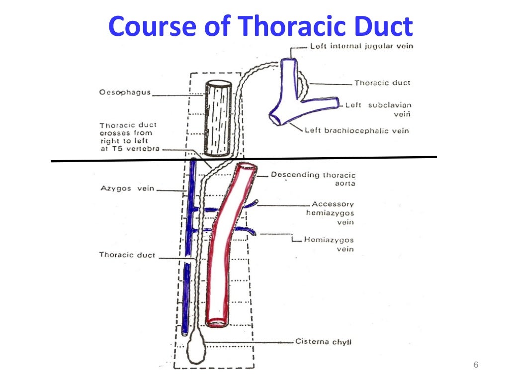
The thoracic duct is 38 to 45 centimeters long and 2 to 5 millimeters in diameter. It runs from the superior aspect of the cisterna chyli, a lymph sac at the L2 vertebral level, to the lower cervical spine. From the cisterna chyli, the duct continues superiorly, running between the aorta and the azygous vein and anterior to the vertebral column. The thoracic duct ascends through the aortic hiatus of the diaphragm entering the posterior mediastinum, still to the right of the vertebral column. It courses posterior to the esophagus at the T7 level and crosses over the midline to the left side of the thorax around the T5 vertebral level. As it continues upward, it runs behind the aorta and to the left of the esophagus ascending 2-3 cm above the clavicle. In the superior mediastinum, it passes behind the left common carotid artery, the vagus nerve, and the internal jugular vein. It then descends to empty into the junction of the left subclavian and internal jugular veins.
What is the superior mediastinum?
In the superior mediastinum, it passes behind the left common carotid artery, the vagus nerve, and the internal jugular vein. It then descends to empty into the junction of the left subclavian and internal jugular veins. The wall of the thoracic duct has three layers: the intima, the media, and the adventitia.
Which muscle contracts to move lymph forward?
The smooth muscle contracts regularly to move lymph flow forward. The thoracic duct also contains valves which may be unicuspid, bicuspid, or tricuspid, but are usually bicuspid. At the junction of the lymphatic and venous system, a bicuspid valve prevents venous backflow into the lymphatic system. [3]
What system does the lymph duct drain into?
Lymph ducts empty into the circulatory system, draining into the:
What chapter is the lymphatic system?
Start studying Chapter 31: Lymphatic System. Learn vocabulary, terms, and more with flashcards, games, and other study tools.
Which two capillary networks are dependent on each other?
b. Lymphatic and blood capilla ry networks are dependent on each other.
Overview
In human anatomy, the thoracic duct is the larger of the two lymph ducts of the lymphatic system. It is also known as the left lymphatic duct, alimentary duct, chyliferous duct, and Van Hoorne's canal. The other duct is the right lymphatic duct. The thoracic duct carries chyle, a liquid containing both lymph and emulsified fats, rather than pure lymph. It also collects most of the lymph in the …
Structure
In adults, the thoracic duct is typically 38–45 cm in length and has an average diameter of about 5 mm. The vessel usually starts from the level of the twelfth thoracic vertebra (T12) and extends to the root of the neck. It drains into the systemic (blood) circulation at the angle of the left subclavian and internal jugular veins as a single trunk, at the commencement of the brachiocephalic vein.
The thoracic duct originates in the abdomen from the confluence of the right and left lumbar trunks and …
Function
The thoracic duct collects most of the lymph in the body other than from the right thorax, arm, head, and neck. These are drained by the right lymphatic duct.
The lymph transport, in the thoracic duct, is mainly caused by the action of breathing, aided by the duct's smooth muscle and by internal valves which prevent the lymph from flowing back down again. There are also two valves at the junc…
Clinical significance
The first sign of a malignancy, especially an intra-abdominal one, may be an enlarged Virchow's node, a lymph node in the left supraclavicular area, in the vicinity where the thoracic duct empties into the left brachiocephalic vein, right between where the left subclavian vein and left internal jugular join (i.e., the left Pirogoff angle). When the thoracic duct is blocked or damaged a large amount of lymph can quickly accumulate in the pleural cavity, this situation is called chylothorax.
Additional images
• Transverse section of thorax, showing relations of pulmonary artery.
• The arch of the aorta, and its branches.
• Deep lymph nodes and vessels of the thorax and abdomen (diagrammatic).
See also
• Lymph duct
• Lymphatic system
External links
• Anatomy figure: 21:05-02 at Human Anatomy Online, SUNY Downstate Medical Center — "The thoracic duct and azygos venous network"
• Anatomy image:8901 at the SUNY Downstate Medical Center
• figures/chapter_24/24-5.HTM: Basic Human Anatomy at Dartmouth Medical School