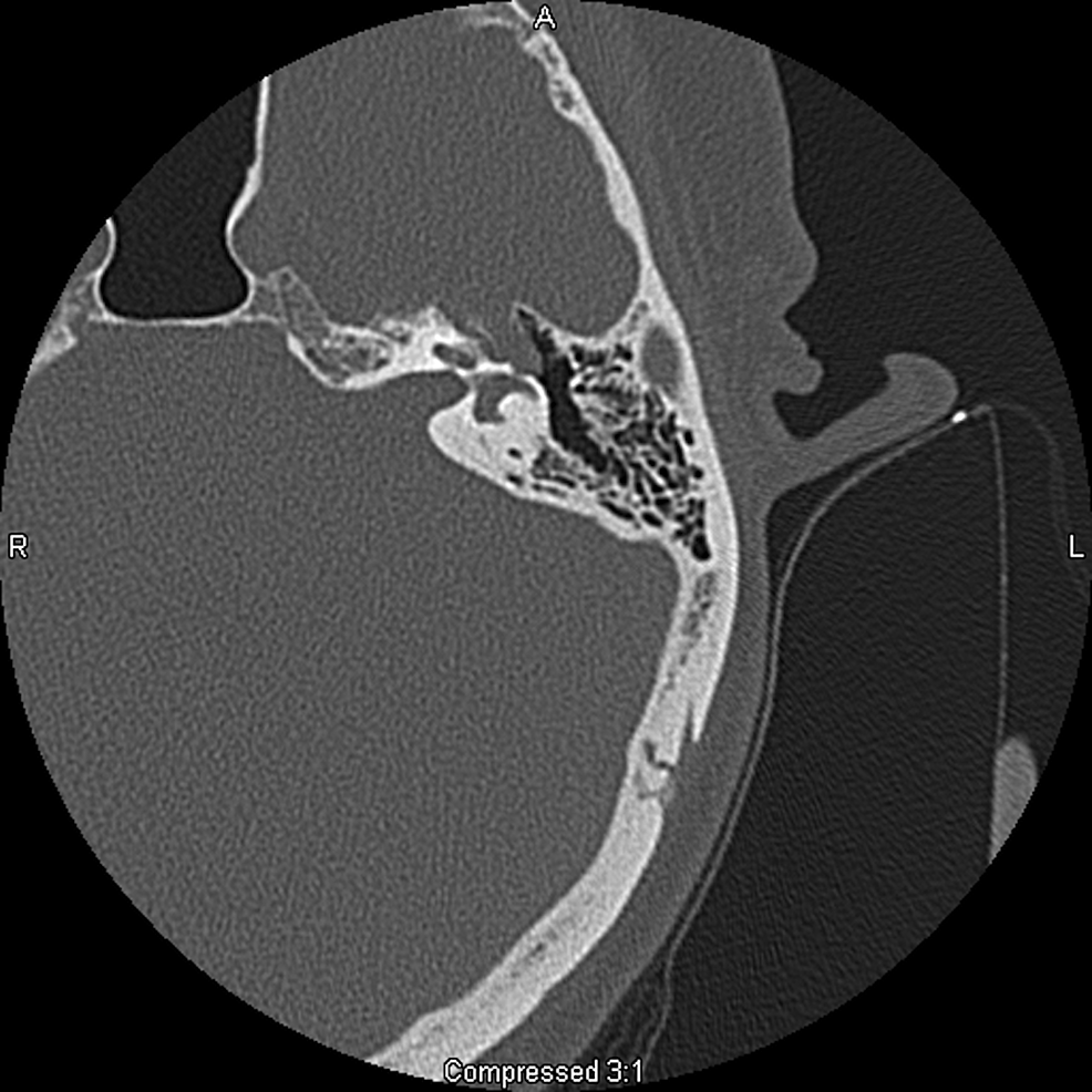
What synapse is at the geniculate ganglion?
The geniculate ganglion (from Latin genu, for "knee") is a collection of pseudounipolar sensory neurons of the facial nerve located in the facial canal of the head. It receives fibers from the facial nerve....Geniculate ganglion.Geniculate ganglion genu structure like formTA26287FMA53414Anatomical terms of neuroanatomy8 more rows
What forms the geniculate ganglion?
In the facial canal, the two roots fuse together. At the first bend of the Z, they form the geniculate ganglion. The ganglion then sends out nerve fibers to several nerve branches, including: Tympanic (ear) segment of the facial nerve.
What and where is the geniculate ganglion?
The geniculate or genicular ganglion contains fibres for taste and somatic sensation and is located in the petrous temporal bone.
Which mixed cranial nerve is associated with the geniculate ganglion?
Facial Nerve. The facial nerve is mixed nerve containing both sensory and motor components. The nerve emanates from the brain stem at the ventral part of the pontomedullary junction. The nerve enters the internal auditory meatus where the sensory part of the nerve forms the geniculate ganglion.
What is the pathway of the facial nerve?
It arises from the brain stem and extends posteriorly to the abducens nerve and anteriorly to the vestibulocochlear nerve. It courses through the facial canal in the temporal bone and exits through the stylomastoid foramen after which it divides into terminal branches at the posterior edge of the parotid gland.
What is the Vidian nerve?
Medical Definition of Vidian nerve : a nerve formed by the union of the greater petrosal and the deep petrosal nerves that passes forward through the pterygoid canal in the sphenoid bone and joins the pterygopalatine ganglion.
Which foramen does the facial nerve pass through?
The facial nerve exits the base of the skull at the stylomastoid foramen, which is an opening in the bone located near the base of the ear.
Is LGN in the thalamus?
The lateral geniculate nucleus (LGN; also called the lateral geniculate body or lateral geniculate complex) is a structure in the thalamus and a key component of the mammalian visual pathway. It is a small, ovoid, ventral projection of the thalamus where the thalamus connects with the optic nerve.
What kind of ganglion is the trigeminal ganglion?
semilunar sensory ganglionThe semilunar sensory ganglion (also known as the trigeminal ganglion or Gasserian ganglion) is a thin, crescent-shaped structure situated in Meckel's cave within the middle cranial fossa.
What does cranial nerve 7 affect?
The two 7th Cranial Nerves (CN VII) are located on either side of the brainstem, at the top of the medulla. They are mixed cranial nerves with BOTH sensory and motor function. CN VII controls the face and is mainly FACE MOVEMENT with some face sensation.
Where does the facial nerve synapse?
The first branch of the facial nerve, the greater petrosal nerve, arises here from the geniculate ganglion. The greater petrosal nerve runs through the pterygoid canal and synapses at the pterygopalatine ganglion.
What nerve does the chorda tympani connected to?
Chorda tympani nerve It merges with the lingual nerve, a branch of the maxillary nerve (V3). This nerve transports the nerves of taste for the anterior two-thirds of the tongue and contains secretory fibers for the sublingual and submaxillary glands. In addition, it sends a branch to the auditory tube.
What kind of ganglion is the trigeminal ganglion?
semilunar sensory ganglionThe semilunar sensory ganglion (also known as the trigeminal ganglion or Gasserian ganglion) is a thin, crescent-shaped structure situated in Meckel's cave within the middle cranial fossa.
What are the 5 muscles of facial expression?
– Lower group- contains depressor anguli oris, depressor labi inferioris and the mentalis. – Upper group- contains risorius, zygomaticus major, zygomaticus minor, levator labii superioris, levator labii superioris alaeque nasi and levator anguli oris.
Where does deep Petrosal come from?
The deep petrosal nerve is a branch from the internal carotid plexus. The plexus is located on the lateral side of the internal carotid as it courses superiorly. The deep petrosal enters the skull through the carotid canal with the internal carotid artery.
What is the ciliary ganglion?
Ciliary ganglion is a peripheral parasympathetic ganglion. It is situated near the apex of orbit between the optic nerve and lateral rectus muscle. It is related medially to the ophthalmic artery and laterally to the lateral rectus muscle.