
What is the function of seminiferous tubules?
The seminiferous tubules are the site of the germination, maturation, and transportation of the sperm cells within the male testes. Seminiferous tubules are made up of columnar Sertoli cells surrounded by spermatogenic cells on the epithelial interior and stem cells exteriorly.
Where are seminiferous tubules located in the male testicles?
The seminiferous tubules are located in the male testicles, which are the two oval-shaped organs on located beneath the penis. In each testicle, there are approximately 800 seminiferous tubules.
What is the path of sperm in the female reproductive system?
The path of sperm can be traced from the seminiferous tubules of the testes where the sperm is produced, to the epididymis where the sperm undergoes concentration and maturation, to the ampulla of the vas deferens, to the prostatic urethra, the urethra and finally ejaculated in to the vaginal canal.
What is the size of the seminiferous tubes?
The seminiferous tubes occupy between 85 and 90% of the volume of the testes, and they fulfill a predominantly exocrine function in the male reproductive system. They are located, specifically, inside the testicular lobes. Each lobes contains between 1 and 5 seminiferous tubes, approximately 70mm long and 0.2mm wide.
What occurs in the seminiferous tubules?
The seminiferous tubules are the site of spermatogenesis where germ cells develop into spermatozoa in close interaction with Sertoli cells.
What structure is contained inside the seminiferous tubules?
The seminiferous tubules contain both germ cells and Sertoli cells and are the site of sperm production (the exocrine function) and the interstitial area contains both the Leydig cells, which produce testosterone, and the blood vascular elements (which carry out the endocrine function).
Where does meiosis occur in seminiferous tubules?
Seminiferous tubules are located within the testes, and are the specific location of meiosis, and the subsequent creation of male gametes, namely spermatozoa.
What does the seminiferous tubules do in male reproductive system?
The seminiferous tubules are the basic units of the testicles where the SSCs proliferate and differentiate through cyclic events (mitosis, meiosis, postmeiotic spermatid development, and spermiogenesis) to generate spermatozoa in a process called spermatogenesis [19].
What structure is contained inside the seminiferous tubules quizlet?
SEMINIFEROUS TUBULES, which contain SPERMATOGENIC CELLS and SERTOLI CELLS, are located within a connective tissue stroma known as the TUNICA VASCULOSA.
Which of the following is not found within the seminiferous tubule?
In testis, which of the following cells are not found in the interstitial spaces between the seminiferous tubules? Sertoli and germinal cells are not found in the interstitial spaces between the seminiferous tubules, whereas Leydig cells are found in the interstitial spaces between the seminiferous tubules.
Where does spermatogenesis take place?
the testesAs mentioned above, spermatogenesis is the process by which sperm cell production occurs; the germ cells give rise to the haploid spermatozoa. Sperm production takes place inside the seminiferous tubules, which is a convoluted cluster of tubes located inside the testes.
What are the 4 steps of spermatogenesis?
The process of germ cell development during spermatogenesis can be divided into five succesive stages: (1) spermatogonia, (2) primary spermatocytes, (3) secondary spermatocytes, (4) spermatids, and (5) spermatozoa.
What are the 3 stages of spermatogenesis?
(A) The three main stages of spermatogenesis: (i) spermatocytogenesis, (ii) meiosis, and (iii) spermiogenesis, including the illustration of two major sources of variation.
Do seminiferous tubules produce hormones?
The main hormone secreted by the testes is testosterone, an androgenic hormone. Testosterone is secreted by cells that lie between the seminiferous tubules, known as the Leydig cells.
Do seminiferous tubules produce sperm?
The seminiferous tubules, in which the sperm are produced, constitute about 90 percent of the testicular... The immature cells (called spermatogonia) are all derived from cells called stem cells in the outer wall of the seminiferous tubules.
What is the role of the seminiferous tubules in pregnancy?
The seminiferous tubules are the site of spermatogenesis, where germ cells develop into spermatozoa in close interaction with Sertoli cells. The Sertoli cells91 form the seminiferous tubules and provide the cytoarchitectural arrangements for the developing germinal cells.
How the structure of the seminiferous tubules are adapted for their function?
The seminiferous tubules are located in the testicles (or the testes) and are long, coiled, tubular structures that carry out the function of spermatogenesis (sperm production). Cells in the seminiferous tubules, called the primary spermatocytes, divide by meiosis to produce sperm.
Where are seminiferous tubules found?
There are around 800 seminiferous tubules in each testicle, and this is where meiosis and subsequent development of spermatozoa occurs. In a mature adult male, each of these tubules creates thousands of sperm every second.
Which part of the epididymis is attached to the body of seminiferous tubules?
The body of the epididymis is attached to the body of seminiferous tubules (rete testis) via the afferent ducts. Spermatozoa are transported to the epididymis for storage and maturation before becoming a part of semen.
Where are Leydig cells located?
Leydig cells can be found around seminiferous tubules forming groups of up to ten cells. They are generally described as polygonal cells with eosinophilic cytoplasm and a large round nucleus with a prominent nucleolus.
Where are seminiferous tubules located?
The seminiferous tubules are tiny channels located in the testes , where the germination, maturation and transport of sperm take place towards the testicular network
What is the outer layer of the seminiferous tubules?
Around the outer surface of the seminiferous tubules is the tunica propria, also called the limiting layer.
How many types of seminiferous tubules are there?
Two types of seminiferous tubules are distinguished, depending on the function they fulfill within the testicular structure:
What are the sustainable cells of Sertoli?
The sustainable cells of Sertoli complement the nutrition and development of the sperm. They also increase the presence of testosterone in the seminiferous tubules.
What process produces sperm?
Basically, these cells produce sperm after going through the processes of mitosis (reproduction of cells) and meiosis (division of cells), respectively.
Where does the spermiogenesis process take place?
From there, the journey to the epididymis continues, where the spermiogenesis process takes place; that is, the structural formation of the sperm through the allocation of the acrosome.
Is sperm production feasible?
In short, thanks to these small conduits, the sperm production process is feasible, and consequently, the reproductive functions that make fertilization and the generation of life possible among human beings.

Overview
Function
Spermatogenesis, the process for producing spermatozoa, takes place in the seminiferous tubules. During spermatogenesis, the DNA of spermatogenic cells in the seminiferous tubules is subject to damage from such sources as reactive oxygen species. The genomic integrity of spermatogenic cells is protected by DNA repair processes. Deficiencies in the enzymes employed in these repair processes may lead to infertility.
Structure
The epithelium of the tubule consists of a type of sustentacular cells known as Sertoli cells, which are tall, columnar type cells that line the tubule.
In between the Sertoli cells are spermatogenic cells, which differentiate through meiosis to sperm cells. Sertoli cells function to nourish the developing sperm cells. They secrete androgen-binding protein, a binding protein which increases the concentration of testosterone inside the seminifer…
Additional images
• Seminiferous tubule (right) with sperm (black, tiny, ovoid). H&E stain.
• Longitudinal section through the left side of the scrotum and the left testis (Seminiferous tubules visible in center, but not labeled).
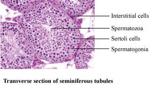
• Seminiferous tubule (transverse section).
See also
• Leydig cells
External links
• Histology image: 17802loa – Histology Learning System at Boston University
• Image
• Diagram