What does the calcaneus articulate with?
The calcaneus is located in the hindfoot with the talus and is the largest bone of the foot. It is commonly referred to as the heel. It articulates with the talus superiorly and the cuboid anteriorly and shares a joint space with the talocalcaneonavicular joint.
What ligament attaches to anterior process of calcaneus?
The bifurcate ligament attaches the anterior process of the calcaneus to the navicular and cuboid bones. Excessive traction to this ligament can result in a fracture of the anterior process of the calcaneus.
Which tarsal does the Achilles tendon attach?
Calcaneus. The largest of the tarsal bones, the calcaneus is cuboidal with an anteriorly directed long axis. The bulbous posterior process bears the attachment of the Achilles tendon.
What attaches to the posterior calcaneus?
The posterior surface of the calcaneus has a circular convex structure with three distinct facets. The middle facet serves as the attachment site for the calcaneal tendon (Achilles tendon), while the superior facet is separated from the calcaneal tendon by the retrocalcaneal bursa.
What is anterior process of calcaneus?
The anterior process of the calcaneus is a prominence on the heel bone (calcaneus) that is located in front and to the outside of the ankle (Figure 1). Fractures of the anterior process of the calcaneus occur following an acute injury to the foot.
Where does the calcaneus attach?
The calcaneus is the largest bone in the foot. It projects posterior to the tibia and fibula and acts as a short lever for the calf muscles (gastrocnemius and soleus) which insert onto its posterior surface via the Achilles tendon.
Is the calcaneal tendon The Achilles tendon?
What is an Achilles tendon? The Achilles (uh-KILL-ease) tendon is a band of tissue in the back of your leg. This tendon links your heel bone (calcaneus, pronounced cal-KAY-nee-us) to your calf muscles. It's also called the calcaneal tendon.
Why does my calcaneus hurt?
Common causes of heel pain include obesity, ill-fitting shoes, running and jumping on hard surfaces, abnormal walking style, injuries and certain diseases. Plantar fasciitis is inflammation of the ligament that runs the length of the foot, commonly caused by overstretching.
Where is the anterior process of the calcaneus?
The anterior process off the calcaneus is located at the anterior (front) portion of the heel bone. X-ray and ultrasound examination can show an anterior process of the calcaneum fracture. Ultrasound examination at The Foot and Ankle Centre can assist in diagnosing this problem.
What attaches to Sustentaculum Tali?
At the upper and forepart of the medial surface of the calcaneus is a horizontal eminence, the sustentaculum tali, which gives attachment to a slip of the tendon of the Tibialis posterior.
Where is bifurcate ligament located?
The bifurcate ligament is a strong band, attached behind to the deep hollow on the upper surface of the calcaneus and dividing in front in a Y-shaped manner into a calcaneocuboid and a calcaneonavicular part.
What is the function of bifurcate ligament?
The bifurcate ligament is a ligament that sits on the outside of your foot and assists in stabilising your midfoot. It is attached to your calcaneus (heel bone) with two arms in a Y shape: one that attaches to your cuboid bone, and the other to your navicular bone.
Where does calcaneal tendon tear occur?
calcaneal tendon rupture: occurs 2-6 cm proximal to the calcaneus; it occurs within the hypovascular watershed zone.
What is calcaneal tendinitis?
calcaneal tendinitis: is inflammation within the tendon itself, which may swollen with abnormal internal signal (MRI) calcaneal tendon ossification. calcaneal peritendinitis: inflammation in the structures surrounding the tendon, usually ≥5 cm above the heel.
What is the shape of the proximal fibers of the calcaneal tendon?
The proximal fibers of the calcaneal tendon have a rounded appearance that becomes relatively flat about four centimeters proximal to the insertion site. This alignment gives the tendon its spiral pattern that helps locomotion.
What is the term for a tendon that is weak and more susceptible to tear?
calcaneal tendinosis: gradual thickening of the tendon without visible inflammatory process; even though the tendon is thick, it is weak and more vulnerable to tear. calcaneal tendinopathy: is broad terminology that encompasses tendinitis and tendinosis. calcaneal enthesopathy.
Which tendon is enclosed in the synovial sheath?
All the tendons that cross the ankle are enclosed within their own synovial sheath except for the calcaneal tendon, which has no covering surrounding synovial sheath but a posterior paratenon (so there is no calcaneal synovitis, but rather tendinitis or peritendinitis instead 1 ).
Which tendon is the strongest?
The calcaneal tendon, commonly known as the Achilles tendon, is the strongest and largest tendon of the human body. It is also one of the commonest tendons to become injured due to its high biomechanical load but poor vascularity 2.
Who first used the term Achilles?
The oldest written record for the term Achilles being used to describe the calcaneal tendon was in 1693 by Flemish/Dutch anatomist Philip Verheyen (1648-1710) in his text Corporis Humani Anatomia 4.
What is the posterior aspect of the calcaneus?
The posterior aspect is rough and concavo-convex in shape. This convexity supports the fibroadipose tissue (Kager's fat pad) between the calcaneal tendon and the ankle joint.
What are the two bones that the calcaneus articulates with?
Role with the talus and cuboid. The calcaneus articulates with two bones: the talus and the cuboid. There are three articular surfaces for the talus. The largest is the slightly convex oval surface for the body of the talus.
What is the main band of the inferior extensor retinaculum?
the main band of the inferior extensor retinaculum. the stem of the bifurcate ligament. There is a prominent medial margin on the medial process of the calcaneal tuberosity which provides attachments for the superficial part of the flexor retinaculum and distally the plantar aponeurosis.
How many articulations does the Achilles tendon have?
Achilles tendon (medial view) There are two articulations (with three facets) with the talus and one saddle-shaped articulation with the cuboid. The sustentaculum tali is a horizontal shelf that arises from the anteromedial portion of the calcaneus.
Which ligament attaches to the medial margin of the sustentaculum tali?
Flexor retinaculum of the foot (posterior view) The plantar calcaneonavicular ligament attaches to the medial margin of the sustentaculum tali.
Which aspect of the calcaneus features many curves to accommodate the talus and the many different?
Anterior aspect. The front of the calcaneus features many curves to accommodate the talus and the many different tarsal bones, which lead to the metatarsals and phalanges. The back of the calacaneus is not as complex, featuring a tuberosity and a medial process.
What is the calcaneal sulcus?
The calcaneal sulcus provides attachments for the: interosseous, talocalcaneal, cervical ligaments and. medial root of the inferior extensor retinaculum. Calcaneal sulcus (superior view) There is also a non-articular area on the calcaneus, distal to the posterior talar facet which provides an attachment surface for:
Which ligaments attach to the calcaneus?
The calcaneus has a number of joint stabilizing ligaments attached to it, such as the calcaneofibular, talocalcaneal, calcaneocuboid, and calcaneonavicular ligaments. The plantar aponeurosis and long plantar ligament support the arch of the foot and attach to the calcaneus as well 4).
What is the calcaneus?
What is calcaneus. Calcaneus or heel bone, is one of seven tarsal bones that forms the heel of the foot. The weight of the body is carried primarily by the two largest tarsal bones, the talus, which articulates with the tibia and fibula superiorly, the strong calcaneus, which forms the heel of the foot. The tibia articulates with the talus ...
Why do calcaneus fractures cause bone to widen?
Because most calcaneus fractures cause the bone to widen and shorten, the goal of treatment is to restore the normal anatomy of the heel. In general, patients whose normal heel anatomy is restored have better outcomes. In most cases, recreating the normal heel anatomy involves surgery.
How long does it take for a calcaneus fracture to heal?
If your calcaneus fracture is severe, however, it may take from 1 to 2 years before recovery is complete. Patients whose x-rays show good healing and normal heel anatomy often have ongoing symptoms after treatment.
What part of the calf muscles touches the ground?
The thick tendon of the calf muscles attaches to the posterior surface of the calcaneus. The part of the calcaneus that touches the ground is the calcaneal tuberosity, and the medial, shelf like projection is the sustentaculum tali (supporter of the talus) or talar shelf.
How long does it take to recover from a fractured calcaneus?
If your calcaneus fracture is severe, however, it may take from 1 to 2 years before recovery is complete.
Why do you need a CT scan for calcaneus?
Because of the complex anatomy of the calcaneus, a CT scan is routinely ordered after a calcaneus bone fracture has been diagnosed on x-ray. A CT scan will produce a more detailed, cross-sectional image of your foot and can provide your doctor with valuable information about the severity of your fracture.
What is the function of tendons in the foot?
There are a number of tendons located in the foot and ankle all responsible for different ankle, foot and toe movements. Tendons also help to provide stability around the foot and ankle.
Which muscle is located on the back of the calf?
The two main calf muscles, gastrocnemius and soleus, run down the back of the calf and join together to form a strong, thick tendon, the Achilles tendon, that attaches to the back of the heel.
What Is Causing My Foot Tendon Pain?
Foot and ankle pain is often due to tendon injuries. The location of the pain will depend on which tendon is damaged
How many extensor tendons are there in the foot?
There are two sets of extensor tendons found in the foot from three muscles, extensor digitorum tendons and hallucis longus.
What causes a tendon to hurt?
Foot tendon pain may be due to: 1 Tendonitis: When the foot or ankle tendons become inflamed causing pain and swelling LEARN MORE > 2 Tendonosis: Wear and tear or degeneration of the foot tendon 3 Tendon Tears: Where the foot or ankle tendon partially or completely ruptures
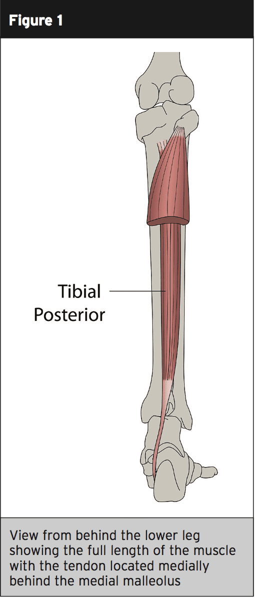
What is the deepest muscle in the back of the leg?
Tibialis posterior is the deepest muscle on the back of the leg. The tendon passes behind the inner ankle bone (medial malleolus) and underneath the foot attaching to the tarsal bones. The tibialis posterior tendon is the main invertor of the foot and also helps the calf muscles to plantarflex the foot.
Why are peroneal tendons important?
Functionally, they are very important for providing stability when running , particularly on uneven ground. RELATED ARTICLE: Peroneal Tendonitis - Causes, Symptoms & Treatment.
What is the condition where the tendon attaches to the heel bone?
Insertional Achilles tendonitis affects the lower portion of your tendon where it attaches to your heel bone.
How to treat Achilles tendonitis?
Many treatments are available for Achilles tendonitis, ranging from home remedies, like rest and anti-inflammatory medication, to more invasive treatments, like steroid injections, platelet-rich plasma (PRP) injections, and surgery. Your doctor might suggest:
How long does it take for Achilles tendonitis to heal?
Recovery and outlook from Achilles tendonitis. Tendonitis usually goes away after a few days, following rest and proper home treatment (including the RICE method). Recovery takes a lot longer if you continue to put pressure on the tendon or don’t change your exercise habits to prevent another injury or rupture.
What causes pain in the back of the heel?
tight calf muscles. limited range of motion when flexing your foot. skin on your heel overly warm to the touch. The main symptom of Achilles tendonitis is pain and swelling in the backside of your heel when you walk or run. Other symptoms include tight calf muscles and limited range ...
Why does my Achilles tendon hurt?
Some causes include: exercising without a proper warmup. straining the calf muscles during repeated exercise or physical activity. playing sports, such as tennis, that require quick stops and changes of direction.
How to stop swelling in Achilles tendon?
elevating your foot to decrease any swelling. wearing a brace or walking boot to prevent heel movement. going to physical therapy. taking anti-inflammatory medication, such as aspirin ( Bufferin) or ibuprofen ( Advil ), for a limited time. wearing a shoe with a built-up heel to take tension off your Achilles tendon.
What are the complications of Achilles tendonitis?
The most common complications of Achilles tendonitis are pain, having trouble walking or exercising, and your tendon or heel bone becoming deformed.
What is the shape of the appendicular bone?
This appendicular bone has a roughly triangular shape.
Which bone is weaker, spongy or compact?
The proximal epiphysis of the femur has a preponderance of spongy bone, which is weaker than compact bone. Bone density decreases with age. There is a reduced ability to balance that often accompanies old age. The proximal epiphysis of the femur has a preponderance of spongy bone, which is weaker than compact bone.
