What veins make up the superior vena cava?
The superior vena cava is formed by the left and right brachiocephalic veins—also referred to as the innominate veins—on the right side of the upper chest, posterior (behind) to the lower border of the first costal cartilage.
Where does the superior vena cava end and the Atrium end?
The superior vena cava ends at the junction of the superior vena cava and the left atrium, emptying into the upper aspect of the right atrium at the level of the third costal cartilage. 3 Anatomical variations of the superior vena cava include:
Why is the superior vena cava a valveless structure?
The superior vena cava is a valveless structure. This allows the pressure in the right atrium to be conducted upwards into the right internal jugular vein.
What are the tributaries of the superior vena?
Tributaries. The superior vena cava contains venous blood from the head, neck, both upper limbs and from structures within the thorax. It is formed by the union of the right and left brachiocephalic veins – which provide venous drainage of the head, neck, and upper limbs. At the level of T4, the superior vena cava receives the azygous vein, ...
Which two veins merge to form the subclavian veins?
CardsTerm Blood Circulation (6)Definition Heart → Arteries →Arterioles →Capillaries → Venules → Veins → HeartTerm What 2 veins merge to form the subclavian vein?Definition Axillary External JugularTerm What two veins constitute the brachiocephalics?Definition Subclavian Internal Jugular117 more rows•Apr 12, 2011
Which veins merge to form the inferior vena cava?
The inferior vena cava (IVC) is a large retroperitoneal vessel formed by the confluence of the right and left common iliac veins.
What forms the superior vena cava?
The right and left brachiocephalic, or innominate, veins converge to form the superior vena cava at the level of the right first costal cartilage. These veins form at the venous angle from a merging of the subclavian and jugular veins posterior to the sternoclavicular joints.
How is vena cava formed?
The inferior vena cava is formed by the coming together of the two major veins from the legs, the common iliac veins, at the level of the fifth lumbar vertebra, just below the small of the back.
Where is the superior vena cava located?
The superior vena cava is located in the thorax (chest), more specifically, it is in the anterior (front) right, ...
How big is the superior vena cava?
The superior vena cava is relatively large and measures .78 inches in diameter and 2.7 inches in length. 1 . All blood vessels—including veins and arteries—have basically the same structure. Namely, they are hollow tubes with a lumen (open inner space).
What is the role of the Vena Cava?
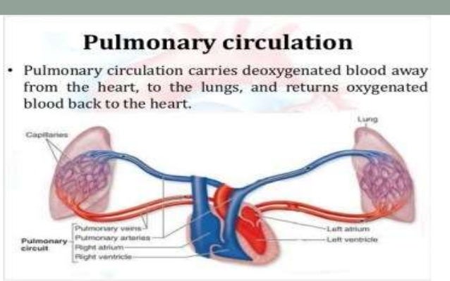
The vena cava plays a vital role in helping to circulate deoxygenated blood from the upper half of the body, draining it into the heart, so the blood can continue to the lungs to be reoxygenated.
What is the function of a vein?
Veins are tubular, hollow structures that form part of the circulatory system of the body; in most instances, veins carry deoxygenated blood toward the heart. The superior vena cava (SVC) is one of the two largest veins in the body and is considered one of the many systemic veins.
What is persistent left superior vena cava?
Persistent left superior vena cava (persistent LSVC): is a common anomaly of the systemic veins. It occurs most commonly with congenital heart disease. (CHD). 9 . Superior vena cava syndrome (SVCA): is a condition involving a compressed or partial blockage of the superior vena cava.
Which chamber of the heart drains blood from the head, eyes, neck, and upper limbs?
The superior vena cava is a vital structure in the human circulatory system that helps drain large amounts of deoxygenated blood from the head, eyes, neck, and upper limbs into the upper left chamber (atrium) of the heart.
Which muscle prevents blood from backing up into the superior vena cava during its contraction period?
The mechanism that prevents blood from backing up into the superior vena cava from the right atrium during its contraction period (called systole ) is part of a muscle that comprises the atrial walls, which wraps around the site of the entrance of the vena cava.
Why is the superior vena cava blocked?
Superior vena cava obstruction can occur either due to external compression or from an occlusion within the vessel lumen itself. The most common cause of SVC obstruction is malignancy, typically from lung cancer, lymphoma, or metastatic disease.
Which veins drain the head?
It is formed by the union of the right and left brachiocephalic veins – which provide venous drainage of the head, neck, and upper limbs. At the level of T4, the superior vena cava receives the azygous vein, which drains the upper lumbar region and thoracic wall.
Where does the SVC enter the pericardium?
At the level of the second costal cartilage, the SVC enters the middle mediastinum and becomes surrounded by the fibrous pericardium.
What causes a raised JVP?
Causes of a raised JVP include right-sided heart failure, pulmonary hypertension, and SVC obstruction. [end-clinical] Tributaries. The superior vena cava contains venous blood from the head, neck, both upper limbs and from structures within the thorax.