Why does my tibialis anterior hurt when I Walk?
swollen veins that are hard or sore when you touch them. Why does my tibialis anterior muscle hurt when I walk? Exertional compartment syndrome occurs when the sheath that contains your tibialis anterior muscle is too small. During exercise, as bloodflow to the muscle increases, the muscle swells up and presses against the sheath.
How to stretch the anterior tibial muscle?
- Stand up. ...
- Bend both knees slightly.
- One foot remains squarely on the ground. ...
- Keeping your toe firmly on the ground, pull the stretching leg forward so you feel a stretch from the top of your stretching foot through your shins.
- Once you feel a good stretch, hold it for 15 to 30 seconds.
- Repeat the stretch with the other foot.
What is the best treatment for posterior tibial tendonitis?
for at least three weeks.
- Single leg heel raise, building up gradually to 50 repeats
- Toe walking, starting at 8-10 yards and building up to 100 yards of continuous toe walking
- Balance board tapping: On a balance board, “tap” the board to the ground and return to a balanced position 20 times each for all five of the positions illustrated in ...
What causes tibia pain?
- Pain on the inside of the shin, usually on the lower third.
- Symptoms often occur after running long distances.
- When pressing in over the area your leg will feel tender and sore.
- You may even have swelling over the site of the fracture.
- If you have a stress fracture you may also feel a particularly tender spot at the exact point of the stress fracture.
Is the tibialis anterior a skeletal muscle?
The tibialis anterior muscle is a muscle in humans that originates along the upper two-thirds of the lateral (outside) surface of the tibia and inserts into the medial cuneiform and first metatarsal bones of the foot. It acts to dorsiflex and invert the foot....Tibialis anterior muscleFMA22532Anatomical terms of muscle14 more rows
What type of muscle is the tibialis posterior?
Relations. Tibialis posterior is the deepest and most central muscle in the posterior compartment of leg. It is located posterior to the tibia, fibula and interosseous membrane of leg. The latter separates tibialis posterior from the anterior leg muscles.
Is the tibialis anterior a flexor or extensor?
flexorThe tibialis anterior muscle is flexor, inverter (in addition to posterior tibial muscle) and adductor (in addition to the long extensor of hallux) of the foot. It also plays a role in suspension of the arch and controls supination of the rearfoot [10].
Is tibialis anterior concentric or eccentric?
Anterior Tibialis Function It shortens concentrically every time you bring your foot toward your shin and lengthens eccentrically right before you place your foot on the ground. Perhaps the eccentric function of anterior tibialis is why shin splints are so prominent in runners.
Is tibialis posterior a Pennate muscle?
The muscle fibers are arranged in pennate fashion (like a bird's feather), which means that it is very strong, but has only small range of functional movement. That small range of functional movement is a significant factor in the pathology of the Tibilais posterior.
Where is tibialis anterior muscle?
The tibialis anterior tendon (TAT) begins at the distal one-third of the tibia. It travels across the anterior ankle and dorsum of the foot to insert vertically on the medial cuneiform and the base of the first metatarsal. It is the most medial tendon of the ankle and foot.
What are the actions of the tibialis anterior quizlet?
Primary Action: Extension of the four lateral toes, assists with dorsiflexion of the foot at the ankle.
What is the shin muscle called?
tibialis anterior muscleThe tibialis anterior muscle is the muscle located in the front part of the shin bone of your lower leg. The muscle courses from an area just below your knee, down the front of your shin, and finally attaches to the top of your foot.
How does anterior tibialis work?
0:091:30Tibialis Anterior Exercises - YouTubeYouTubeStart of suggested clipEnd of suggested clipMaybe seven eight or ten pounds place that on top of your foot you can hold it for stability. AndMoreMaybe seven eight or ten pounds place that on top of your foot you can hold it for stability. And then just lift and lower your foot. Ten or fifteen times.
What is a eccentric muscle contraction?
Introduction. An eccentric (lengthening) muscle contraction occurs when a force applied to the muscle exceeds the momentary force produced by the muscle itself, resulting in the forced lengthening of the muscle-tendon system while contracting (Lindstedt et al., 2001).
What is an example of an eccentric contraction?
Eccentric contraction occurs when the total length of the muscle increases as tension is produced. For example, the lowering phase of a biceps curl constitutes an eccentric contraction. Muscles are capable of generating greater forces under eccentric conditions than under either isometric or concentric contractions.
What is concentric and eccentric muscle contraction?
In a concentric contraction, the muscle tension rises to meet the resistance then remains stable as the muscle shortens. During eccentric contraction, the muscle lengthens as the resistance becomes greater than the force the muscle is producing.
Is tibialis posterior a calf muscle?
The tibialis posterior muscle is the most central of all the leg muscles, and is located in the deep posterior compartment of the leg. It is the key stabilizing muscle of the lower leg....Tibialis posterior muscleInsertionNavicular and medial cuneiform boneArteryPosterior tibial arteryNerveTibial nerve11 more rows
What muscles are used to Dorsiflex the foot?
The tibialis anterior muscle, found in the anterior compartment of the leg, is the primary muscle that facilitates dorsiflexion of the ankle joint.
Which nerve innervates the tibialis anterior muscle?
Innervation. The deep fibular (peroneal) nerve (L4, L5), a branch of the common fibular nerve, innervates the tibialis anterior muscle.
What is the function of the tibialis anterior dorsiflex?
It plays an important role in the activities of walking, hiking and kicking the ball by stabilizing the ankle joint as the foot hits the floor and pull it clear of the ground as the leg continues moving.
How long does it take to read a tibialis?
Reading time: 4 minutes . Tibialis anterior muscle (Musculus tibialis anterior) Tibialis anterior is a fusiform muscle found in the anterior part of the leg. Lying superficially in the leg, this muscle is easily palpable lateral to the anterior border of tibia. Along with fibularis (peroneus) tertius, extensor digitorum longus ...
Which muscle tendon is located beneath the extensor retinaculum?
The tendon of tibialis anterior usually passes beneath the extensor retinaculum which holds it in place. However, in some cases, the superficial and deep layers of the extensor retinaculum form a separate tunnel for the muscle’s tendon.
Which muscle is the most medial?
Tibialis anterior muscle lies medial to extensor digitorum longus and extensor hallucis longus, which makes it the most medial muscle in the anterior compartment of the leg. It also covers the anterior tibial vessels and deep fibular nerve in the proximal part of the leg.
Which muscle is the main dorsiflexor of the foot?
Along with fibularis (peroneus) tertius, extensor digitorum longus and extensor hallucis longus, it comprises the anterior (or extensor) compartment of the leg . This muscle acts as the main foot dorsiflexor on the talocrural joint, but it also inverses the foot at the subtalar joint. Both actions play important roles in the gait cycle.
Which artery supplies the tendon?
The body of the muscle is entirely supplied by the branches of anterior tibial artery; anterior muscular, medial muscular branches and anterior tibial recurrent artery. The tendon is mainly supplied by the branches of anterior tibial artery but also by the branches of posterior tibial artery.
Where is the tibialis anterior located?
Treatments. The tibialis anterior muscle is the muscle located in the front part of the shin bone of your lower leg. The muscle courses from an area just below your knee, down the front of your shin, and finally attaches to the top of your foot.
How to help anterior tibialis?
A physical therapist may use various treatments to help improve the function and mobility of your anterior tibialis muscle. Typical treatments may include: Anterior tibialis stretching. Strengthening exercises for your anterior tibialis. Kinesiology tape 2 . Massage to the muscle. Neuromuscular electrical stimulation to help improve neuromuscular ...
Why is my anterior tibialis weak?
Your anterior tibialis muscle may also become weak if you suffer a sprained ankle or ankle fracture that requires a long period of immobilization. 4 . If you are having weakness, pain, or tightness in your lower leg or shin, you may benefit from a visit to your physician or physical therapist to assess your specific situation. ...
Which muscle attaches to the top of the foot?
Since the anterior tibial muscle attaches to the top of your foot, it also helps to raise the arch of your foot . Hero Images / Getty Images.
What muscle is used to pull your foot in?
Function of the Tibialis Anterior Muscle. Your anterior tibialis muscle serves to help flex your ankle and foot off the ground, as occurs when tapping your foot. The muscle also helps to pull your foot in, a motion called inversion.
What is the tibialis anterior muscle?
Anatomical terms of muscle. The tibialis anterior muscle is a muscle in humans that originates along the upper two-thirds of the lateral (outside) surface of the tibia and inserts into the medial cuneiform and first metatarsal bones of the foot. It acts to dorsiflex and invert the foot.
Where is the tibialis anterior?
The tibialis anterior overlaps the anterior tibial vessels and deep peroneal nerve in the upper part of the leg.
What is the function of the tibialis anterior?
It also functions to 'lock' the ankle, as in toe-kicking a ball, when held in an isometric contraction. Antagonists are plantar-flexors of the posterior compartment such as soleus and gastrocnemius . The movements of tibialis anterior are dorsiflexion and inversion of the ankle.
What muscle is responsible for keeping the leg vertical when walking on uneven ground?
However, actions of tibialis anterior are dependent on whether the foot is weight bearing or not (closed or open kinetic chain). When the foot is on the ground, the muscle helps to balance the leg and talus on the other tarsal bones so that the leg is kept vertical even when walking on uneven ground.
Which muscle is responsible for dorsiflexing and inverting the foot?
The tibialis anterior muscle is the most medial muscle of the anterior compartment of the leg. It is responsible for dorsiflexing and inverting the foot, and is the largest dorsiflexor of the foot.
What muscle is the dorsiflex of the foot?
Flexor muscle in humans that dorsiflexes the foot. Tibialis anterior muscle. Tibialis anterior. Animation. Details. Pronunciation. / ˌtɪbiˈeɪlɪs / or / ˌtɪbiˈælɪs /. Origin. From the upper 1/2 or 2/3 of the lateral surface of the tibia and the adjacent interosseous membrane.
Which antagonist of the tibialis anterior is most accurate?
However, the most accurate antagonist of the tibialis anterior is the peroneus longus. The tibialis anterior aides in the activities of walking, running, hiking, kicking a ball, or any activity that requires moving the leg or keeping the leg vertical.
Where is the tibialis anterior located?
The Tibialis anterior (Tibialis anticus) is situated on the lateral side of the tibia; it is thick and fleshy above, tendinous below. The fibers run vertically downward, and end in a tendon, which is apparent on the anterior surface of the muscle at the lower third of the leg.
What is the function of the tibialis anterior?
Function. Tibialis anterior is the primary dorsiflexor of the ankle with synergistic action of extensor hallicus longus, extensor digitorium longus and peroneous tertius. Inversion of the foot. Adduction of the foot. Contributor of maintaining the medial arch of the foot.
Which muscles are underactive?
Integrated Anatomy. Tibialis anterior is one of the muscles that tend to be inhibited and underactive this leads to overactive of synergistic muscles; extensor hallicus longus, extensor digitorium longus and peroneous tertius.
Which muscle is stronger, the tibialis or the dorsiflexor?
The action of the tibialis anterior muscle is considerably stronger than that of the other three dorsiflexor muscles of the foot.
What is the term for pain along the path of a muscle?
Pain along the path of this muscle is often referred to as " Shin splints ". Also called medial tibial stress syndrome (MTSS)
Where is the Tibiofascialis anterior?
The Tibiofascialis anterior, a small muscle from the lower part of the tibia to the transverse or cruciate crural ligaments or deep fascia.
How to palpate the distal tendon?
Palpate the distal tendon by strumming perpendicular across it. Continue palpating the tibialis anterior proximally to lateral tibial condyle by strumming perpendicular to the fibers.
Where is the tibialis anterior muscle located?
Location of the Tibialis Anterior Muscle. The tibialis anterior muscle is a lower leg muscle that lies anteriorly, as the name suggests. In other words, this muscle lies on the anterior portion (compartment) of the lower leg on the lateral side of the tibia. The tibia is the larger, stronger, and anterior ...
What is the function of the tibialis anterior?
Functions of the Tibialis Anterior. This lower leg muscle is responsible for: dorsiflexion (flexing the ankle so that the toes move toward the shins) and. inversion of the ankle (movement of the sole towards the median plane) stabilizing the ankle during foot contact with the ground. Dorsiflexion is an important movement for walking, jogging, ...
What is a shin splint?
Shin Splints or Tibial Stress Syndrome. Common term for pain in the anterior compartment of the leg caused by irritation of the tibialis anterior muscle as might follow extreme or unusual exercise without adequate prior conditioning.
How to tell the border between the tibialis anterior and the adjacent extensor digitorum?
To clearly discern the border between the tibialis anterior and the adjacent extensor digitorum longus (EDL), use inversion and eversion. Inversion will engage the tibialis anterior but not the EDL; eversion will engage the EDL but not the tibialis anterior.
What are the muscles of the lower leg?
Three lower leg muscles are particularly well known. The first two are the calf muscles: gastrocnemius and the soleus. They are without any doubt the most powerful muscles in the lower leg. The third one is the tibialis anterior muscle. Other lower leg muscles include: tibialis posterior (ankle inversion); peroneus longus and peroneus brevis (ankle ...
What muscles are involved in the ankle?
This lower leg muscle is responsible for: 1 dorsiflexion (flexing the ankle so that the toes move toward the shins) and 2 inversion of the ankle (movement of the sole towards the median plane) 3 stabilizing the ankle during foot contact with the ground
Which nerve innervates the tibialis anterior muscle?
The tibialis anterior muscle is innervated by the deep peroneal nerve, a branch of the common peroneal nerve.
Where does the tibialis anterior muscle originate?
The tibialis anterior muscle lies in the anterior compartment of the leg and originates from the proximal lateral tibial metaphysis and proximal two thirds of the tibial shaft and interosseous membrane. The tendon twists and crosses the extensor hallucis longus tendon at the level of the ankle at which it enters a synovial tendon sheath. The tendon courses dorsomedially across the foot and rotates 90 degrees from the myotendinous junction to the broad insertion.18 The majority of tendons insert at the plantar medial border of the first metatarsal and medial cuneiform. Approximately 10% of tendons have variations to the insertion, the most common being a bifid insertion to the cuneiform and first metatarsal, or insertions reaching proximally or distally along the medial column of the foot. 19,20 Accessory tibialis anterior tendons have been reported, but are rare and have not been reported to cause pathology.
Where is the tibialis anterior?
The tibialis anterior is bounded proximally to the ankle by the superior extensor retinaculum, and variably may enter a synovial tendon sheath within the retinacular fibers at this level. More distally, the tibialis anterior tendon predictably enters a synovial sheath as it passes into the inferior extensor retinaculum complex. An early study using a modified Spaltehoz technique failed to reveal any zones of hypovascularity within the tendon, 21 but a later study using immunohistochemical methods suggests that such a zone exists within the inferior retinacular system where tendon rupture is most likely to occur. 22,23
What is the largest dorsiflexor muscle?
This injury is common in running and jumping. The tibialis anterior muscle is the largest of the dorsiflexor muscles. It originates from the lateral condyle of the tibia and inserts into the medial and plantar surfaces of the medial cuneiform bone. The tibialis anterior muscle is responsible for ankle dorsiflexion and inversion of the foot. Its innervation is supplied by the deep peroneal nerve (L4 and L5).
What is the function of the tibialis anterior?
Its primary function is to decelerate the foot during the initial plantarflexion that occurs immediately following heel strike, and to clear the foot during toe-off. Absence of this muscle in active individuals is poorly tolerated, causing a slapping of the foot during heel strike and a steppage gait, with difficulty clearing the foot during swing.
What muscle is the third toe?
Stiffness and spasms of the third toe and cramping in the tibialis anterior muscle.
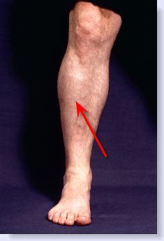
What causes pain on the medial side of the tibia?
Repetitive stress on the muscle causes inflammation, swelling, and a dull, aching pain on the medial side of the tibia.
Where is the LIV-4 muscle located?
LIV-4 is located between the tendon and the prominence of the medial malleolus, in a depression over the palpable joint space.
What is the tibialis anterior muscle?
What is Tibialis Anterior Muscle and What is its Function? [1-2] The Tibialis Anterior Muscle is located on the anterior compartment of the leg. Anterior compartment consists of 4 muscles and Tibialis anterior muscle is the largest in size among these four muscles.1 The tibialis anterior muscle is the strongest dorsiflexor and helps to lift ...
What is the Treatment for Tibialis Anterior Muscle Strain?
The pain and muscle inflammation of the Tibialis Anterior Muscle Strain responds to rapid conservative therapy. The severe pain and muscle strain associated with muscle tear may not respond to conservative therapy. The MRI and ultrasound images help to differentiate the type of injury such as presence or absence of muscle tear. Conservative treatment is recommended for sprain that is not associated with a muscle tear.
What causes medial tibial stress syndrome?
Injury or stress of tibialis anterior muscle causes medial tibial stress syndrome.4 Injury resulting in tibialis anterior muscle strain is observed after fall, repeated use of lower leg and direct impact over the muscle by moving object. Tibialis Anterior Muscle Strain or Injury causes the following symptoms:
What happens if you strain your tibialis anterior muscle?
Any type of Strain or Injury to the Tibialis Anterior Muscle will result in the inability of the patient to flex the foot and extend the toes resulting in difficulty with ambulation. An injury to this muscle can also put excess pressure on the ankles resulting in them getting weak causing imbalance when walking.
Why is physical therapy recommended for muscle atrophy?
Physical therapy is recommended for pain and prevents muscle stiffness during the initial phase. In the later phase of the disease, once the pain is less severe than physical therapy is advised for muscle atrophy and muscle weaknesses.
Can running on uneven surfaces cause tibialis?
Running or walking on uneven surfaces may result in a Tibialis Anterior Muscle Strain.
Can a muscle tear be conservative?
The severe pain and muscle strain associated with muscle tear may not respond to conservative therapy. The MRI and ultrasound images help to differentiate the type of injury such as presence or absence of muscle tear. Conservative treatment is recommended for sprain that is not associated with a muscle tear.

Overview
Clinical significance
Some clinicians attempt to treat tibialis anterior muscle issues with acupuncture techniques, such as dry needling. There is significant bias in studies evaluating the efficacy of acupuncture versus medical treatments, and the decision to use acupuncture should be made carefully.
A tibialis anterior hernia is a rare type of hernia in which fat or other material protrudes through a defect in the tibialis anterior muscle. It may be caused by trauma, such as an inadvertent kick to …
Structure
The tibialis anterior muscle arises from:
• the lateral condyle of the tibia.
• the upper 2/3 of the lateral surface of the tibia.
• the adjoining part of the interosseous membrane.
Function
The tibialis anterior muscle is the most medial muscle of the anterior compartment of the leg. It is responsible for dorsiflexing and inverting the foot, and is the largest dorsiflexor of the foot. The muscle has two origins, one being the lateral tibial condyle and the other being the upper lateral surface of the tibia, and inserts on the medial surface of the medial cuneiform and adjoining part of base of the first metatarsal of the foot allowing the toe to be pulled up and held in a locked po…
Additional images
medial view of dissected ankle has two muscles
• Lateral aspect of right leg.
• Tibialis anterior muscle
• Cross-section through top third and second third of right leg.
See also
• Tibialis posterior muscle
External links
• Anatomy photo:15:st-0415 at the SUNY Downstate Medical Center
• Tibialis Anterior from Wheeless' Textbook of Orthopaedics
Description

Origin
- It arises from: 1. Lateral condyle and upper half or two-thirds of the lateral surface of the body of the tibia 2. Adjoining part of the interosseous membrane 3. Deep surface of the fascia 4. Intermuscular septum between it and the Extensor digitorum longus. Image: Tibialis anterior (highlighted in green) - anterior view
Function
- Tibialis anterior is the primary dorsiflexor of the ankle with synergistic action of extensor hallicus longus, extensor digitorium longus and peroneous tertius.
- Inversion of the foot.
- Adduction of the foot.
- Contributor of maintaining the medial arch of the foot.
Integrated Anatomy
- Tibialis anterior is one of the muscles that tend to be inhibited and underactive this leads to overactive of synergistic muscles; extensor hallicus longus, extensor digitorium longus and peroneous tertius. People with an inhibited or weak Tibialis anterior, for example those with hemiplegia or parkinson's, will have an abnormality in their anticipatory postural adjustment (AP…
Clinical Relevance
- Pain along the path of this muscle is often referred to as "Shin splints". Also called medial tibial stress syndrome (MTSS)
Assessment
- Palpation
The client is supine. Place your resistance hand on the medial side of the distal foot. Resist the client from dorsiflexing and inverting the foot. Look the distal tendon of the tibialis anterior on the medial side of the ankle joint and foot; it is usually visible. Palpate the distal tendon by strummin… - Power
The action of the tibialis anterior muscle is considerably stronger than that of the other three dorsiflexor muscles of the foot.