
Functions of MHC class I:
- Major function of MHC-I is to bind peptide antigens and present to CD8+ T cells (T helper cells)
- CD8 T cells are specific for MHC-I antigen
- MHC-I binds endogenous antigen and present to T helper cells.
- MHC-I molecules are found on surface of all nucleated cells.
What are the MHC class 1 molecules?
Structure of MHC Class-I Molecule :
- Extracellular segment,
- Transmembrane segment,
- Cytoplasmic tail segment.
What the Heck is a MHC molecule?
The major histocompatibility complex (MHC) is a large locus on vertebrate DNA containing a set of closely linked polymorphic genes that code for cell surface proteins essential for the adaptive immune system.These cell surface proteins are called MHC molecules.. This locus got its name because it was discovered via the study of transplanted tissue compatibility.
What with MHC molecules are associated?
The prime histocompatibility complex is a set of cell surface for recognition by the suitable T-cells. MHC molecules liaise interactions of leukocytes, which are also called white blood cells, which are immune cells.
What cells express MHC class II molecules?
MHC II molecules are expressed on immune cells such as B cells, monocytes, macrophages and dendritic cells and on epithelial cells following inflammatory signals, while MHC I molecules are expressed more ubiquitously.

What cells are MHC found on?
MHC class I molecules are one of two primary classes of major histocompatibility complex (MHC) molecules (the other being MHC class II) and are found on the cell surface of all nucleated cells in the bodies of vertebrates. They also occur on platelets, but not on red blood cells.
Where are MHC antigens found?
chromosome 6The major histocompatibility antigens are located on chromosome 6, and red cells antigens are located on a number of other chromosomes. For this reason, HLA-compatible donors may differ for red cell antigens including ABO, Rh, and other potentially hemolytic antigens.
Is MHC found in humans?
The Major Histocompatibility complex (MHC) system known as the human leukocyte antigen (HLA) in humans is located on the short arm of chromosome 6 (6p21. 3) and contains the most polymorphic gene cluster of the entire human genome.
Why MHC is formed in immune system?
This is an important process in the immune system response for destroying invading pathogens. The MHC complex on the cell surface is necessary for cell self-recognition and the prevention of the immune system targeting its own cells.
How many MHC molecules are present in human?
Thus, with three MHC class I genes and a possible four sets of MHC class II genes on each chromosome 6, a human typically expresses six different MHC class I molecules and eight different MHC class II molecules on his or her cells.
What is MHC and its function?
MHC is the tissue-antigen that allows the immune system (more specifically T cells) to bind to, recognize, and tolerate itself (autorecognition). MHC is also the chaperone for intracellular peptides that are complexed with MHCs and presented to T cell receptors (TCRs) as potential foreign antigens.
Do all cells have MHC 1?
Classical MHC class I molecules are ubiquitously expressed on all mammalian cells including cells of epithelial origin while MHC class II molecules are selectively expressed on antigen-presenting cells (APC) including dendritic cells (DC), macrophages, and B cells.
Do red blood cells have MHC?
red blood cells do not have noticeable MHC 1 molecules. But rather the have high levels of CD47 markers which are recognized by the immune system as self. On the other hand red blood cells during development are nucleated they may contain small traces of MHC 1 molecules.
Why are T cells MHC restricted?
The biological reason of MHC restriction is to prevent supernumerary wandering lymphocytes generation, hence energy saving and economy of cell-building materials. T-cells are a type of lymphocyte that is significant in the immune system to activate other immune cells.
What does MHC interact with?
Intracellular tumor-related antigens can be presented as peptides in the major histocompatibility complex (MHC) on the cell surface, which interact with the T cell receptors (TCR) on antigen-specific T cells to stimulate an anti-tumor response.
What are the functions of MHC I and MHC II?
Major histocompatibility complex (MHC) class I and class II proteins play a pivotal role in the adaptive branch of the immune system. Both classes of proteins share the task of presenting peptides on the cell surface for recognition by T cells.
What would happen to a person who fails to make MHC molecules?
What would happen to a person who failed to make MHC molecules? ?? An acquired immune response will not develop in the absence of MHC molecules, which are required for antigen presentation to activate B and T lymphocytes.
What is the difference between MHC 1 and MHC 2?
MHC class 1 molecules present endogenous antigens to cytotoxic T cells. MHC class 2 molecules present exogenous antigens to helper T cells. Thus, the main difference between MHC class 1 and 2 molecules is the type of antigens presented by each type of MHC molecules.
What is the difference between MHC and HLA?
The main difference between the two groups is that MHC is often found in vertebrates, while HLA is only found in humans. To simplify, HLA is the human body's version of MHC. Part of the responsibility of these antigens is to detect cells entering the body.
How were MHC molecules discovered and what is their function?
Major histocompatibility complex (MHC) molecules are cell surface proteins that present antigenic peptides to T cells. Human MHC proteins are called HLA molecules. They were initially discovered as products of polymorphic genes that mediate transplant rejection. Their physiologic function is antigen presentation.
Why are T cells MHC restricted?
The biological reason of MHC restriction is to prevent supernumerary wandering lymphocytes generation, hence energy saving and economy of cell-building materials. T-cells are a type of lymphocyte that is significant in the immune system to activate other immune cells.
What is the MHC in vertebrate DNA?
( PDB: 1hsa ) The major histocompatibility complex ( MHC) is a large locus on vertebrate DNA containing a set of closely linked polymorphic genes that code for cell surface proteins essential for the adaptive immune system .
Which receptors do MHC molecules bind to?
Antigen presentation: MHC molecules bind to both T cell receptor and CD4 / CD8 co-receptors on T lymphocytes, and the antigen epitope held in the peptide-binding groove of the MHC molecule interacts with the variable Ig-Like domain of the TCR to trigger T-cell activation
Why is MHC diversity important?
MHC diversity has also been suggested as a possible indicator for conservation, because large, stable populations tend to display greater MHC diversity, than smaller, isolated populations. Small, fragmented populations that have experienced a population bottleneck typically have lower MHC diversity. For example, relatively low MHC diversity has been observed in the cheetah ( Acinonyx jubatus ), Eurasian beaver ( Castor fiber ), and giant panda ( Ailuropoda melanoleuca ). In 2007 low MHC diversity was attributed a role in disease susceptibility in the Tasmanian devil ( Sarcophilus harrisii ), native to the isolated island of Tasmania, such that an antigen of a transmissible tumor, involved in devil facial tumour disease, appears to be recognized as a self antigen. To offset inbreeding, efforts to sustain genetic diversity in populations of endangered species and of captive animals have been suggested.
What is the MHC in mice called?
However, historically, the MHC in mice is called the Histocompatibility system 2 or just the H-2, in rats - RT1, and in chicken - B-locus.
How many genes are in the MHC locus?
Usual MHC contains about a hundred genes and pseudogenes, not all of them are involved in immunity. In humans, the MHC region occurs on chromosome 6, between the flanking genetic markers MOG and COL11A2 (from 6p22.1 to 6p21.3 about 29Mb to 33Mb on the hg38 assembly), and contains 224 genes spanning 3.6 mega base pairs (3 600 000 bases). About half have known immune functions. The human MHC is also called the HLA ( human leukocyte antigen) complex (often just the HLA). Similarly, there is SLA (Swine leukocyte antigens), BoLA (Bovine leukocyte antigens), DLA for dogs, etc. However, historically, the MHC in mice is called the Histocompatibility system 2 or just the H-2, in rats - RT1, and in chicken - B-locus.
What is the role of MHC in TCRs?
Tissue allorecognition: MHC molecules in complex with peptide epitopes are essentially ligands for TCRs. T cells become activated by binding to the peptide-binding grooves of any MHC molecule that they were not trained to recognize during positive selection in the thymus .
How did the MHC locus get its name?
This locus got its name because it was discovered via the study of transplanted tissue compatibility. Later studies revealed that tissue rejection due to incompatibility is an experimental artifact masking the real function of MHC molecules: binding an antigen derived from self-proteins, or from pathogens, and bringing the antigen presentation to the cell surface for recognition by the appropriate T-cells. MHC molecules mediate the interactions of leukocytes, also called white blood cells (WBCs), with other leukocytes or with body cells. The MHC determines donor compatibility for organ transplant, as well as one's susceptibility to autoimmune diseases .
What is the class of MHC molecules?
There are two classes of MHC molecules: MHC-I and MHC-II.
What does MHC do to T lymphocytes?
MHC molecules enable T-lymphocytes to recognize epitopes of antigens and discriminate self from non-self. Unlike B-cell receptors on B-lymphocytes that are able to directly bind epitopes on antigens, the T-cell receptors (TCRs) of T-lymphocytes can only recognize epitopes - typically short chains of amino acids called peptides - after they are ...
What is the most polymorphic gene in the human genome?
The MHC genes are the most polymorphic genes in the human genome, possessing many alleles for each gene. The MHC genes are co-dominantly expressed so that an individual expresses the alleles inherited from each parent. In this way, the number of MHC molecules that bind peptide for presentation to T-lymphocytes is maximized. In addition, each MHC molecule is able to bind a wide variety of different peptides, both self-peptides and foreign peptides. There are two classes of MHC molecules: MHC-I and MHC-II.
Where are antigens engulfed?
Antigens are engulfed by dendritic cells and placed in a phagosome. Some of the proteins escape from the phagosome into the cytosol of the dendritic cell where they become endogenous antigens.
Where are endogenous antigens located?
Figure 12.3 A. 3: Binding of Peptide Epitopes from Endogenous Antigens to MHC-I Molecules by a Dendritic Cell.Endogenous antigens are those located within the cytosol of the cells of the body. Examples include: a. viral proteins produced during viral replication, b. proteins produced by intracellular bacteria such as Rickettsias and Chlamydias during their replication, c. proteins that have escaped into the cytosol from the phagosome of phagocytes such as antigen-presenting cells, d. tumor antigens produced by cancer cells, e. and self peptides from human cell proteins.
Which peptides are transported by the Golgi complex?
The Golgi complex, in turn, transports the MHC-I/peptide complexes by way of an exocytic vesicle to the cytoplasmic membrane where they become anchored. Here, the peptide and MHC-I/peptide complexes can be recognized by naive T8-lymphocytes by way of TCRs and CD8 molecules having a complementary shape.
Where are peptides transported?
The peptides are transported into the rough endoplasmic reticulum (ER) by a transporter protein called TAP. The peptides then bind to the grooves of newly synthesized MHC-I molecules. The endoplasmic reticulum transports the MHC-I molecules with bound peptides to the Golgi complex.
Where does MHC move?
MHC and bound peptide move in Golgi to cell surface where they interact with CD8+
How many combinations of MHC are there?
MHC molecules coded by multiple independent genes, more than 1 loci for each protein, 36 combinations for Class II, 6 for Class I
Which class of MHC might activate CD8+?
MHC Class I and peptide might activate CD8+
Which cells are CD8+?
CD8+ cells, expressed on all nucleated cells, thymic epithelial cells
How many subregions are there in each set of genes?
3, each set of genes has 2 subregions which code for 2 different chains (a, b)
Where are MHC class I molecules found?
MHC class I molecules are one of two primary classes of major histocompatibility complex (MHC) molecules (the other being MHC class II) and are found on the cell surface of all nucleated cells in the bodies of vertebrates. They also occur on platelets, but not on red blood cells. Their function is to display peptide fragments of proteins from within the cell to cytotoxic T cells; this will trigger an immediate response from the immune system against a particular non-self antigen displayed with the help of an MHC class I protein. Because MHC class I molecules present peptides derived from cytosolic proteins, the pathway of MHC class I presentation is often called cytosolic or endogenous pathway.
How does MHC class 1 molecule get to the cell surface?
Once the peptide is loaded onto the MHC class I molecule, the complex dissociates and it leaves the ER through the secretory pathway to reach the cell surface. The transport of the MHC class I molecules through the secretory pathway involves several posttranslational modifications of the MHC molecule. Some of the posttranslational modifications occur in the ER and involve change to the N-glycan regions of the protein, followed by extensive changes to the N-glycans in the Golgi apparatus. The N-glycans mature fully before they reach the cell surface.
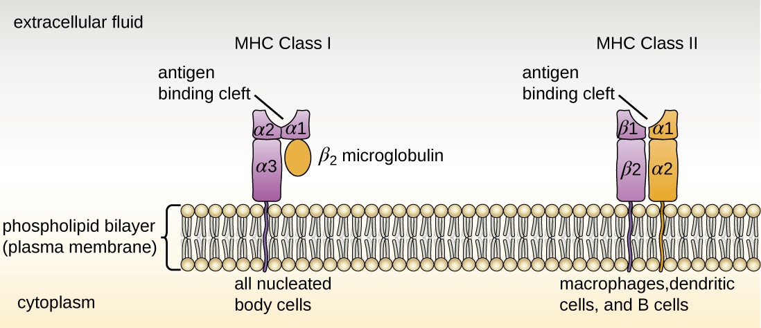
What are the two peptides in a class I MHC?
MHC class I molecules are heterodimers that consist of two polypeptide chains, α and β 2 -microglobulin (B2M). The two chains are linked noncovalently via interaction of B2M and the α 3 domain. Only the α chain is polymorphic and encoded by a HLA gene, while the B2M subunit is not polymorphic and encoded by the Beta-2 microglobulin gene. The α 3 domain is plasma membrane-spanning and interacts with the CD8 co-receptor of T-cells. The α 3 -CD8 interaction holds the MHC I molecule in place while the T cell receptor (TCR) on the surface of the cytotoxic T cell binds its α 1 -α 2 heterodimer ligand, and checks the coupled peptide for antigenicity. The α 1 and α 2 domains fold to make up a groove for peptides to bind. MHC class I molecules bind peptides that are predominantly 8-10 amino acid in length (Parham 87), but the binding of longer peptides have also been reported.
Why are trans-species polymorphisms common in MHC class I genes?
There are, however, documented cases of trans-species polymorphisms in MHC class I genes, where a particular allele in an evolutionary related MHC class I gene remains in two species, likely due to strong pathogen-mediated balancing selection by pathogens that can infect both species.
What is the function of class I MHC?
Thus, the function of the class I MHC is to display intracellular proteins to cytotoxic T cells (CTLs). However, class I MHC can also present peptides generated from exogenous proteins, in a process known as cross-presentation .
Where are MHC class I peptides removed from the ER?
Peptides that fail to bind MHC class I molecules in the lumen of the endoplasmic reticulum (ER) are removed from the ER via the sec61 channel into the cytosol, where they might undergo further trimming in size, and might be translocated by TAP back into ER for binding to a MHC class I molecule.
Which transporter is responsible for peptide translocation?
The peptide translocation from the cytosol into the lumen of the ER is accomplished by the transporter associated with antigen processing (TAP). TAP is a member of the ABC transporter family and is a heterodimeric multimembrane-spanning polypeptide consisting of TAP1 and TAP2. The two subunits form a peptide binding site and two ATP binding sites that face the cytosol. TAP binds peptides on the cytoplasmic side and translocates them under ATP consumption into the lumen of the ER. The MHC class I molecule is then, in turn, loaded with peptides in the lumen of the ER.
What is the molecule of MHC II?
MHC-II molecules are dimers consisting of a 133 KDa α-chain and 28KDa β-chain which are associated by non-covalent interactions.
Where are MHC class II proteins found?
Unlike class I proteins, they have a restricted tissue distribution and are chiefly found on macrophages, dendritic cells, B cells, and other Antigen Presenting Cells ( APCs) only.
What is the purpose of the II in MHC?
The Ii prevents endogenous peptides from binding to the groove of MHC class II molecules. After removal of Ii in the acidic endosomal compartments, peptides are able to bind to the MHC groove. A particular peptide exhibiting immunodominance loads onto MHC class II molecules.
What is the major histocompatibility complex?
Major Histocompatibility Complex (MHC) is a part of the genome of all vertebrates that code for molecules which are important in immune recognition. In humans, the MHC is a cluster of genes located on chromosome 6 which code for MHC proteins also called Human Leukocyte Antigen ...
Why is MHC class II important?
Since they sample and present antigens from exogenous sources, MHC class II molecules are critical for the initiation of the antigen-specific immune response.
What is the function of MHC proteins?
MHC proteins are a set of cell surface proteins essential for the acquired immune system to recognize foreign molecules which in turn determines histocompatibility.
Which class of MHC has a 1 and 2 subunit?
Class II MHC molecules have β1 and β2 subunits and thus can be recognized by CD4 co-receptors.
Do mhcs recognize autoantigens?
They are able to recognize MHC and do not recognize autoantigens
Do red blood cells produce MHC?
red blood cells do not produce MHC and, therefore, do not display the fact that they have been infected by presenting antigen.
Overview
The major histocompatibility complex (MHC) is a large locus on vertebrate DNA containing a set of closely linked polymorphic genes that code for cell surface proteins essential for the adaptive immune system. These cell surface proteins are called MHC molecules.
This locus got its name because it was discovered via the study of transplante…
Discovery
The first descriptions of the MHC were made by British immunologist Peter Gorer in 1936. MHC genes were first identified in inbred mice strains. Clarence Little transplanted tumors across different strains and found rejection of transplanted tumors according to strains of host versus donor. George Snell selectively bred two mouse strains, attained a new strain nearly identical to one of the progenitor strains, but differing crucially in histocompatibility—that is, tissue compatibili…
Genes
The MHC locus is present in all jawed vertebrates, it is assumed to have arisen about 450 million years ago. Despite the difference in the number of genes included in the MHC of different species, the overall organization of the locus is rather similar. Usual MHC contains about a hundred genes and pseudogenes, not all of them are involved in immunity. In humans, the MHC region occurs on chromosome 6, between the flanking genetic markers MOG and COL11A2 (from 6p22.1 to 6p21.…
Proteins
MHC class I molecules are expressed in all nucleated cells and also in platelets—in essence all cells but red blood cells. It presents epitopes to killer T cells, also called cytotoxic T lymphocytes (CTLs). A CTL expresses CD8 receptors, in addition to T-cell receptors (TCR)s. When a CTL's CD8 receptor docks to a MHC class I molecule, if the CTL's TCR fits the epitope within the MHC class I mole…
Antigen processing and presentation
Peptides are processed and presented by two classical pathways:
• In MHC class II, phagocytes such as macrophages and immature dendritic cells take up entities by phagocytosis into phagosomes—though B cells exhibit the more general endocytosis into endosomes—which fuse with lysosomes whose acidic enzymes cleave the uptaken protein into many different peptides. Via physicoc…
T lymphocyte recognition restrictions
In their development in the thymus, T lymphocytes are selected to recognize MHC molecules of the host, but not recognize other self antigens. Following selection, each T lymphocyte shows dual specificity: The TCR recognizes self MHC, but only non-self antigens.
MHC restriction occurs during lymphocyte development in the thymus through a process known as positive selection. T cells that do not receive a positive survival signal — mediated mainly by t…
In sexual mate selection
MHC molecules enable immune system surveillance of the population of protein molecules in a host cell, and greater MHC diversity permits greater diversity of antigen presentation. In 1976, Yamazaki et al demonstrated a sexual selection mate choice by male mice for females of a different MHC. Similar results have been obtained with fish. Some data find lower rates of early pregnancy loss in human couples of dissimilar MHC genes.
Evolutionary diversity
Most mammals have MHC variants similar to those of humans, who bear great allelic diversity, especially among the nine classical genes—seemingly due largely to gene duplication—though human MHC regions have many pseudogenes. The most diverse loci, namely HLA-A, HLA-B, and HLA-C, have roughly 6000, 7200, and 5800 known alleles, respectively. Many HLA alleles are ancient, sometimes of closer homology to a chimpanzee MHC alleles than to some other human …