Where are synaptic vessels located?
Types. In the central nervous system, a synapse is a small gap at the end of a neuron that allows a signal to pass from one neuron to the next. Synapses are found where nerve cells connect with other nerve cells. Synapses are key to the brain's function, especially when it comes to memory. 1 .
Where is synapse vesicle located?
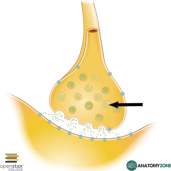
Synaptic Vesicle. The structure indicated is a synaptic vesicle located in a presynaptic bouton. Synaptic vesicles are structures found at synaptic terminals which contain various different types of neurotransmitter, the chemicals which mediate neurotransmission.Synaptic vesicles are contained within pools within the nerve terminal.
Where are secretory vesicles produced?
- Mitochondria: Synthesis of macromolecules and exocytosis takes ATP. So there must be many mitochondria.
- Endoplasmic reticulum: Rough ER modifies polypeptides and smooth ER synthesizes lipids. It depends on what is being secreted.
- Golgi Body: Further modification of proteins, including the synthesis of glycoproteins and lipoproteins.
Where do the vesicles originate that are involved in secretion?
Where do secretory vesicles originate? Secretory vesicles form from the trans Golgi network, and they release their contents to the cell exterior by exocytosis in response to extracellular signals.The secreted product can be either a small molecule (such as histamine) or a protein (such as a hormone or digestive enzyme).
Are synaptic vesicles located in cell body?
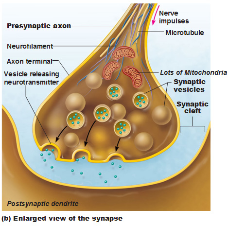
Explanation: Synaptic vesicles are located in the axon terminals (in the synaptic bulbs), close to the presynaptic membrane ready to deliver the neurotransmitters by exocytosis.
Where are synaptic vesicles located in a neuron quizlet?
Synaptic vesicles (membrane organelles) found in the presynaptic terminal, containing neurotransmitters whick communicate with the postsynaptic neuron.
Are synaptic vesicles located in the axon terminal?
Neurotransmitters are stored in synaptic vesicles. These vesicles are located in the axon terminal of a presynaptic neuron of the central or peripheral nervous system.
What part of the neuron contains synaptic vesicles?
axon' Synaptic Vesicles: small secretory vesicles that contain a neurotransmitter, are found inside an axon near the presynaptic membrane, and release their contents into the “synaptic cleft” after fusing with the membrane.
What are synaptic vesicles quizlet?
synaptic vesicle. fluid-filed space at a synapse through which neurotransmitters diffuse. synaptic cleft. in the nervous system, the neuron that sends the message.
What is the location of the vesicles containing neurotransmitter molecules quizlet?
Neurotransmitter molecules are stored in vesicles in the synaptic knob of the presynaptic (transmitting) neuron.
Where is synapse located?
In the central nervous system, a synapse is a small gap at the end of a neuron that allows a signal to pass from one neuron to the next. Synapses are found where nerve cells connect with other nerve cells.
What is found in axon terminals?
aka axon terminals, synaptic boutons are small swellings that are found at the terminal ends of axons. They are typically the sites where synapses with other neurons are found, and neurotransmitters are stored there to communicate with other neurons via these synapses.
Where are vesicles stored?
Neurotransmitter Release. At rest, neurotransmitter-containing vesicles are stored at the terminal of the neuron in one of two places. A small number of vesicles are positioned along the pre-synaptic membrane in places called "active zones." This is where neurotransmitter release occurs.
Where are synaptic vesicles formed?
the Golgi apparatusSynaptic vesicles are initially formed in the Golgi apparatus, where proteins critical for their function are synthesized and inserted into the plasma membrane.
Are synaptic vesicles in dendrites?
Dendritic dopamine release. Dopamine has been observed in a variety of dendritic organelles including large dense core vesicles, small synaptic vesicles, and tubulovesicular structures resembling smooth ER in dopaminergic neurons (Bjorklund and Lindvall, 1975; Nirenberg et al., 1996).
Where is the presynaptic membrane located?
axon terminalA presynaptic membrane is a specialized area of membrane of the axon terminal that faces the plasma membrane of the neuron or muscle fiber with which the axon terminal establishes a synaptic junction.
Where would you find neurotransmitters stored in vesicles?
Neurotransmitter is stored inside small sacs called synaptic vesicles, and is released into the synaptic cleft of the synapse when a vesicle fuses with the cell membrane. This process, which is known as exocytosis, can release neurotransmitter in less than a millisecond.
What do synaptic vesicles contain?
Synaptic vesicles contain small ribonucleic acids (sRNAs) including transfer RNA fragments (trfRNA) and microRNAs (miRNA).
Which of the following substances are stored in synaptic vesicles quizlet?
Molecules of what substances are stored in synaptic terminals? Synaptic terminals contain vesicles of neurotransmitters that are released when action potentials reach the synaptic terminals.
What is the name of the space between the presynaptic and postsynaptic membrane?
There is a small gap between the axon terminal of the presynaptic neuron and the membrane of the postsynaptic cell, and this gap is called the synaptic cleft.
What is the function of synapsin in the synaptic vesicle?
The finding that synapsin is phosphorylated after strong depolarization of the nerve terminal, as well as the finding that it associates with synaptic vesicles, inspired interest in how synapsin affects synaptic transmission . Because synapsin was known to be phosphorylated in a calcium-dependent manner in response to neuronal activity, researchers investigated its role in transmission by injecting different forms of synapsin (phosphorylated or dephosphorylated) into the squid giant synapse nerve terminal ( Llinas et al., 1991; Greengard et al., 1993 ). After injection, the axon was stimulated to release transmitter at a low frequency, and the amount of released neurotransmitter was monitored over a 10-minute period. Results from these experiments showed that injecting the dephosphorylated form of synapsin caused a gradual decrease in the magnitude of transmitter released, but injecting the phosphorylated form of synapsin had no effect on transmitter release. Importantly, when these experimenters injected a kinase (calcium-calmodulin-dependent protein kinase II; CaMKII) into the nerve terminal, causing the phosphorylation of endogenous synapsin proteins, they observed a gradual increase in transmitter release.
How are synaptic vesicles reused?
Once in the nerve terminal, small clear synaptic vesicles are reused many times for transmitter release, and thus are recycled within the nerve terminal without the immediate need for the neuron to make more vesicles. To move synaptic vesicles within the nerve terminal environment, tethering proteins interact to chaperone (aid in the movement of) vesicles between storage pools, recycling pools, and the docking sites for transmitter release (see Chapter 9, for more information on these synaptic vesicle pools).
How is glutamate loaded into the synaptic vesicles?
Glutamate is loaded into synaptic vesicles via the vesicular glutamate transporter (VGLUT; see Fig. 18.3 ). There are three known types of VGLUTs, called VGLUT 1, 2, and 3. Like the vesicular acetylcholine transporter, vesicular glutamate transporters are driven by a proton gradient that is maintained by a vesicular proton-dependent ATPase (also called a “proton pump”; but see Box 16.2 for more details). Since VGLUTs are found only in glutamatergic neurons, they (unlike glutamate and its synthetic enzymes) can be used to uniquely identify these neurons using histological labeling techniques.
What are the proteins in the vesicles of a torpedo?
Synaptic vesicles from Torpedo electric organ contain, in addition to acetylcholine, a glycosaminoglycan named vesiculin, several unidentified proteins, cholesterol, phospholipids, Ca 2+, and ATP ( Ceccarelli and Hurlbut, 1980; Rephaeli and Parsons, 1982; Oorschot and Jones, 1987 ). Vesiculin exists in Torpedo vesicles in a molar ratio to acetylcholine of about 1:12 ( Whittaker, 1971; Whittaker et al., 1974), and the protein is released by stimulation, although not in parallel with the release of acetylcholine ( Zimmermann and Whittaker, 1974a, b). A possibly similar protein has been shown to be released from the isolated phrenic nerve–diaphragm preparations of the mouse, at least one-third of it apparently being released from the nerve by stimulation (Musick and Hubbard, 1972; Musick, 1979 ).
Which proteins assist in moving synaptic vesicles into a vesicle docking site?
Figure 8.14. Cytoskeletal cytomatrix proteins assist in moving synaptic vesicles into a vesicle docking site in the nerve terminal.
Where is SV2C expressed?
SV2C is extensively expressed in the basal ganglia and preferentially localized in dopaminergic cells, as observed in mice, in comparison to other counterparts of the SV2 family (SV2A and SV2B).
Which neuronal vesicles take up serotonin?
Monoaminergic neuronal synaptic vesicles, adrenal medulla chromaffin granules, and platelet dense granules, take up serotonin and catecholamines by a common ATP-dependent process which involves a proton pump ATPase and a vesicular monoamine transporter activated by the proton gradient generated by the pump.
Formation
Synaptic vesicles are made of a lipid bilayer in which transport proteins specific to each type of neurotransmitter are inserted. Neurotransmitters are moved from the cell's cytoplasm into the vesicles by vesicular transporters that rely on active transport mechanisms involving an exchange of protons (H + ions).
Neurotransmitter release
Neurotransmitters are released when vesicles fuse with the cell's plasma membrane at "active zones" of a synapse. Vesicles are scattered throughout the synapse, with some docked at the active zones, and the majority away from the active zone.
Effects of neurotoxins
Some neurotoxins, such as batrachotoxin, are known to destroy synaptic vesicles. The tetanus toxin damages VAMP, a type of v-SNARE (Kandel et al, 2000). Botulinum toxins damage t-SNARES and v-SNARES and thus inhibit synaptic transmission (Kandel et al, 2000).