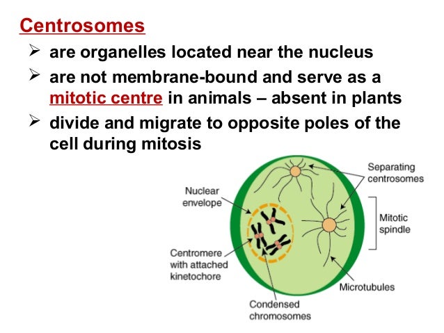
What happens to the centrosome before cell division?
Before cell division, the centrosome duplicates and then, as division begins, the two centrosomes move to opposite ends of the cell. Proteins called microtubules assemble into a spindle between the two centrosomes and help separate the replicated chromosomes into the daughter cells.
How do chromosomes move to the opposite poles during meiosis?
Chromosomes move to the opposite cell poles. Similar to mitosis, microtubules such as the kinetochore fibers interact to pull the chromosomes to the cell poles. Unlike in mitosis, sister chromatids remain together after the homologous chromosomes move to opposite poles. At the end of anaphase I of meiosis, the cell enters into telophase I.
Where do chromosomes line up in meiosis 2?
The chromosomes line up at the metaphase II plate at the cell's center. The kinetochore fibers of the sister chromatids point toward opposite poles. At the end of metaphase II of meiosis, the cell enters into anaphase II. Sister chromatids separate and begin moving to opposite ends (poles) of the cell.
What is the function of centrioles and centrosomes in animal cells?
Centrioles and centrosomes function in the organization of microtubules during cell division. What are animal cell centrosomes? The centrosome also called the cytocenter is an organelle in cell biology that serves as the animal cell’s main microtubule-organizing center (MTOC) as well as a regulator of cell-cycle progression.
How do centrosomes organize cells?
What is a centrosome?
About this website

What happens to centrosomes during meiosis?
Centrosome Before cell division, the centrosome duplicates and then, as division begins, the two centrosomes move to opposite ends of the cell. Proteins called microtubules assemble into a spindle between the two centrosomes and help separate the replicated chromosomes into the daughter cells.
Where do the centrosomes centrioles migrate?
Prophase and Asters and the Mitotic Spindle In prophase, each centrosome with centrioles migrates toward opposite ends of the cell. A single pair of centrioles is positioned at each cell pole. The mitotic spindle initially appears as structures called asters which surround each centriole pair.
What phase do centrosomes migrate?
The mitotic spindle also begins to develop during prophase. As the cell's two centrosomes move toward opposite poles, microtubules gradually assemble between them, forming the network that will later pull the duplicated chromosomes apart.
Where are centrosomes located during mitosis?
The centrosome is located in the cytoplasm usually close to the nucleus. It consists of two centrioles — oriented at right angles to each other — embedded in a mass of amorphous material containing more than 100 different proteins.It is duplicated during S phase of the cell cycle.
What is a centrosome in mitosis?
A centrosome is an organelle located near the nucleus in the cytoplasm that divides and migrates to opposite poles of the cell during mitosis and is involved in the formation of the mitotic spindle, assembly of microtubules, and regulation of cell cycle progression.
What phase do centrioles move to opposite ends of the cell?
ProphaseThus, the correct answer is 'Prophase. '
What are the stages of meiosis division?
In each round of division, cells go through four stages: prophase, metaphase, anaphase, and telophase.
Do centrosomes replicate in mitosis?
Duplication of the single centrosome is initiated at the G1/S transition and completed before mitosis, where the duplicated centrosomes play a role in organizing the poles of the mitotic spindle. The centrosomes are segregated at mitosis such that each of the two cells resulting from division receives only one.
What is the location of centrosome?
The centrosome is positioned in the cytoplasm outside the nucleus but often near to it. A single centriole is also to be found at the basal end of cilia and flagella. In this context it is called a 'basal body' and is connected with the growth and operation of the microtubules in a cilium or flagellum.
Where is centrosome located in the cell?
cytoplasmThe centrosome of animal cells is located in the cytoplasm just outside the nuclear envelope. The centrosome contains a pair of structures, called centrioles, positioned at right angles, or orthogonally, to one another. Each centriole comprises a cylindrical array of short microtubules.
What is the difference between mitosis and meiosis?
Mitosis is a process where a single cell divides into two identical daughter cells (cell division). facts What is meiosis? Meiosis is a process where a single cell divides twice to produce four cells containing half the original amount of genetic information.
What is the location of centrosome?
The centrosome is positioned in the cytoplasm outside the nucleus but often near to it. A single centriole is also to be found at the basal end of cilia and flagella. In this context it is called a 'basal body' and is connected with the growth and operation of the microtubules in a cilium or flagellum.
What is the function of centrosome and centrioles?
Centrosomes are structures found inside of cells. They are made from two centrioles. Centrioles are microtubule rings. The main purpose of a centrosome is to organize microtubules and provide structure for the cell, as well as work to pull chromatids apart during cell division.
Is the centrosome the centriole?
The centrosome is responsible for the formation of the spindle apparatus during the cell division. The main difference between centriole and centrosome is that centriole is the microtubule unit which forms the centrosome whereas centrosome is an organelle in the cytoplasm which is made up of two centrioles.
What is the difference between Centriole and centrosome?
A centrosome is an organelle that consists of two centrioles. A centriole is a structure made of microtubule proteins arranged in a particular way. A centriole is always smaller than a centrosome and also forms flagella and cilia. Both centrosomes and centrioles are found in animal cells and some protists.
Centrosome - an overview | ScienceDirect Topics
H. Schatten, in Brenner's Encyclopedia of Genetics (Second Edition), 2013 Centrosomes. Centrosomes (MTOCs) play critical roles in the nucleation and organization of microtubules into mitotic configurations that are very important for accurate chromosome alignment and chromosome movement to the mitotic poles. A typical mammalian cell centrosome consists of a pair of perpendicularly oriented ...
Centrosome Definition & Meaning - Merriam-Webster
The meaning of CENTROSOME is centriole. Recent Examples on the Web The story should stop there, because the virus needs a special key to move past the centrosome. — USA Today, 23 May 2022 The story should stop there, because the virus needs a special key to move past the centrosome. — USA Today, 23 May 2022 The story should stop there, because the virus needs a special key to move past the ...
Centrosome - Definition, Structure, Functions and Centriole - VEDANTU
Cell division is an urgent branch of cell science. Centrosomes assume a significant role in this procedure. Recall that the two centrioles of a solitary centrosome are situated at right edges to one another, implying that the microtubules in these centrioles will be shown in one of two commonly opposite headings.
Centrosome - Wikipedia
In cell biology, the centrosome (Latin centrum 'center' + Greek sōma 'body') (archaically cytocentre) is an organelle that serves as the main microtubule organizing center (MTOC) of the animal cell, as well as a regulator of cell-cycle progression. The centrosome provides structure for the cell. The centrosome is thought to have evolved only in the metazoan lineage of eukaryotic cells.
How do centrosomes help Drosophila?
Centrosomes of Drosophila oocytes help to transport nutrients from the surrounding nurse cells before being degenerated. In the beginning they nucleate microtubules, along which the inactive centrioles of the surrounding nurse cells migrate into the oocyte [ 114] and organize a large MTOC [ 115 ]. The microtubules emanating from the aggregated centrioles grow into nurse cells through the ring canals [ 114 ], directing the nutrients and mRNA flow into the oocyte from the nurse cells [ 114, 115 ]. In the course of degeneration, the centriolar aggregate eventually loses pericentriolar material and ceases microtubule-nucleating function [ 116 ]. Finally in the GV stage, MTOCs disperse into the cytoplasm and disappear beyond detection. The final fate of centrioles has not been investigated at the ultrastructural level, but they are presumed to disintegrate completely before the oocytes enter meiosis [ 116, 117 ].
What are centrosomes in spermatids?
Animal spermatids and primary oocytes initially have typical centrosomes comprising pairs of centrioles and pericentriolar fibrous centrosomal proteins. These somatic cell–like centrosomes are partially or completely degenerated during gametogenesis. Centrosome reduction during spermiogenesis comprises attenuation of microtubule nucleation function, loss of pericentriolar material, and centriole degeneration. Centrosome reduction during oogenesis is due to complete degeneration of centrioles, which leads to dispersal of the pericentriolar centrosomal proteins, loss of replicating capacity of the spindle poles, and switching to acentrosomal mode of spindle organization. Oocyte centrosome reduction plays an important role in preventing parthenogenetic embryogenesis and balancing centrosome number in the embryonic cells.
What are the proteins in the centriolar cylinder?
The centriolar cylinder and the fibrous pericentriolar material comprise more than 100 different types of proteins [ 7 – 9 ]. Among them, the γ-tubulin ring complexes (γTuRC), comprising γ-tubulin and accessory proteins, are directly involved in microtubule nucleation [ 10, 11 ]. γTuRC are embedded in the pericentriolar matrix [ 12 ], possibly anchored by the centrosome protein pericentrin [reviewed in 13 ]. Several cytoplasmic and nuclear proteins associate with centrosomes in a microtubule-dependent or independent manner. Dynein and proteins coupled with dynein move to the centrosome through microtubular tracks by minus end–directed motor activity [ 14 ]. Some proteins localize to centrosomes only during dividing stages [ 8 ]. Nuclear-mitotic apparatus protein (NuMA) is a nuclear protein during interphase, but it associates with centrosomes during spindle assembly after nuclear envelope breakdown [ 15 ]. Some newly discovered centriolar/centrosomal proteins have been described in recent reviews [ 16, 17 ].
How many centrioles are in a gamete?
A widely accepted hypothesis about centrosomal inheritance during animal fertilization assumes that the male gamete contributes two centrioles that organize a functional zygotic centrosome by recruiting centrosomal proteins from the oocyte cytoplasm [ 63, 64, 88 ]. The centrioles duplicate during the pronuclear stage, making two pairs of centrioles, which are equivalent to two centrosomes [ 89, 90 ]. The duplicated centrosomes with centriolar duplexes organize the cleavage spindles [ 91 ]. Hence the sperm centrioles are the progenitor of all centrioles of the offspring.
What are the centrioles of a centrosome?
A typical centrosome consists of a pair of centrioles associated with fibrous pericentriolar material [ 4 ]. Typical centrioles are cylinder-shaped structures made up of nine symmetrically oriented microtubular triplets measuring 0.5 μm in length and 0.2 μm in diameter [ 5, 6 ]. The two centrioles are associated with each other in an orthogonal orientation with the axis of the newly formed (daughter) centriole crossing the axis of the older (mother) centriole. The mother centrioles have flap-like appendages at the distal end and cone-shaped and striated depositions called the appendages on the outer wall, whereas the daughter centrioles have “cartwheel” organization in the proximal lumen [ 6 ]. The fibrous material is confined around the mother centrioles performing the microtubule nucleating function.
Why do centrosomes degenerate during spermiogenesis?
Centrosome degeneration has been studied in some experimental models. In cultured cells, centrosomes degenerate when they are physically damaged by X-rays [ 72 ], laser irradiation [ 73 ], or treated with antimitotic drugs [ 74 ]. Centrosome degeneration in cultured cells exposed to antiglutamylated tubulin antibody closely mimics spermatogenic centrosome reduction. The antibody binds to the glutamylated sites of tubulin, making them inaccessible to the centriolar organizer proteins resulting in disintegration of centrioles. Centriole loss is accompanied by the disjunction of centrosomal material from the pericentriolar region and scattering into the cytoplasm. Analogous centrosome degeneration in gametes may be related to depletion of cytoplasmic reserves of centrosomal constituents, since the synthetic activities of the nuclei are totally shut down during the late spermiogenesis stages. In Chlamydomonas and Paramecium, microtubular triplets of the basal bodies/centrioles degenerate as the result of a deficiency of δ-tubulin [ 75, 76 ]; ε-tubulin [ 77, 78 ]; Bld10-p [ 79 ]; and Vfl1 [ 80 ], etc. [ 17 ]. It is very likely that centriole degeneration during animal spermiogeneses could be related to deficiency of homologous molecules. The role of a ubiquitin-proteasome system in centriole degeneration is also an interesting field to be investigated [ 81 ]. Pericentrin and Spd-2 are involved in recruiting the centrosomal materials around the pericentriolar lattice in frog egg extract and nematode embryonic cells [ 82, 83 ]. Conversely, a lack of these proteins could be implicated in disjunction and/or loss of pericentriolar material from the degenerating centrioles of gametogenic cells. The existence of divergent molecular pathways of pericentriolar material disjunction and centriolar disintegration would explain why these two events are temporally separated during spermiogenesis. Centrioles of various differentiating somatic cells lose pericentriolar material and cease to function as MTOC before degeneration [ 49 ].
What are the proteins that make up spermatocytes?
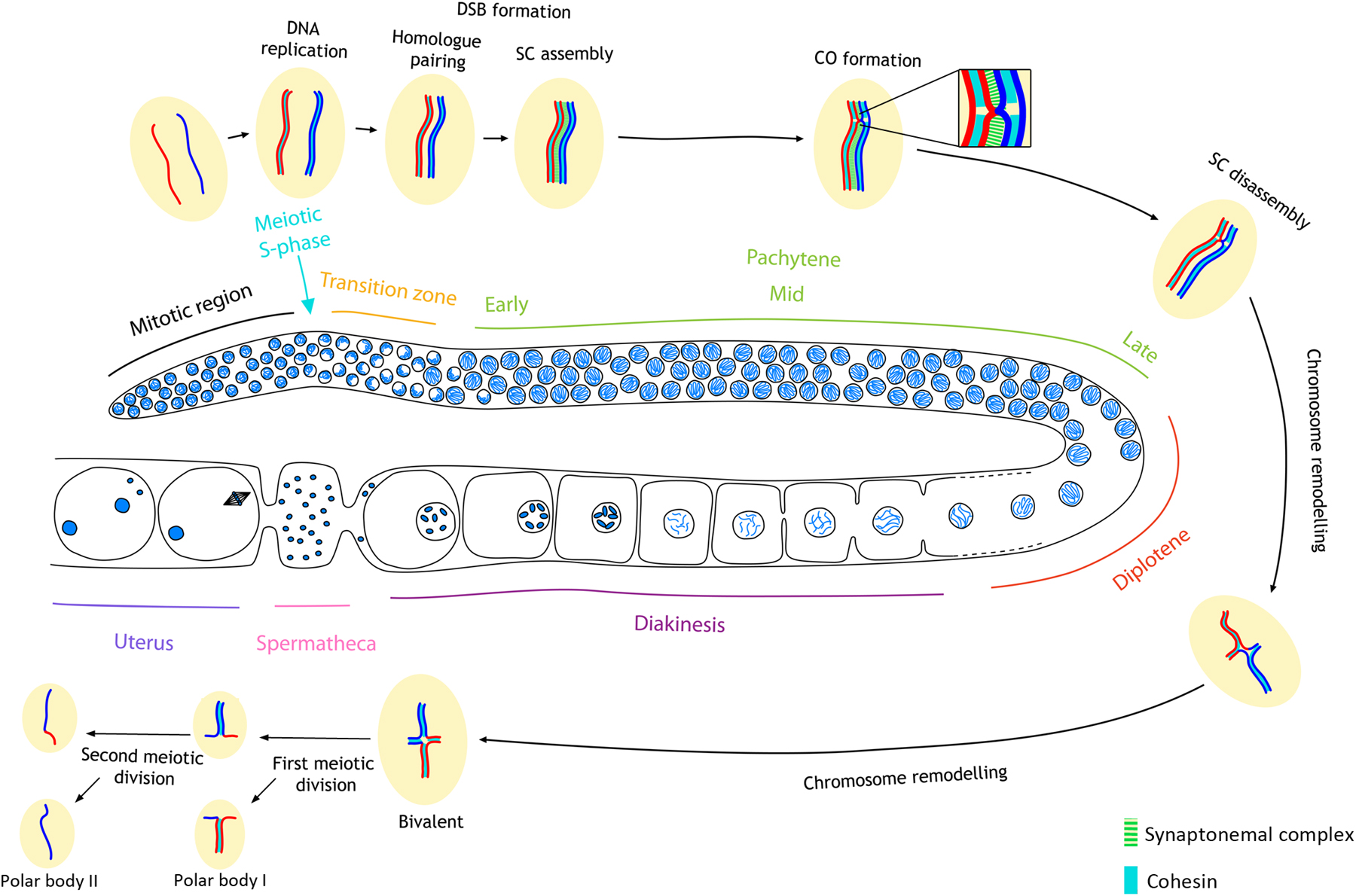
The primary and secondary spermatocytes show typical somatic cytoplasmic organization. Mouse spermatocytes possess the major centrosomal proteins such as centrin [ 44 ], γ-tubulin, and pericentrin (G. Manandhar and G. Schatten, unpublished observations). Male meiotic spindles of most animal species display two centrioles in each pole [ 45 ]. Centrosomes are structurally and functionally intact until the end of meiosis.
Where do chromosomes line up in meiosis?
The chromosomes line up at the metaphase II plate at the cell's center. The kinetochore fibers of the sister chromatids point toward opposite poles. At the end of metaphase II of meiosis, the cell enters into anaphase II.
What happens to chromosomes in meiosis?
Chromosomes thicken and detach from the nuclear envelope. Similar to mitosis, the centrioles migrate away from one another and both the nuclear envelope and nucleoli break down. Likewise, the chromosomes begin their migration to the metaphase plate. At the end of prophase I of meiosis, the cell enters into metaphase I.
What is the function of microtubules in meiosis?
Similar to mitosis, microtubules such as the kinetochore fibers interact to pull the chromosomes to the cell poles. Unlike in mitosis, sister chromatids remain together after the homologous chromosomes move to opposite poles. At the end of anaphase I of meiosis, the cell enters into telophase I.
What happens at the end of metaphase I of meiosis?
At the end of metaphase I of meiosis, the cell enters into anaphase I.
What phase of meiosis is the nucleus bounded by?
At the end of interphase, the cell enters the next phase of meiosis: Prophase I.
What happens at the end of telophase?
At the end of telophase I and cytokinesis, two daughter cells are produced, each with one-half the number of chromosomes of the original parent cell. Depending on the kind of cell, various processes occur in preparation for meiosis II. There is, however, a constant: The genetic material does not replicate again.
What happens to the sister chromatids in meiosis?
In anaphase II of meiosis, the following events occur: Sister chromatids separate and begin moving to opposite ends (poles) of the cell. Spindle fibers not connected to chromatids lengthen and elongate the cell. Once the paired sister chromatids separate from one another, each is considered a full chromosome.
What is the function of centrosomes?
Centrosomes behave as the major MTOCs in most animal cells , whereby polymerisation of MTs is implicated in cell division as well as functions such as the formation of the basal body of cilia and flagella, and the maintenance of cell shape and polarity (Azimzadeh & Marshall, 2010 ).
What happens when bivalents are aligned at metaphase I?
When bivalents are aligned at the metaphase I plate, centrosomes have completed their migration towards opposite poles in control spermatocytes, and bipolar spindles are unambiguously observed. In these spermatocytes, SYCP3 appears as intense centromeric signals and faint interchromatid domain patches (Parra et al, 2004 ), and kinetochore MTs reach the aligned bivalents at the metaphase I plate (Fig 7A ). Aligned bipolar spindles II are observed in control metaphase II (Fig 7B ). PCNT is detected at the opposite poles in these bipolar spindles (Fig 7C and D ). In contrast, Plk1 (Δ/Δ) presented cells with non-migrated poles with a level of chromatin condensation corresponding to prometaphase I/metaphase I and prometaphase II/metaphase II, respectively. We will classify these cells as monopolar spindles I (Fig 7E) and monopolar spindles II (Fig 7F ). Plk1 (Δ/Δ) monopolar spindles I and II show two PCNT signals very proximal to each other, corroborating a single pole per cell (Fig 7G and H ). The known target of PLK1, centromere protein U (CENP-U) (Kang et al, 2011 ), is not detected in its phosphorylated form (CENP-UP) in Plk1 (Δ/Δ) monopolar spindles (Fig EV5B ), in contrast to control (Fig EV5A ). In these monopolar spindles I and II, bivalents and chromosomes, respectively, are not located to form a correct metaphase plate but arranged into an irregular crown around the MTs emanating from the monopolar spindle. Testis cryosections counterstained with DAPI also showed dividing spermatocytes in a crown arrangement that seem to correspond to monopolar metaphases (Fig EV3C ). The squashing technique allowed us to verify in a three-dimensional view the control bipolar spindles I (Movie EV1) versus Plk1 (Δ/Δ) monopolar spindles I (Movie EV2 ).
How do centrosomes organize cells?
The centrosome is an important part of how the cell organizes the cell division. There are a lot of processes that need to be coordinated together when you have two cells, both their nucleus and the cytoplasm, moving away from each other. Microtubules create a spindle, and that's really the structural elements of the cell that coordinate the cells moving away from each other. And the centrosomes organize the microtubules, so it's called the microtubules organizing center. The centrosomes duplicate before cell division, so they then help to organize the microtubules and the cell division process.
What is a centrosome?
Centrosome. Centrosome. =. A centrosome is a cellular structure involved in the process of cell division.