Where do the cranial nerves primarily arise from?
Cranial Nerves. The cranial nerves are composed of twelve pairs of nerves that emanate from the nervous tissue of the brain.In order to reach their targets they must ultimately exit/enter the cranium through openings in the skull.Hence, their name is derived from their association with the cranium. The following are the list of cranial nerves, their functions, and tumor examples:
Where does the spinal cord enter the skull?
Spinal cord: The spinal cord proper extends from the foramen magnum of the skull to the first or second lumbar vertebrae. It creates a two-way pathway between the brain and the body and divides into four regions - cervical, thoracic, lumbar, and sacral.
Which cranial nerves go through the canals of the skull?
The trigeminal nerve has three divisions, which are:
- Ophthalmic. The ophthalmic division sends sensory information from the upper part of your face, including your forehead, scalp, and upper eyelids.
- Maxillary. This division communicates sensory information from the middle part of your face, including your cheeks, upper lip, and nasal cavity.
- Mandibular. ...
Where does C1 spinal nerve exit?
The spinal nerve emerges from the spinal column through an opening ( intervertebral foramen) between adjacent vertebrae. This is true for all spinal nerves except for the first spinal nerve pair (C1), which emerges between the occipital bone and the atlas (the first vertebra).
What are the functions of the cranial nerves?
Some of these functions include directing sense and motor impulses, equilibrium control, eye movement and vision, hearing, respiration, swallowing, smelling, facial sensation, and tasting. The names and major functions of these nerves are listed below.
How many cranial nerves are there?
There are 12 paired cranial nerves that arise from the brainstem. Aspects of vision, like peripheral vision, are under the control of the optic cranial nerve (II). Medical professionals can test visual acuity using a Snellen chart. The trigeminal cranial nerve is the largest of the cranial nerves.
Which cranial nerve is responsible for the sense of smell?
Olfactory Nerve : Sense of smell. Optic Nerve: Vision. Oculomotor Nerve: Eyeball and eyelid movement. Trochlear Nerve: Eye movement. Trigeminal Nerve: This is the largest cranial nerve and is divided into three branches consisting of the ophthalmic, maxillary and mandibular nerves.
Which nerves are involved in the peripheral nervous system?
Peripheral nervous system connections with various organs and structures of the body are established through cranial nerves and spinal nerves. While some cranial nerves contain only sensory neurons, most cranial nerves and all spinal nerves contain both motor and sensory neurons.
Which nerve is responsible for swallowing?
This nerve is commonly tested by observing for facial symmetry. 1 The glossopharyngeal nerve plays a role in swallowing, sense of taste, and saliva secretion. The vagus nerve is involved in smooth muscle sensory and motor control in the throat, lungs, heart, and digestive system.
Which nerve controls the sense of taste?
Facial Nerve: Facial expressions and sense of taste. Vestibulocochlear Nerve : Equilibrium and hearing. Glossopharyngeal Nerve: Swallowing, sense of taste, and saliva secretion. Vagus Nerve: Smooth muscle sensory and motor control in throat, lungs, heart, and digestive system.
Where do the olfactory and optic nerves come from?
The olfactory and optic nerves arise from the anterior portion of the brain called the cerebrum. The oculomotor and trochlear cranial nerves stem from the midbrain. The trigeminal, abducens, and facial nerves arise in the pons. The vestibulocochlear nerve arises in the inner ears and goes to the pons.
What are the functions of the cranial nerves?
Their functions are usually categorized as being either sensory or motor. Sensory nerves are involved with your senses, such as smell, hearing, and touch. Motor nerves control the movement and function of muscles or glands. Keep reading to learn more about each of the 12 cranial nerves and how they function.
Which nerve is responsible for tracking the movement of the head?
This generates nerve impulses that are transmitted to the cochlear nerve. Vestibular portion. Another set of special cells in this portion can track both linear and rotational movements of your head. This information is transmitted to the vestibular nerve and used to adjust your balance and equilibrium.
What is the function of the oculomotor nerve?
The oculomotor nerve has two different motor functions: muscle function and pupil response. Muscle function. Your oculomotor nerve provides motor function to four of the six muscles around your eyes. These muscles help your eyes move and focus on objects.
How many cranial nerves are there?
What are cranial nerves? Your cranial nerves are pairs of nerves that connect your brain to different parts of your head, neck, and trunk. There are 12 of them, each named for their function or structure. Each nerve also has a corresponding Roman numeral between I and XII.
What nerves are involved in smell?
I. Olfactory nerve. The olfactory nerve transmits sensory information to your brain regarding smells that you encounter. When you inhale aromatic molecules, they dissolve in a moist lining at the roof of your nasal cavity, called the olfactory epithelium.
What nerve sends sensations to the heart?
The vagus nerve is a very diverse nerve. It has both sensory and motor functions, including: communicating sensation information from your ear canal and parts of your throat. sending sensory information from organs in your chest and trunk, such as your heart and intestines.
What nerve controls the muscles in your neck?
Your accessory nerve is a motor nerve that controls the muscles in your neck. These muscles allow you to rotate, flex, and extend your neck and shoulders. It’s divided into two parts: spinal and cranial. The spinal portion originates in the upper part of your spinal cord.
Which cranial nerves exit through canals?
Tip: Cranial nerves with the number 2 in them (e.g. 2-optic and 12-hypoglossal) exit through a canal of the same name. They are the only cranial nerves to pass through canals. Modalities. Simplistically, each cranial nerve can be described as being sensory, motor or both.
Where do cranial nerves come from?
Cranial nerves III – XII arise from the brain stem (Figure 1). They can arise from a specific part of the brain stem (midbrain, pons or medulla), or from a junction between two parts: Midbrain – the trochlear nerve (IV) comes from the posterior side of the midbrain.
How many cranial nerves are there?
The cranial nerves are a set of 12 paired nerves that arise directly from the brain. The first two nerves ( olfactory and optic) arise from the cerebrum, whereas the remaining ten emerge from the brain stem.
How many paired nerves are there in the cranial nerve?
Summary of the Cranial Nerves. The cranial nerves are a set of 12 paired nerves that arise directly from the brain. The first two nerves ( olfactory and optic) arise from the cerebrum, whereas the remaining ten emerge from the brain stem.
How many types of information can a cranial nerve transmit?
Simplistically, each cranial nerve can be described as being sensory, motor or both. They can more specifically transmit seven types of information; three are unique to cranial nerves (SSS, SVS and SVM). See table 1 for a summary of the cranial nerves, their modalities and functions.
Where do olfactory and optic nerves originate?
The olfactory nerve (CN I) and optic nerve (CN II) originate from the cerebrum. Cranial nerves III - XII arise from the brain stem (Figure 1). They can arise from a specific part of the brain stem (midbrain, pons or medulla), or from a junction between two parts: Midbrain - the trochlear nerve (IV) comes from the posterior side of the midbrain.
What are the functions of the cranial nerves?
Summary of the function of each Cranial Nerve: 1 1st Cranial Nerve (olfactory nerve) – Responsible for smell. If injured by tumor or surgery for tumor removal, food taste is also altered. 2 2nd Cranial Nerve (optic nerve) – Responsible for vision. A partial injury to this nerve may result is a “field cut” or partial vision loss. 3 3rd, 4th, and 6th Cranial Nerves (oculomotor, trochlear, and abducens) – Controlling movement of the eyeball. Injury can cause double vision. The third cranial nerve also controls pupil dilation. 4 5th Cranial Nerve (trigeminal nerve) – Controls both function and sensation of the face. An injury can result in difficulty chewing and diminished facial sensation or facial numbness. 5 7th Cranial Nerve (facial nerve) – Controls facial movements. An injury can result in a facial “droop”. 6 8th Cranial Nerve (auditory or acoustic nerve) – Responsible for hearing. Skull-base surgery can sometimes leave hearing intact once a tumor on this nerve is removed. 7 9th Cranial Nerve (glossopharyngeal) – Responsible for sensation to the back of the throat. 8 10th Cranial Nerve (vagus nerve) – Protects against choking, and allows for normal swallowing and speech. 9 11th Cranial Nerve (spinal accessory nerve) – Responsible for shrugging shoulders. 10 12th Cranial Nerve (hypoglossal nerve) – Responsible for tongue movement.
How many cranial nerves are there?
There are 12 pairs of cranial nerves, all emerging from the base of the skull and the brain stem. Each pair of nerves is responsible for a specific, basic function such as hearing, smelling, swallowing, blinking, or focusing the eyes. One of each pair of cranial nerves provides feeling and function, or innervates, ...
What nerves are involved in double vision?
A partial injury to this nerve may result is a “field cut” or partial vision loss. 3rd, 4th, and 6th Cranial Nerves (oculomotor, trochlear, and abducens) – Controlling movement of the eyeball. Injury can cause double vision. The third cranial nerve also controls pupil dilation.
What is the skull base?
The skull base provides the base on which the brain rests inside the skull. The skull base region describes the area of and directly around this bony structure. Contained within the skull base region are the eye orbits, ear canals, two carotid arteries, two vertebral arteries, 12 cranial nerves and the blood drainage system of the brain.
What are the two types of lesions that make up the skull base?
Recent advances in skull base surgery. Two kinds of disorders or lesions may make skull-base surgery necessary: benign or malignant tumors, and vascular lesions , such as aneurysms, malformations of the veins and arteries, and fistulas (an abnormal connection between vessels). Traditionally, many lesions at the base of the skull have been inoperable.
Which side of the body does a tumor of the skull base affect?
One of each pair of cranial nerves provides feeling and function, or innervates, the right side of the body and the other nerve in the pair innervates the left side. Tumors of the skull base often affect the cranial nerves, both by their presence and by the steps the surgeon must take to remove the tumor.
Where do neurosurgeons find lesions?
Neurosurgeons often reach lesions in this area of the skull base through the mastoid region and/or labyrinth of the ear. As such, the base of the skull and posterior fossa are clearly exposed to aid in removing lesions within this area.
Which nerve passes through the foramen?
The maxillary nerve (branch of the trigeminal nerve, CN V) passes through this foramen. Foramen Ovale. The foramen ovale is another opening located at the base of the greater wing of the sphenoid. It is positioned posterolateral to the foramen rotundum within the middle cranial fossa.
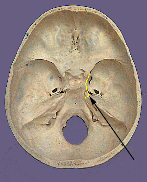
What is the cranial foramen?
The Cranial Foramina. A foramen (pl. foramina) is an opening that allows the passage of structures from one region to another. In the skull base, there are numerous foramina that transmit cranial nerves, blood vessels and other structures – these are collectively referred to as the cranial foramina.
What is the foramina of the skull?
The foramina of the skull are most commonly considered in the context of the cranial nerves. In this section, we will discuss the foramina that transmit cranial nerves. The cribriform foramina refer to numerous perforations in the cribriform plate of the ethmoid bone.
What is the canal in the ear?
The canal connects the posterior cranial fossa and the inner ear, transporting neurovascular structures to the auditory and vestibular apparatus. The facial and vestibulocochlear nerves pass through the internal acoustic meatus, alongside the vestibular ganglion and labyrinthine artery. Jugular Foramen.
What is the foramen?
Log In. A foramen (pl. foramina) is an opening that allows the passage of structures from one region to another. In the skull base, there are numerous foramina that transmit cranial nerves, blood vessels and other structures - these are collectively referred to as the cranial foramina. In this article, we shall look at some ...
Where does the foramen spinosum exit?
Foramen Spinosum. The foramen spinosum is located within the middle cranial fossa , laterally to the foramen ovale. It allows the passage of the middle meningeal artery, the middle meningeal vein and the meningeal branch of CN V 3.
Where is the accessory nerve located?
It lies in the occipital bone within the posterior cranial fossa, and allows the passage of the medulla and meninges, the vertebral arteries, the anterior and posterior spinal arteries and the dural veins. The spinal division of the accessory nerve ascends through the foramen magnum to join the cranial division.