What veins drain blood from the liver?
[edit on Wikidata] The hepatic veins are the veins that drain de-oxygenated blood from the liver into the inferior vena cava. There are usually three upper hepatic veins draining from the left, middle, and right parts of the liver.
What is the pathway of hepatic venous drainage?
The final common pathway of hepatic venous drainage is via the three hepatic veins which enter into the IVC at the dome of the liver. Tributaries include the right hepatic vein, middle hepatic vein, and left hepatic vein.
What are the hepatic veins?
The hepatic veins are three large vessels that drain the venous blood from the liver into the inferior vena cava. The main hepatic veins are the right, intermediate and left hepatic veins.
What is the path of blood flow through the liver?
Blood from these central veins will ultimately converge in the right and left hepatic veins, which exit the superior surface of the liver and empty into the inferior vena cava to be distributed to the rest of the body.
Where do hepatic veins come from?
Structure & Location. The hepatic veins arise from the core vein central liver lobule —a subsection of the liver—and drain blood to the IVC. These veins vary in size between 6 and 15 millimeters (mm) in diameter, and they’re named after the corresponding part of the liver that they cover. These include: 1 .
What are the hepatic veins?
The hepatic veins arise from the core vein central liver lobule—a subsection of the liver—and drain blood to the IVC. These veins vary in size between 6 and 15 millimeters (mm) in diameter, and they’re named after the corresponding part of the liver that they cover. These include: 1 1 Right hepatic vein: The longest of the hepatic veins, the right hepatic vein and lies in the right portal fissure, which divides the liver into an anterior (front-facing) and posterior (rear-facing) sections. 2 Middle hepatic vein: This vein runs at the middle portal fissure, dividing the liver into right and left lobes. It runs just behind the IVC. 3 Left hepatic vein: This vein is found in the left portal fissure, splitting up the left lobe of the liver into a more medial and lateral sections. 4 Caudate lobe veins: These terminal veins perform the function of draining blood directly to the IVC. They run from the caudate lobe, which is connected to the right lobe of the liver via a narrow structure called the caudate process.
What is the function of the hepatic veins?
The primary function of the hepatic veins is to serve as an important cog of the circulatory system. They deliver deoxygenated blood from the liver and other lower digestive organs like the colon, small intestine, stomach, and pancreas, back to the heart; this is done via the IVC. 2 Since the liver serves the important function ...
What is the condition where blood clots in the hepatic veins cause swelling?
Clots of the hepatic veins lead to a rare disorder called Budd-Chiari syndrome. 3 This disease is characterized by swelling in the liver, and spleen, caused by the interrupted blood flow as a result of these blockages. It also increases pressure on these veins, and fluid may build up in the abdomen.
What percentage of the population has hepatic veins?
Anatomical Variations. Variations to the anatomy of the hepatic veins are not uncommon and occur in approximately 30% of the population. 1 In most cases, the right hepatic vein will be what’s affected.
What are the three major hepatic veins?
Relatively larger in size, there are three major hepatic veins—the left, middle, and right—corre sponding to the left, middle, and right portions of the liver. 1 These structures originate in the liver’s lobule and also serve to transport blood from the colon, pancreas, small intestine, and stomach. Anatomically, they’re often used as landmarks ...
What is the condition where blood is unable to drain from the liver?
When a blockage occurs of these veins and blood is unable to drain from the liver, a rare disease, Budd-Chiari syndrome can result. 3 These veins can also develop hypertension—high blood pressure in these veins—can also arise in cases of chronic liver disease. Notably, this is often a feature of liver cirrhosis . Mehau Kulyuk/Getty Images.
Where is the hepatic portal vein located?
Generally, the hepatic portal vein is about 8 centimeters (3 inches) long in adults, and is located in the upper right quadrant of the abdomen, which originates behind the neck of the pancreas and is part of the hepatic portal system.
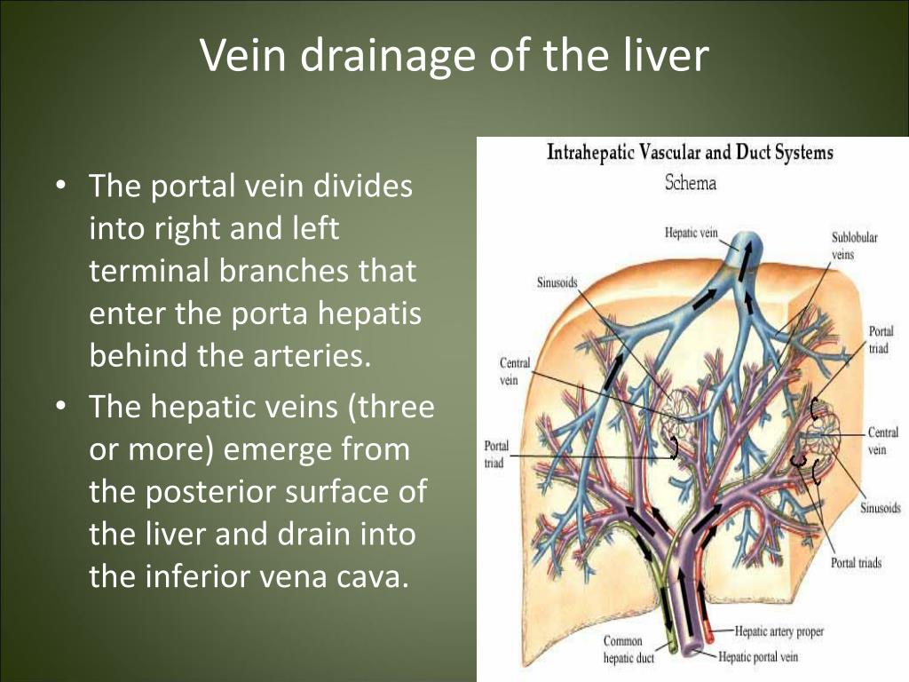
Where does the portal vein go before the liver?
However, before reaching the liver, the portal vein bisects into the left and right, with each side further dividing from venous branches into portal venules. These portal venule branches run alongside hepatic arterioles in the spaces between the liver lobules, and these two vessels, along with a common bile duct, form the hepatic portal triad.
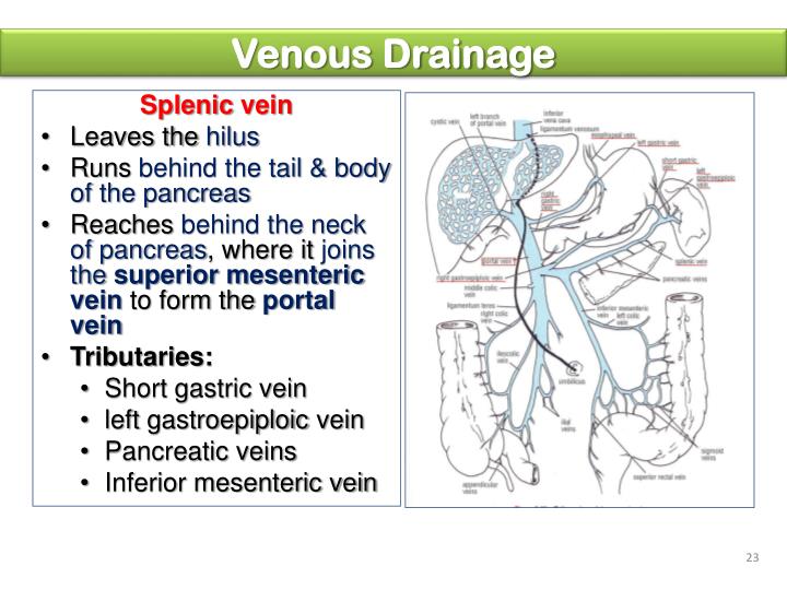
What is the confluence of the splenic and mesenteric veins?
While there may be some variations between individuals, the hepatic portal vein is usually formed by the convergence of the superior mesenteric vein and the splenic vein, referred to as the splenic-mesenteric confluence. In some individuals, the hepatic portal vein also directly joins with the inferior mesenteric vein.
What is the hepatic portal vein?
This vein is part of the hepatic portal system that receives all of the blood draining from the abdominal digestive tract, as well as from the pancreas, gallbladder, and spleen. ‘Hepatic’ means of or relating to the liver, therefore the hepatic portal vein is a blood vessel that sends nutrient-rich blood from the gastrointestinal tract ...
What is the role of the hepatic portal system in the body?
Additionally, the hepatic portal system plays a key role in cleansing the blood of the bacteria and toxins that are picked up by the blood while it is being perfused through the intestines.
Which veins are associated with the intestinal veins?
Other than the previously mentioned hepatic and related veins, the principal associated intestinal veins are the inferior mesenteric vein, superior mesenteric vein, and the splenic vein (which converges with the pancreatic veins before it meets the inferior mesenteric vein, and ultimately meets the superior mesenteric vein). The left and right gastric veins, which form an arc along the lesser curvature of the stomach, also empty into the hepatic portal vein. Broadly, the hepatocytes that process the blood play a large role in protein synthesis, carbohydrate metabolism, lipid metabolism, and detoxification.Solidify your knowledge about the hepatic portal vein
Which vein joins the inferior mesenteric vein?
In some individuals, the hepatic portal vein also directly joins with the inferior mesenteric vein. Even less common, but also possible anastomoses are the cystic and gastric veins.
Where do the hepatic veins drain from?
There are usually three upper hepatic veins draining from the left, middle, and right parts of the liver.
Which veins drain from the right, middle, and left regions of the liver?
The hepatic veins are divided into an upper and a lower group. The upper three drain the central veins from the right, middle, and left regions of the liver and are larger than the lower group of veins.
What are the veins in the liver?
The hepatic veins are the veins of the liver, two of which are shown in this diagram. The hepatic veins are the veins that drain de-oxygenated blood from the liver into the inferior vena cava. There are usually three upper hepatic veins draining from the left, middle, and right parts of the liver. These are larger than the group ...
Where do the veins in the liver come from?
The lower group of from six to twenty smaller hepatic veins come from the right lobe and the caudate lobe, are in contact with the hepatic tissue, and are valveless. All the veins empty into the inferior vena cava at the back of the liver.
Can hepatic veins be connected to portal veins?
The syndrome can be fulminant, acute, chronic, or asymptomatic. The hepatic veins may be connected with the portal veins in a TIPS procedure.
Where do hepatic veins drain?
Hepatic veins collected from different segments of the liver drain into the inferior vena cava at the posterior surface of the liver below the diaphragm.
How to access hepatic vein?
A hepatic vein (most commonly, the right hepatic vein) is accessed using a transjugular approach (jugular vein to superior vena cava, through right atrium to inferior vena cava, and into right hepatic vein), and a stiff wire is placed . A stiff metal cannula is placed over the wire into the hepatic vein. An 18G or 19G core biopsy needle is placed through the cannula and used to obtain multiple samples of liver tissue. Because the right hepatic vein courses posteriorly through the right lobe, the needle is directed anteriorly to avoid transgression of the liver capsule. Because the middle hepatic vein courses more anteriorly, however, lateral or posterior sampling may be safer with this approach.
Why does sacrificing the middle hepatic vein rarely affect segment IV?
Sacrificing the middle hepatic vein seldom affects segment IV because of the collateral drainage provided by the umbilical fissure vein. Extrahepatic control of the middle hepatic vein may not be necessary, unless the tumor is near its trunk.
What waveform is used to trace the middle hepatic vein?
Spectral tracing of the middle hepatic vein shows a typical triphasic waveform with A, S, and D waves representing reflection of cardiac motion in the hepatic veins.
What waveform is used for spectral tracing of the right hepatic vein?
Spectral tracing of the right hepatic vein shows a typical triphasic waveform with A, S, and D waves representing reflection of cardiac motion in the hepatic veins.
Why are hepatic veins useful?
The hepatic veins can be useful in placing an abnormality within a segment (Fig. 73-1 ).
What phase of contrast enhancement shows dark veins?
First of 6 axial T1-weighted MR images obtained during the arterial (portal venous inflow) phase of contrast-enhancement shows the hepatic veins as dark, unenhanced structures, while the aorta, arteries, and portal venous branches are bright due to contrast enhancement.
Where are the veins located in the liver?
The veins are formed by branches of the segmental and subsegmental veins of the liver, and are originally formed at the center of the liver lobule where the hepatic venous radicle is called the central vein.
Where does hepatic venous drainage enter the IVC?
Venous Drainage. The final common pathway of hepatic venous drainage is via the three hepatic veins which enter into the IVC at the dome of the liver. Tributaries include the right hepatic vein, middle hepatic vein, and left hepatic vein.
What is the image of the confluence of the hepatic veins?
This cross sectional ultrasound image of the confluence of the hepatic veins has been likened to the face of a “bunny”. (Image courtesy of Ashley Davidoff M.D.)
What is the branch of the hepatic vein terminating in a liver lobule?
(Image courtesy of Ashley Davidoff M.D.) There are a multiplicity of lobules, each with a central vein and each delivering the “goods” to venules, which collectively join to form the hepatic veins and then into the IVC.
Which vein enters the IVC at a point more caudal than the major hepatic vein?
The venous drainage of the caudate lobe enters independently into the IVC at a point more caudal than the major hepatic veins.
Which vein is open and solitary?
Hepatic veins tend to be open and solitary, allowing them to be distinguished from the branches of the portal vein, which are more or less collapsed and always accompanied by an artery and duct. Lobule. A branch of the hepatic vein terminating in a liver lobule. (Image courtesy of Ashley Davidoff M.D.) Lobules.
Where is the catheter in a venogram?
The venogram shows a catheter in the middle hepatic vein filling the venules of a subsegment. The destination of the blood is the inferior vena cava, which is the structure filled with contrast above the diaphragm. (red color overlay) (Image courtesy of Ashley Davidoff M.D.)
Where does the hepatic portal system drain blood?
It drains blood from the spleen and the gastrointestinal tract to the liver. The hepatic veins begin at the junction of splenic veins and superior mesenteric. The blood from the cystic veins and the inferior mesenteric gastric veins is also drained by the hepatic vein. The ailments that affect the hepatic portal system should be identified ...
Which vein carries 75% of the hepatic blood flow?
The hepatic portal ve in carries 75% of the hepatic blood flow and hence is crucial. It is not a true vein as it does not conduct blood directly to the heart.
What is the hepatic portal vein?
The hepatic portal vein is the part of the hepatic portal system. It carries blood from the intestines, gallbladder, pancreas and spleen and delivers it to the liver. It contains blood with nutrients and toxins after digestion.
What is the portal vein?
However, there are other systems of veins in the body that are referred to as the portal venous system. The hepatic portal vein is the largest vein in the abdominal cavity.
Which system of veins transports blood from the digestive tract to the liver?
The hepatic portal system is the system of veins that transports blood from the digestive tract to the liver. It consists of the hepatic portal ve in and other veins that drain into the hepatic portal vein , viz. the superior mesenteric vein, the inferior mesenteric vein, and the splenic vein. The hepatic portal vein carries blood rich in nutrients and toxins that are processed in the liver and then the hepatic vein carries the blood to the heart through the systemic circulation.
Which vein carries blood to the liver?
The hepatic portal vein carries nutrient-rich blood from the intestine and other parts such as the gallbladder, pancreas and spleen to the liver, whereas the hepatic vein carries deoxygenated blood from the liver to the vena cava. The hepatic portal vein brings the blood to the liver, whereas the hepatic vein transports blood from the liver to the systemic circulation and back to the heart.
Which organ processes food before entering the systemic circulation?
It also ensures that food ingested is processed by the liver first before entering the systemic circulation. This way, ingested toxins are detoxified by hepatocytes.
Where does the portal vein go before entering the liver?
Immediately before entering the liver, the portal vein divides into right and left branches which then enter the parenchyma of the liver separately.
Which vein drains into the left renal vein?
Right testicular/ovarian vein – drain the right testes or ovary respectively in men and women (the left testicular/ovarian vein drains into the left renal vein).
What are the structures of the inferior vena cava?
During its long course, the inferior vena cava shares an anatomical relationship with numerous abdominal structures – including the right common iliac artery, the root of the mesentery, the head of the pancreas, the bile duct, the portal vein and the liver.
What is the name of the vein between the portal tributaries of the mesenteric veins and the?
Retroperitoneal – Between the portal tributaries of the mesenteric veins and the retroperitoneal veins.
Where does the superior mesenteric vein drain blood from?
The superior mesenteric vein drains blood from the small intestine, cecum, ascending colon and transverse colon. It begins in the right iliac fossa, as a convergence of the veins draining the terminal ileum, cecum and appendix. It ascends within the mesentery of the small intestine, and then travels posteriorly to the neck of the pancreas to join the splenic vein.
Where does venous return to the GI tract?
There are no tributaries from the spleen, pancreas, gallbladder or the abdominal part of the GI tract – as these structures are first drained into the portal venous system. However, venous return from these structures ultimately enters the inferior vena cava via the hepatic veins (after being processed by the liver).
Which organ is responsible for the venous drainage of all structures below the diaphragm?
The inferior vena cava is responsible for the venous drainage of all structures below the diaphragm. It receives tributaries from:
Overview
- Structure & Location
The hepatic veins arise from the core vein central liver lobule—a subsection of the liver—and drain blood to the IVC. These veins vary in size between 6 and 15 millimeters (mm) in diameter, and they’re named after the corresponding part of the liver that they cover. These include:1 1. Righ… - Anatomical Variations
Variations to the anatomy of the hepatic veins are not uncommon and occur in approximately 30% of the population.1 In most cases, the right hepaticvein will be what’s affected. Doctors have observed early bifurcation (splitting into two) or trifurcation (splitting into three) of this vein—wit…
Structure
Clinical significance
Additional images
In human anatomy, the hepatic veins are the veins that drain de-oxygenated blood from the liver into the inferior vena cava. There are usually three upper hepatic veins draining from the left, middle, and right parts of the liver. These are larger than the group of lower hepatic veins that can number from six to twenty. All of the hepatic veins drain into the inferior vena cava.
External links
The hepatic veins are divided into an upper and a lower group. The upper three drain the central veins from the right, middle, and left regions of the liver and are larger than the lower group of veins.
The lower group of from six to twenty smaller hepatic veins come from the right lobe and the caudate lobe, are in contact with the hepatic tissue, and are valveless. All the veins empty into th…