Where does the obturator nerve exit?
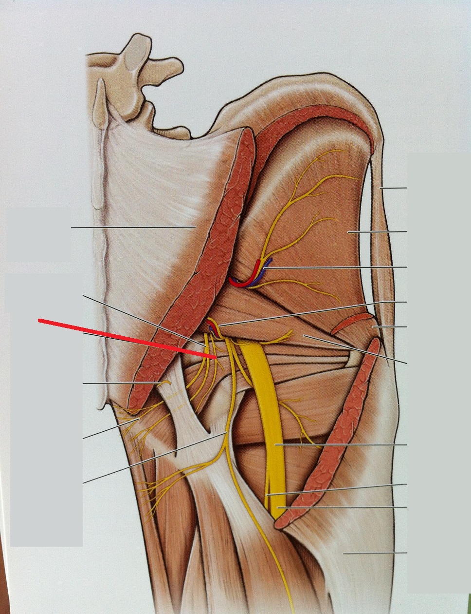
The obturator nerve emerges from the medial side of the psoas muscle, crosses the lesser pelvis, and passes through the obturator foramen into the medial thigh, innervating the adductor longus, brevis, and magnus; gracilis; obturator externus; and pectineus muscles, whose action is to adduct the thigh.
Where does the obturator nerve exit the pelvis?
Anatomy. Pelvis: The nerve descends medial to psoas major to the obturator canal where it divides into anterior and posterior divisions. The anterior division exits from the obturator canal to enter the medial compartment of the thigh. The posterior division exits through obturator externus.
Where does the obturator nerve arise from?
The obturator nerve is one of the largest branches of the lumbar plexus. It is a mixed nerve which arises from the ventral (anterior) rami of the spinal nerves L2-L4.
What is the distribution of the obturator nerve?
The obturator nerve divides into anterior and posterior divisions of the obturator nerve. These divisions both provide skeletal musculature innervation, and the anterior division terminates as the cutaneous branch of the obturator nerve.
Where does the obturator nerve begin and end?
The obturator nerve is derived from L2-4 and travels along the medial border of the iliopsoas muscle; it is both a motor and a sensory nerve. It travels through the obturator foramen with the obturator artery and vein into the thigh. The obturator nerve divides into anterior and posterior branches.
What nerve exits the obturator foramen?
Obturator nerve1. Obturator nerve (L2, L3, L4). It leaves the pelvis through the obturator foramen and supplies the adductors of the thigh.
What is the correct origin and termination for the obturator nerve?
The obturator nerve originates from the anterior divisions of the L2, L3, and L4 spinal nerve roots. It descends through the fibers of the psoas major, and emerges from its medial border near the brim of the pelvis.
Where is the obturator located?
The internal obturator muscle arises from the inner surface of the antero-lateral wall of the pelvis. It surrounds the obturator foramen. It is attached to the inferior pubic ramus and ischium, and at the side to the inner surface of the hip bone below and behind the pelvic brim.
What is the difference between femoral nerve and obturator nerve?
This is the femoral nerve, this is the obturator nerve. The white structure between them is the psoas major tendon. Both these nerves arise from the lumbar plexus, which lies up here within the thickness of the psoas major muscle. The femoral nerve emerges lateral to psoas major, the obturator nerve medial to it.
Which muscle is partially paralyzed with the obturator nerve damage?
The nerve has no sensory function. Partial or complete paralysis affects the adductor muscles, with consequent loss of function.
What are the symptoms of obturator nerve damage?
Muscle weakness in your thigh. Numbness in your thigh. Pain that gets worse with side-to-side leg movements. Sensation of pins and needles in your groin.
Overview
Your obturator nerve is one of many peripheral nerves that run through your groin. It’s part of your peripheral nervous system. This system helps your brain communicate with the rest of your body.
Function
This nerve provides motor (muscle movement) and sensory (sensation) innervation to your inner thigh.
Anatomy
Nerve fibers that make up your obturator nerve start in the lower part of your spine. This includes spine bones (vertebrae) L2, L3 and L4.
Conditions and Disorders
One of the main issues of an obturator nerve injury is neuropathic pain. Conditions that cause it include:
Care
It might not be possible to prevent some causes of obturator neuropathy. Trauma from childbirth or crush injuries can be out of your control.
Where does the obturator nerve begin?
The obturator nerve begins at the medial border of the psoas major muscle. It travels through the obturator foramen (an opening in the pelvic bone) before entering the thigh, where it branches into two parts, an anterior branch and posterior branch. The obturator nerve is part of the group of nerves called the anterior lumbar plexus.
What nerve is responsible for the abductor muscles?
The obturator nerve is part of the group of nerves called the anterior lumbar plexus. The nerve provides sensory perception to the skin on the medial side of the thigh. It also provides motor function to the hip and knee joints and the abductor muscles and gracilis. The obturator nerve can be damaged through injury to the nerve itself ...
Can a damaged obturator nerve cause pain?
A damaged obturator nerve can cause pain, numbness, and weakness of the thigh. Mild damage to the obturator nerve can be treated with physical therapy. More severe cases may require surgery. The nerve has the ability to regenerate itself at a rate of about one inch per month. Last medically reviewed on January 21, 2018.
Where does the obturator nerve travel?
It then travels posteriorly to the common iliac arteries and laterally along the pelvic wall – towards the obturator foramen of the pelvis.
What is the obturator nerve?
The Obturator Nerve. The obturator nerve is a major peripheral nerve of the lower limb. In this article, we shall look at the anatomy of the obturator nerve – its anatomical course, functions and clinical correlations.
What nerve is in the middle of the thigh?
The cutaneous branch of the obturator nerve supplies the skin of the middle part of the medial thigh. The obturator nerve can be damaged during surgery involving the pelvis or abdomen. Symptoms include numbness and paraesthesia on the medial aspect of the thigh and weakness in adduction of the thigh.
What nerve innervates the hamstring?
Motor Functions. The obturator nerve innervates all the muscles in the medial compartment of the thigh - except the hamstring part of the adductor magnus (innervated by the tibial nerve). They are collectively known as the hip adductors: Adductor longus - adducts thigh.
What is the anterior division of the adductor?
Anterior division (anterior to the adductor brevis): Descends in a plane between the adductor longus and adductor brevis (towards the femoral artery). Here, it supplies motor fibres to the adductor longus, adductor brevis and gracilis. It can also supply the pectineus muscle.
Which nerve pierces fascia lata?
It then pierces the fascia lata to become the cutaneous branch of the obturator nerve. Pierces the obturator externus muscle, and then descends in a plane between the adductor brevis and adductor magnus. Innervates the obturator externus and adductor magnus muscles.
Which nerve innervates the medial thigh?
The obturator nerve innervates all the muscles in the medial compartment of the thigh – except the hamstring part of the adductor magnus (innervated by the tibial nerve). Fig 4 – Muscles of the medial thigh. The overlying muscles in the anterior compartment have been removed.
Where does the obturator nerve come from?
The obturator nerve arises from the lumbar plexus on the posterior abdominal wall and descends within the psoas muscle, emerging from the medial margin of the muscle to enter the pelvis. The nerve path continues by following along the lateral wall of the pelvis, passing through the obturator canal, to enter the medial compartment of the thigh.
Which muscle is innervated by the anterior branch of the adductor brevis muscle?
On the anterior surface of the adductor brevis muscle the anterior branch travels underneath the pectineus and adductor longus muscles to innervate the adductor longus, gracilis, and adductor brevis muscles. This branch also often contribute to the pectineus muscle. The cutaneous branches innervate the skin on the medial thigh.
Which muscle is the anterior and posterior branch of the adductor?
From here the nerve divides into the anterior and posterior branch which are separated by the adductor brevis muscle . The posterior branch travels underneath the adductor muscle along the anterior surface of the adductor magnus muscle, innervating the obturator externus, adductor brevis, as well as part of the adductor magnus muscle ...
Can a obturator nerve be damaged?
Injury to the nerve is rare as it lies deep within the pelvis and medial thigh. It can be damaged through direct injury to the nerve or to surrounding muscle tissue. Mild damage to the obturator nerve can be treated with physiotherapy. More severe cases may require surgery.
Where is the obturator nerve located?
The obturator nerve in human anatomy arises from the ventral divisions of the second, third, and fourth lumbar nerves in the lumbar plexus; the branch from the third is the largest, while that from the second is often very small.
What is the function of the obturator nerve?
Function. The obturator nerve is responsible for the sensory innervation of the skin of the medial aspect of the thigh . The nerve is also responsible for the motor innervation of the adductor muscles of the lower limb ( external obturator. adductor longus, adductor brevis, adductor magnus, gracilis) and the pectineus (inconstant).
Where does the obturator nerve go?
The obturator nerve (Figs 6.15, 6.16) supplies all the muscles in the medial compartment of the thigh. It enters the thigh by passing through the obturator foramen accompanied by the obturator artery. As it goes through the foramen it divides into anterior and posterior branches.
Where does the obturator nerve enter the medial thigh?
The obturator nerve emerges from the medial side of the psoas muscle, crosses the lesser pelvis, and passes through the obturator foramen into the medial thigh, innervating the adductor longus, brevis, and magnus; gracilis; obturator externus; and pectineus muscles, whose action is to adduct the thigh.
What is the purpose of obturator nerve block?
Obturator nerve block was used extensively to relieve adductor spasm to improve the personal hygiene of patients with spasticity. Oral and intrathecal dosing with baclofen has very significantly reduced the use of neurolytic obturator nerve block for this purpose but it remains a useful technique for those patients in whom pharmacologic methods are not tolerated or do not produce the desired results.
What nerves are entrapped in the obturator canal?
The obturator nerve can become entrapped as it passes through the obturator canal. The anterior branch of the obturator nerve innervates the adductor longus, adductor brevis, and gracilis muscles, as well as giving innervation to the hip joint. The posterior branch innervates the external obturator and adductor magnus (which is dually innervated by the obturator and sciatic nerves) muscles as well as supplying innervation to the knee joint. The obturator nerve originates from posterior divisions of L2, L3, and L4 spinal roots. The obturator artery and vein travel with the obturator nerve in the obturator canal.
Why block a nerve in the hip?
Because the hip joint derives significant innervation from the obturator nerve, block ade of this nerve was one of the main indications in patients who had degenerative hip disease.8 Since the advent of total joint replacement, however, the number of patients who require this type of block has significantly decreased.
What nerves are blocked during prostate surgery?
It is blocked along with the femoral, lateral femoral cutaneous, and sciatic nerves for this purpose.3 One of the most important surgical indications is due to the anatomic relationship of the obturator nerve as it runs close to the neck of the bladder and the prostate.4–7 Because of the proximity of the nerve to the prostate, this nerve can be electrically stimulated during the transurethral resection. This stimulation can produce significant contraction of the adductors, which can interfere with the surgical procedure, and on occasion can even result in the perforation of the bladder. This can occur even with adequate spinal analgesia that blocks the nerve roots proximal to the site of stimulation. Local anesthetic block of the obturator nerve has been well documented to abolish the spasms and facilitate the prostatic surgery.
Which nerve controls adduction and rotation of the thigh?
9.39 Obturator Nerve. The obturator nerve (L2–L4) supplies the pectineus; adductor (longus, brevis, and magnus); gracilis; and external obturator muscles. This nerve controls adduction and rotation of the thigh. A small cutaneous zone on the internal thigh is supplied by sensory fibers.