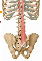
What does anterior longitudinal ligament mean?
The anterior longitudinal ligament is a long dense band of connective tissue—all ligaments are made of some type of connective tissue—that goes from your first vertebra (the atlas) and the front of the base of your skull to the front of your sacrum. It is located on the front side of the vertebral bodies.
What is posterior longitudinal ossification?
Ossification of the posterior longitudinal ligament, also referred to as OPLL, is a spinal condition where the posterior longitudinal ligament becomes calcified and less flexible. The posterior longitudinal ligament runs the entire length of the spine from the neck to the end of the spine and stabilizes the spinal column bones.
Is divided into a posterior and anterior segment?
There are posterior and anterior chambers in the eye. The eyeball is divided into a posterior segment, encompassing most of the spherical back part of the eye where the retina is, and the anterior segment, which is up front and consists of the cornea, the iris and the lens, forming a posterior and anterior chamber. The anterior chamber is the space between the cornea and the iris of the eye.
Are lateral and posterior the same?
Lateral: Same as posterior PLUS no active hip abduction. What are the range-of-motion precautions for Anterior total hip surgical approach? No extreme hip extension combined with external rotation such as kneeling on the operated leg with foot turned in, then moving body weight forward onto the opposite foot.

Where does the anterior longitudinal ligament attach?
The Anterior Longitudinal Ligament attaches to the front (anterior) of each vertebra. This ligament runs up and down the spine (vertical or longitudinal). The Posterior Longitudinal Ligament runs up and down behind (posterior) the spine and inside the spinal canal.
What is the extension of the posterior longitudinal ligament?
The posterior longitudinal ligament extends from the tectorial membrane of the basion to the posterior surface of each vertebra and disc, down to the coccyx.
What does the posterior longitudinal ligament prevent?
Posterior Longitudinal Ligament This ligament is situated within the vertebral canal, and it prevents hyperflexion, which is when bend your spine too far forward.
What does posterior longitudinal ligament turn into?
The posterior longitudinal ligament is a ligament connecting the posterior surfaces of the vertebral bodies of all of the vertebrae. It weakly prevents hyperflexion of the vertebral column....Posterior longitudinal ligamentFMA31894Anatomical terminology8 more rows
What is the posterior longitudinal ligament innervated by?
sinuvertebral nervesThe posterior aspects of the discs and the posterior longitudinal ligament are innervated by the sinuvertebral nerves.
How is OPLL diagnosed?
OPLL can be diagnosed in a clinic visit with your doctor with a physical exam and diagnostic imaging such as X-ray, MRI, or CT scan. While most patients with OPLL can be treated with medications, physical therapy, or lifestyle changes, some may require surgery.
Can OPLL be reversed?
Several options for treating cervical OPLL have been established involving anterior and posterior surgery. Anterior decompression surgery can directly decompress the cervical spinal cord by removing the ossified ligament and always results in better outcomes and neurological improvement [4, 5].
What ligament prevents hyperextension of the knee?
Conclusion: The oblique popliteal ligament was found to be the primary ligamentous restraint to knee hyperextension.
What ligament prevents hyperextension of the spine?
The anterior longitudinal ligamentThe anterior longitudinal ligament attaches to both the vertebra and the intervertebral discs. This ligament helps to prevent hyperextensions of the spine.
Why anterior longitudinal ligament is stronger than posterior?
While anteriorly the ligament is thin due to the elastic fibers, the posterior capsule of each posterior joint is thicker due to the collagenous content.
Which of the following is the direct continuation of the anterior longitudinal ligament?
The anterior atlanto-occipital membrane attaches from the anterior arch of the atlas to the anterior aspect of the clivus. It is a continuation of the anterior longitudinal ligament and serves to prevent excessive neck extension[12,13].
What is the primary function of the ligamentum flavum ligament?
It resists excessive separation of the adjacent vertebral lamina and prevents buckling of the ligament into the spinal canal during extension, preventing canal compression.
Where is the posterior longitudinal ligament located?
The posterior longitudinal ligament is situated within the vertebral canal, and extends along the posterior surfaces of the bodies of the vertebrae, from the body of the axis, where it is continuous with the tectorial membrane of atlanto-axial joint, to the sacrum.
What is the name of the ligaments that run vertically at the center of the vertebrae?
Posterior longitudinal ligament. Posterior longitudinal ligament, in the thoracic region. (Posterior longitudinal ligament runs vertically at center.) Median sagittal section of two lumbar vertebrae and their ligaments.
What is the narrow ligament that covers the basivertebral veins?
The posterior longitudinal ligament is narrow at the vertebral bodies, where it covers the basivertebral veins, and widens at the intervertebral disc space. It is generally quite wide and thin.
Which ligament weakly prevents hyperflexion of the vertebral column?
The posterior longitudinal ligament weakly prevents hyperflexion of the vertebral column. It also limits spinal disc herniation, although it is much narrower than the anterior longitudinal ligament.
Which ligament is narrow at the vertebral bodies?
In the thoracic and lumbar regions, it presents a series of dentations with intervening concave margins. The posterior longitudinal ligament is narrow at the vertebral bodies, where it covers the basivertebral veins, and widens at the intervertebral disc space.
Which ligaments cause back pain?
The posterior longitudinal ligament contains a higher density of nociceptors than many ligaments, so can cause back pain. It may ossify, particularly around cervical vertebrae.
Where is the median sagittal section of the lumbar vertebrae?
(Posterior longitudinal ligament runs vertically at center left.) The posterior longitudinal ligament is situated within the vertebral canal, and extends along the posterior surfaces of the bodies of the vertebrae, from the body of the axis, where it is continuous with ...
What is the posterior longitudinal ligament?
The posterior longitudinal ligament (PLL) is the inferior continuation of the tectorial membrane (see Figs. 5-17 and 5-22). It courses from the posterior aspect of the body of C2, inferiorly to the sacrum, and possibly to the coccyx (Behrsin & Briggs, 1988 ). The ALL and PLL have similar tensile properties ( Przybylski et al., 1996). That is, they can withstand similar loads applied to the spine, although the ALL limits forces applied in extension and the PLL resists forces applied in flexion. The PLL is wide and regularly shaped in the cervical and upper thoracic regions and is also three to four times thicker, from anterior to posterior, in the cervical region than in the thoracic or lumbar regions (Bland, 1989). Its superficial fibers span several vertebrae, and its deep fibers course between adjacent vertebrae. Panjabi and colleagues (1991b) found the cervical PLL to be firmly attached to both the vertebral bodies and the IVDs, whereas Bland (1989) found the PLL to have a stronger discal attachment. In either case, the PLL probably functions to help prevent posterior IVD protrusion. Although the PLL is attached to the entire length of the vertebral bodies in the cervical region ( Przybylski et al., 1998 ), it is more loosely attached to the central region of the vertebral bodies to allow the exit of the basivertebral veins from the vertebral bodies ( Standring et al., 2008 ). The PLL in the middle and lower thoracic and lumbar regions differs from the PLL in the cervical region in that it becomes narrow over the vertebral bodies and then widens considerably over the IVDs in the thoracic and lumbar areas.
What is the PLL of the lumbar region?
The PLL in the lumbar region is denticulated in appearance (Figs. 7-19 and 7-20). That is, it is narrow over the posterior aspect of the vertebral bodies and flares laterally at each IVD, where it attaches to the posterior aspect of the anulus fibrosus. The lumbar PLL is composed of two strata of fibers, superficial and deep. The superficial fibers form a distinct midline band that spans several vertebral levels. The deep fibers are much shorter, and they converge on the IVD and extend laterally to the wide attachment sites of the PLL to the anulus fibrosus of the IVD (Parke & Schiff, 1993 ).
Where is the PLL located?
The PLL extends along the posterior surface of the vertebral bodies from the clivus to the sacrum. Although OPLL may involve any portion of the PLL, by far the most common anatomic location is the cervical spine—accounting for approximately 75% of cases. The process typically involves 2.5 to 4 levels beginning at approximately C3/C4 and progressing distally to involve C4/C5 and C5/C6, although generally sparing C6/C7. Thoracolumbar involvement is less common, accounting for roughly 25% of cases, usually involving the upper thoracic spine rather than the lumbar segments.
Is ossification of the posterior longitudinal ligament a HO?
Ossification of the posterior longitudinal ligament (OPLL) of the spine is considered as a special type of HO . Contrary to decreased serum leptin levels in typical HO patients (Chauveau et al., 2008), increased serum leptin levels were found in OPLL females. Serum leptin levels were significantly higher in patients in whom OPLL extended to the thoracic and/or lumbar spine than in patients in whom OPLL was limited to the cervical spine, and correlated positively with the number of involved vertebrae. These results suggest that hyperleptinemia may contribute to the development of HO of the spinal ligament in female patients with OPLL (Ikeda et al., 2011 ).
Where is the posterior longitudinal ligament located?
Posterior longitudinal ligament. The posterior longitudinal ligament (PLL) is a long and important ligament located immediately posterior to the vertebral bodies (to which it attaches loosely) and intervertebral discs (to which it is firmly attached). It extends from the back of the sacrum inferiorly and gradually broadens as it ascends.
Where does the tectorial membrane extend?
It extends from the back of the sacrum inferiorly and gradually broadens as it ascends. At the level of C2 (the axis) it spreads out and becomes the tectorial membrane that eventually inserts into the base of skull 1,2.
Where is the anterior longitudinal ligament located?
It is located on the front side of the vertebral bodies.
What ligaments provide stability to the column?
Spinal ligaments also provide stability to the column. They do this by limiting the degree of movement in the direction opposite their location. For example, your anterior longitudinal ligament (see below for details) is located in front of your vertebral bodies. When you arch back, it prevents you from going too far.
What is the intertransverse ligament?
Intertransverse ligaments go from a superior (remember, superior refers to an above location, relatively speaking) transverse process of a vertebra to the transverse process of the vertebra below it . The intertransverse ligaments connect these processes together and help limit the action of side bending (lateral flexion). They also form a sort of border between the bodies in front and the bony rings in the back of the vertebrae.
Where does the interspinous ligament connect to the spinous process?
The interspinous ligaments connect the whole of each spinous process vertically. The interspinous ligament starts at the root of the spinous process, where it emerges from the ring of bone located at the back of the body of its respective vertebra, and extends all the way out to the tip.
Which ligaments are more fibrous?
In the thoracic (mid-back) area, the intertransverse ligaments are tougher and more fibrous. Now you know your ligament ABCs. These are the spinal ligaments that affect all or at least large portions of the spine. Other spinal ligaments are specific to an area such as the neck or the sacrum and sacroiliac joints.
Where is the ligament flavum located?
It is located between the laminae of the vertebra. At each vertebral level, fibers originate from a superior lamina (the term superior refers to a location above, relatively speaking) and connect to the inferior lamina (i.e. the lamina just below). The ligamentum flavum limits spinal flexion (bending forward), especially abrupt flexion. This function enables the ligamentum flavum to protect your discs from injury.
Which ligaments limit forward bending?
The supraspinous and interspinous ligaments both limit flexion (forward bending). Located in back, the supraspinous ligament is a strong rope like tissue that connects the tips of the spinous processes from your sacrum up to C7 (otherwise known as the base of the neck).
Where does the Alar ligament run?
Alar ligament: Runs from the posterior aspect of the dens of C2 to the lateral margins of the foramen magnum.
Which ligaments connect the dens to the medial aspect of each occipital condyle?
On the posterior aspect of the dens are two facets for attachment of the alar ligaments . These ligaments connect the dens to the medial aspect of each occipital condyle and help restrict excessive rotation of the head.
What are the structures of vertebrae?
Structure of typical vertebrae. Vertebral body, transverse process, spinous process, vertebral foramen, vertebral arch, articular processes. Intervertebral discs. Consisting of an inner component called the nucleus pulposus surrounded by the annulus fibrosus, they allow the vertebrae to move and act as shock absorbers.
Which part of the cervical spine allows the spinal nerves to exit the vertebral canal?
The intervertebral foramen allows the spinal nerves at each vertebral level to exit the vertebral canal. Finally, the almost horizontal orientation of the articular facets in the cervical spine is, in part, responsible for giving the cervical spine the greatest variety and range of movement.
Where are the intervertebral discs located?
Intervertebral discs. Although not technically a bony component, the intervertebral discs lie in between all cervical vertebrae with the exception of C1 and C2. These discs can be quite significant clinically as they make up the inferior half of the anterior border of the intervertebral and vertebral foramina.
Why is the lumbar spinal cord triangular?
It is triangular in shape and comparatively large to accommodate the expansion of the cervical component of the spinal cord that provides all innervation to the upper limb. A similar enlargement is seen in the lumbar spinal cord to accommodate innervation of the lower limb.
What are the structures that are associated with the spinal column?
To understand this intricate region, we will consider the bony structures first, and then discuss the ligaments, nerves, and musculature that are associated with this region of the spinal column, concluding with some clinical implications of damage to some of these structures.

Overview
Structure
The posterior longitudinal ligament is situated within the vertebral canal. It extends along the posterior surfaces of the bodies of the vertebrae, from the body of the axis to the sacrum and possibly the coccyx. It is continuous with the tectorial membrane of atlanto-axial joint. The ligament is thicker in the thoracic than in the cervical and lumbar regions. In the thoracic and lumbar regions, it presents a series of dentations with intervening concave margins.
Function
The posterior longitudinal ligament weakly prevents hyperflexion of the vertebral column. It also limits spinal disc herniation, although it is much narrower than the anterior longitudinal ligament.
Clinical significance
The posterior longitudinal ligament is much narrower than the anterior longitudinal ligament. Because of this, spinal disc herniations usually occur in a posterolateral direction.
The posterior longitudinal ligament contains a higher density of nociceptors than many ligaments, so can cause back pain. It may ossify, particularly around cervical vertebrae.
The posterior longitudinal ligament has a high density of vasomotor fibres, allowing for increase…
See also
• Anterior longitudinal ligament
• Intervertebral disc
Additional images
• F: Posterior longitudinal ligament
• Membrana tectoria, transverse, and alar ligaments.
External links
• Atlas image: back_bone25 at the University of Michigan Health System - "Vertebral Column, Dissection, Anterior & Posterior Views"
• lesson7 at The Anatomy Lesson by Wesley Norman (Georgetown University) - "Lateral Pharyngeal Region"