
Where is the patella nerve located?
This nerve goes around the outside of your knee before traveling down the outside of your lower leg. At the bottom of your knee, it lies between the bone and skin, which makes it vulnerable to compression by anything that puts pressure on the outside of your knee.
Where are the suprascapular nerves located?
The suprascapular nerve runs through the posterior triangle of the neck, anterior of the trapezius muscle and dorsal of the omohyoid muscle, in direction of the scapula.
Where is the intrinsic nerve plexuses located?
intrinsic nerve plexuses situated within the walls of the alimentary canal submucosal and myenteric nerve plexuses Nerve pleuses of the GI tract that interconnect like chicken-wire & regulate digestive activity
Where is the nerve which is connected to the heart?
The vagus nerve (also known as the 10th cranial nerve or CN X) is a very long nerve that originates in the brain stem and extends down through the neck and into the chest and abdomen. It carries both motor and sensory information, and it supplies innervation to the heart, major blood vessels, airways, lungs, esophagus, stomach, and intestines.
What causes sural nerve entrapment?
What is the sural nerve?
Where does the sural nerve enter the foot?
Where is the sural nerve located?
Which nerve innervates the skin over the posterolateral aspect of the distal third of the leg?
Which nerve supplies the lateral side of the dorsum of the foot?
See 3 more
About this website

Where does the sural nerve go?
Your sural nerve is just below your skin's surface in the back of your lower leg (calf). It enables you to detect foot position and sensations, including touch, temperature and pain. Your sural nerve can help diagnose and treat complex nerve issues.
What does the sural nerve run with?
Upon arising, the sural nerve descends between the heads of the gastrocnemius muscle. It then passes near the lateral margin of the calcaneal tendon, ( running alongside the small saphenous vein until it reaches the ankle.
What does sural nerve pain feel like?
Sural neuritis typically causes a burning sensation, which is sometimes accompanied by tenderness and swelling along the side of the ankle and foot. If you experience this type of pain, it's a good idea to make an appointment with a podiatrist.
What happens if the sural nerve is damaged?
Damage or compression of the sural nerve can result in burning pain and diminished sensation or loss of sensation (numbness). This nerve passes down from the back of the knee along the outside of the lower leg. It's located along the surface of the lower one-third of the leg.
How do you test for sural nerve damage?
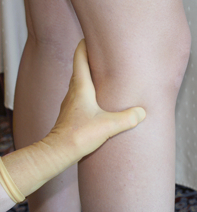
To perform this test, the patient's leg is grasped by the therapist's hands so that the leg is supported and the foot is held in dorsiflexion and inversion. The leg is then passively raised into hip flexion. This is usually felt in the posterolateral calf and/or posterolateral ankle.
What affects the sural nerve?
Sural Nerve Entrapment [7] Entrapment of the sural nerve is most often caused by fascial thickening at the site where the nerve becomes superficial to the gastrocnemius, called the superficial sural aponeurosis. A patient with sural nerve entrapment will present with sensory changes in the area the nerve innervates.
How long does it take for the sural nerve to heal?
Regeneration time depends on how seriously your nerve was injured and the type of injury that you sustained. If your nerve is bruised or traumatized but is not cut, it should recover over 6-12 weeks.
How do you stretch the sural nerve?
Sural Nerve Flossing Gliding Exercises Lay on your back. Pull the affected knee towards the chest, supported by your hands. Gently straighten leg by lifting foot towards the ceiling until a light stretch is felt. Slowly turn your foot towards the inside and bend your ankle towards you.
Where does sural nerve get entrapped?
Entrapment involving the sural nerve typically occurs at the musculotendinous junction of the gastrocnemius muscle and the Achilles tendon within the calf, as the nerve travels through a fibrous arcade (which has been termed the “superficial sural aponeurosis”) [8] (Fig. 71.9a), at the ankle (Fig.
Does the sural nerve heal itself?
In many cases, the condition will resolve on its own with time. Non-surgical treatment includes aggressive massage for nerve desensitization, oral medications to reduce irritation, and avoidance of direct pressure on the nerve.
What causes sural neuritis?
Sural neuritis, also referred to as sural neuralgia, develops when an area of the sural nerve becomes entrapped, irritated or damaged. The most common cause of sural nerve damage is as a result of trauma to the leg, but it can also develop after a surgery if scar tissue ends up enveloping part of the nerve.
What nerve runs down the outside of your foot?
The common peroneal nerve branches from the sciatic nerve and provides sensation to the front and sides of the legs and to the top of the feet. This nerve also controls the muscles in the leg that lift the ankle and toes upward.
What type of nerve is the sural nerve?
The sural nerve (L4-S1) is a cutaneous sensory nerve of the posterolateral calf with cutaneous innervation to the distal one-third of the lower leg.
How do you stretch the sural nerve?
Sural Nerve Flossing Gliding Exercises Lay on your back. Pull the affected knee towards the chest, supported by your hands. Gently straighten leg by lifting foot towards the ceiling until a light stretch is felt. Slowly turn your foot towards the inside and bend your ankle towards you.
How do you perform a sural nerve biopsy?
An incision is made, and the lesser saphenous vein is identified. The vein is then retracted superficially to expose the sural nerve. For a complete nerve biopsy, untied sutures are placed into both ends of the nerve, and the nerve is transected above these sutures.
What does the medial sural nerve innervate?
The medial sural cutaneous nerve (L4-S3) is a sensory nerve of the leg. It supplies cutaneous innervation the posteromedial leg.
What Is Your Sural Nerve? - Cleveland Clinic
Conditions and Disorders What conditions affect the sural nerve? Conditions affecting this nerve include: Diabetes-related neuropathy: Nerve damage that occurs as a result of ongoing high blood sugar. Your sural nerve is one of the most commonly affected nerves by this condition.
Sural nerve - Wikipedia
The sural nerve (L4-S1) is generally considered a pure cutaneous nerve of the posterolateral leg to the lateral ankle. The sural nerve originates from a combination of either the sural communicating branch and medial sural cutaneous nerve, or the lateral sural cutaneous nerve.This group of nerves is termed the sural nerve complex.There are eight documented variations of the sural nerve complex.
Overview
The sural nerve is below your skin’s surface in the back of your lower leg (calf). It’s part of your peripheral nervous system, which helps your brain communicate with the rest of your body.
Anatomy
The sural nerve starts in areas of your upper calf where two nerves join. It travels down the back outer part of your leg, curves at your ankle and ends before your toes.
Conditions and Disorders
Diabetic neuropathy: Nerve damage that occurs as a result of ongoing high blood sugar. Your sural nerve is one of the most commonly affected nerves by this condition.
What nerve supplies the lateral ankle?
Areas of skin sensation supplied by nerves in the leg. Areas of skin supplied by nerves of the leg - the sural nerve supplies the lateral ankle. Deep nerves of the front of the leg. Course of nerves at the bottom of the foot.
What causes sural mononeuropathy?
Sural mononeuropathy is uncommon. If affected, it can be due to a mass lesion such as a ganglion or to trauma , which is the most common cause. Sometimes inflammatory or vasculitic diseases will selectively involve the sural nerve. In addition, the sural nerve will be involved in any kind of generalized peripheral sensory or sensorimotor neuropathy. Sensory changes from sural neuropathy are variable but usually occur in the postero-lateral aspect of the leg and the dorso-lateral foot. These can sometimes be painful with paresthesias and dysesthesias. Nerve conduction studies can be used to delineate sural nerve lesions. Treatment will depend on the cause of the neuropathy. Occasionally biopsy of the nerve is performed for diagnostic purposes. For example, ganglions are usually resected. Traumatic neuropathy is usually treated non-surgically.
What is the sural cutaneous nerve?
The sural cutaneous nerve consists of the fusion of the medial sural cutaneous nerve (MSCN) which is a terminal branch of the tibial nerve and the lateral sur al cutaneous nerve (LSCN) which is one of the terminal branches of the common fibular nerve. These two branches, MSCN and LSCN, are connected by the sural communicating branch and form ...
What nerve is the lateral foot?
Sural nerve. Supplies sensation to the skin of the lateral foot and lateral lower ankle. The sural nerve is a sensory nerve in the calf region ( sura) of the leg. It is made up of branches of the tibial nerve and common fibular nerve, the medial cutaneous branch from the tibial nerve, and the lateral cutaneous branch from the common fibular nerve.
What nerve is responsible for the sensation of the lateral foot and lower ankle?
Function. The sural nerve supplies sensation to the skin of the lateral foot and lateral lower ankle. The nerve transmits sensory signals from the posterior lateral corner of the leg and the lateral foot and 5th toe towards the spinal cord and brain.
Why is sural nerve surgery easier than other procedures?
Because this technique requires few injections to reach adequate anesthesia, a smaller volume of anesthetic is needed. The sural nerve is rather superficial, which makes it more accessible to surgeons. Therefore, it is relatively easier than other procedures.
What is the significance of sural nerve?
Clinical significance. The sural nerve has a purely sensory function, and so its removal results in only a relatively minor deficit. For this reason, it is often used for nerve biopsy. It is often the donor nerve when a nerve allograft is performed.
What nerve is in the distal third of the leg?
The pathway of the nerve includes a non-extensible anatomic fibrous arcade, beyond which it runs superficially in the distal third of the leg. This arch is wide, thick, and unyielding and may fit tightly around the nerve, causing chronic, frictional irritation. Symptoms most commonly associated with irritation of the sural nerve include chronic (because the diagnosis is usually missed for a long time) pain in the posterior aspect of the leg, usually exacerbated with physical exertion. Radiating pain/tingling into the foot may occur distally, which may or may not be accompanied by referred pain proximally in the calf.
What nerve provides sensation?
Description. The Sural Nerve is a cutaneous nerve it provides only sensation, the areas being. Posterolateral aspect of the distal third of the leg. Lateral aspect of the foot, heel, and ankle.
What is the pain in the posterior aspect of the leg?
Symptoms most commonly associated with irritation of the sural nerve include chronic (because the diagnosis is usually missed for a long time) pain in the posterior aspect of the leg, usually exacerbated with physical exertion.
What can be used to reduce frictional irritation in the area?
As with any nerve irritation syndrome manual soft tissue techniques can be used to decrease frictional irritation in the area.
Which nerve supplies sensation to the lower lateral leg, lateral heel, ankle and dorsal lateral foot?
The sural nerve is purely sensory and it supplies sensation to the lower lateral leg, lateral heel, ankle and dorsal lateral foot.
Where is neural irritation at the fibrous arcade?
In the case of neural irritation at the location of the fibrous arcade clinical examination may reveals tenderness with palpation posterior and lateral to the myotendonous junction of the Achilles (at the location of the fibrous arcade).
Is a peronial nerve biopsy a complication?
For this reason, it is often used for nerve biopsy, as well as the donor nerve for nerve grafts. However, there is also evidence of a higher complication rate than peronial nerve biopsy leading to post-operative pain, dysaesthesia and parasthesia during follow ups.
What is the most common injury to the sural nerve?
Damage to your sural nerve can come from a number of different sources, but the most common way a person injuries their sural nerve is during an ankle sprain.
What is the goal of sural nerve treatment?
The goal of sural nerve treatment is to free the nerve from the underlying issue that is causing the irritation. In most cases, sural nerve problems can be managed with non-surgical treatment techniques. Some common non-operative treatment options include:
What are the symptoms of sural nerve irritation?
Symptoms of sural nerve irritation include: Pain on the outside of the foot. A burning sensation. Symptoms that are aggravated by direct pressure. Hypersensitivity. Localized tenderness.
How to diagnose sural nerve damage?
Treating sural nerve issues begins with an accurate diagnosis, and this is best achieved through a physical exam with an orthopedic specialist. Your doctor will ask about your symptoms and take a closer look at your foot. They may also conduct some physical manipulation tests to see if certain movements cause the onset of symptoms. One such test is known as the Tinel test, which involves percussion at the nerve site to see if pain or electrical-like sensations are felt. Doctors will also look for swelling or tenderness in the area. If they suspect that sural nerve damage has occurred, they will likely order some imaging tests.
Where is the sural nerve located?
Your sural nerve runs down the back of your leg, behind the outside edge of your ankle and down the outside of your foot. It provides sensation to those areas and to your two outside toes, and problems can occur if the nerve is irritated or damaged.
What is the test for a nerve?
They may also conduct some physical manipulation tests to see if certain movements cause the onset of symptoms. One such test is known as the Tinel test , which involves percussion at the nerve site to see if pain or electrical-like sensations are felt. Doctors will also look for swelling or tenderness in the area.
What is the best way to break up scar tissue?
Desensitization – This involves direct massage to the area, which may help to break up scar tissue and desensitize the area over time.
What nerve controls movement and sensation in the leg and foot?
Function. The sciatic nerve controls most movement and sensation throughout the leg and foot. The motor branches of the sciatic nerve receive messages from the anterior fibers in the spine and the spinal roots. They travel down the leg, with smaller nerve branches extending to muscles in the leg and foot throughout the course of the nerve.
How many spinal nerves are in the sciatic nerve?
The sciatic nerve is formed by a total of five spinal nerves that combine with each other.
What is the largest nerve in the body?
Anatomy. Function. Associated Conditions. Rehabilitation. The sciatic nerves, located in the legs, are the largest nerves in the body. 1 Each sciatic nerve stimulates movement of leg muscles and carries sensory messages from the leg to the spine.
Why does sciatica hurt?
A herniated disc that pinches a nerve root is the most common of several causes of sciatica. 2 Since the sciatic nerve mediates sensation and movement, an injury or compression of the nerve can cause you to have pain, decreased sensation, and/or weakness.
Which nerve stimulates the leg?
Motor branches of the sciatic nerve stimulate muscles of the leg. These motor nerves stimulate several movements, including extension of the hip, flexion of the leg at the knee, and flexion of the foot and toes.
Which nerve is primarily a motor nerve?
The tibial and fibular nerves travel down the leg to the foot, dividing into smaller motor and sensory branches along the way. The tibial nerve is primarily a motor nerve, and the fibular nerve is primarily a sensory nerve.
Why does my foramen feel compressed?
Problems such as arthritis, inflammation, and bone deterioration can affect the size and shape of the foramen through which the spinal roots travel, resulting in compression of the nerve root (pinched nerve), with resulting symptoms of sciatic dysfunction or pain. Some women experience symptoms of a pinched nerve during pregnancy, and it usually resolves after the baby is born. 4
What causes sural nerve entrapment?
Sural nerve entrapment is a rare clinical condition that occurs when the nerve is compressed or fixed. One of the most common causes is fascial thickening which occurs when the rigid anatomic fibrous arcade formed at the fascial opening becomes thickened and doubled. This forms a fibrous arch that may compress the sural nerve. It can also be caused by fractures of the sural area, intrinsic factors (ganglions and lipomas), prolonged external compression, repetitive ankle sprains, and gastrocnemius muscle injury.
What is the sural nerve?
The sural nerve is a purely sensory nerve. Its main function is to provide the sensory supply for the posterolateral aspect of the distal third of the leg, lateral aspect of the foot, heel and ankle.
Where does the sural nerve enter the foot?
The sural nerve then passes between the lateral malleolus and the calcaneus and enters into the foot. Upon reaching the lateral aspect of the foot, the nerve terminates as the lateral dorsal cutaneous nerve .
Where is the sural nerve located?
The sural nerve (S1, S2) is a peripheral nerve that arises in the posterior compartment of the leg (calf or sural region). It is formed by the union of two smaller sensory nerves: the medial sural cutaneous nerve (a branch of the tibial nerve ), and lateral sural cutaneous nerve (branch of the common fibular nerve ). In the posterior leg, the sural nerve courses alongside the small saphenous vein .
Which nerve innervates the skin over the posterolateral aspect of the distal third of the leg?
In the leg, the sural nerve directly innervates the skin over the posterolateral aspect of the distal third of the leg.
Which nerve supplies the lateral side of the dorsum of the foot?
Lateral dorsal cutaneous nerve of foot, which supplies the lateral side of the dorsum of the foot. This nerve ends as a dorsal digital nerve for the sensory supply of the lateral aspect of the fifth toe.
Overview
The sural nerve (L4-S1) is generally considered a pure cutaneous nerve of the posterolateral leg to the lateral ankle. The sural nerve originates from a combination of either the sural communicating branch and medial sural cutaneous nerve, or the lateral sural cutaneous nerve. This group of nerves is termed the sural nerve complex. There are eight documented variations of the …
Anatomy
The sural nerve (L4-S1) is a cutaneous sensory nerve of the posterolateral calf with cutaneous innervation to the distal one-third of the lower leg. Formation of the sural nerve is the result of either anastomosis of the medial sural cutaneous nerve and the sural communicating nerve, or it may be found as a continuation of the lateral sural cutaneous nerve traveling parallel to the medial sural cutaneous nerve. The sural nerve specifically innervates cutaneous sensorium over the post…
Innervation
The sural nerve provides cutaneous innervation to the skin of the posterior to posterolateral leg. This nerve is part of the sciatic nerve sensorium. It only provides autonomic and sensory nerve fibers to the skin of the posterolateral leg and ankle. These fibers originate from perikaryon located in the spinal ganglia and travel via the lumbosacral plexus via nerve roots L4-S1. When testing for deficits understand that often multiple nerves (lateral calcaneal nerve, sural nerve, and lateral dorsal cut…
Anatomic Course
Grossly, the course of this nerve leads it from its highly varied anastomotic formation to its more predictable terminal course down the remaining posterior leg. The anastomosis forming the sural nerve typically occurs in the deep fascia above or within the surrounding space above the gastrocnemius muscle. Once formed, the sural nerve then pierces out of a fascial crura and travels from its posterior midline position wrapping laterally around the lateral malleolus; once a…
Anatomic Formation
The nerves contributing to the formation of the sural nerve (medial sural cutaneous nerve, lateral sural cutaneous nerve, sural communicating nerve) are deemed the sural nerve complex by some anatomists and surgeons. Eight formations of the sural nerve complex have been described in cadaveric studies.
The pathway of the sural nerve (once formed) is consistent as it travels superficially in the poste…
Clinical significance
The sural nerve has a purely sensory function, and so its removal results in only a relatively minor consequential deficit. Due to its large size and significant length it has had a significant contribution in medicine twofold; the sural nerve is the most frequently accessed donor nerve site for peripheral nerve grafting and serves as the primary diagnostic site for nerve conduction studies for understanding of peripheral nerve pathologies. It is frequently a site of iatrogenic ner…
External links
Referenced papers:
• Steele et al. 208 sample cadaveric review 2021
• Ramakrishnan et al.systematic review on sural nerve formation 2015
• Anatomy photo:11:07-0202 at the SUNY Downstate Medical Center