Where does the thoracic duct exit the diaphragm?
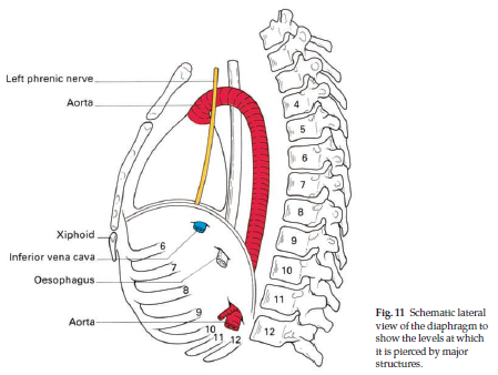
The thoracic duct ascends through the aortic hiatus of the diaphragm entering the posterior mediastinum, still to the right of the vertebral column. It courses posterior to the esophagus at the T7 level and crosses over the midline to the left side of the thorax around the T5 vertebral level.
What structures pass through the diaphragm?
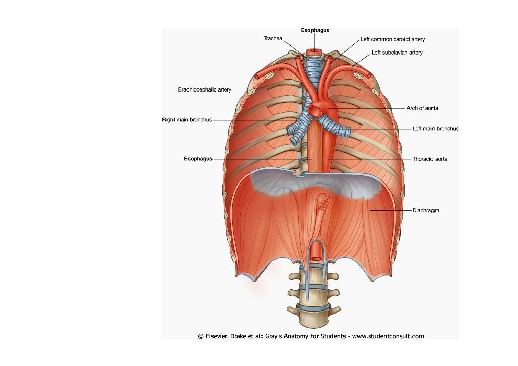
Structures passing between the thoracic and abdominal cavities pass through the diaphragm, or through openings between some of the peripheral attachments. Aortic hiatus: The thoracic aorta passes through this opening between the crura of the diaphragm. The azygos vein and the thoracic duct also pass through here.
What is the thoracic surface of the diaphragm?
Thoracic surface of the diaphragm (diagram) The diaphragm has two surfaces: thoracic and abdominal. The thoracic diaphragm is in contacts with the serous membranes of the heart and lungs ; namely, the pericardium and pleura. The abdominal diaphragm is in direct contact with the liver, stomach , and spleen .
What passes through the central tendon of the diaphragm?
Caval opening: The inferior vena cava passes through the central tendon of the diaphragm at the level of the 8th thoracic vertebra. Esophageal hiatus: The esophagus passes through muscular fibres of the diaphragm slightly to the left of the midline at the level of the T10 vertebra.
See more
What is the thoracic duct and where does it drain?
The thoracic duct is the main lymphatic vessel for the return of chyle/lymph to the systemic venous system. It drains lymph from both lower limbs, abdomen (except the convex area of the liver), left hemithorax, left upper limb and left side of face and neck.
Where does the thoracic duct enter the thorax?
It enters the thorax through the aortic opening of the diaphragm between the aorta and the azygos vein. In the posterior mediastinum, the thoracic duct lies anterior to the vertebral column, the right intercostal arteries, and the hemiazygos veins as they cross to open into the azygos vein.
What goes into thoracic duct?
The thoracic duct drains lymph from the right and left descending thoracic lymph trunks, originating from the lower 6 intercostal spaces (6 to 11). The duct also receives lymph from intercostal spaces 1 to 5 via the upper intercostal lymph trunks. Additional tributaries include the: mediastinal lymph trunks.
Which of the following areas of the body will drain into the thoracic duct?
the thoracic duct: It begins near the lower part of the spine and collects lymph from the pelvis, abdomen, and lower chest. The thoracic duct runs up through the chest and empties into the blood through a large vein near the left side of the neck.
Where do the lymphatic ducts empty into?
The lymphatic vessels drain into collecting ducts, which empty their contents into the two subclavian veins, located under the collarbones. These veins join to form the superior vena cava, the large vein that drains blood from the upper body into the heart.
Which statement is true of the thoracic duct?
Which statement is true of the thoracic duct? It drains the lymph from the entire left side of the body and the right abdomen and leg.
Where is cisterna chyli located?
retrocrural spaceThe cisterna chyli is the abdominal origin of the thoracic duct, and it receives the bilateral lumbar lymphatic trunks. It is located in the retrocrural space, to the right side and behind of the abdominal aorta.
Where is right lymphatic duct located?
neckThe right lymphatic duct is a terminal lymphatic vessel located in the neck, anterior to the anterior scalene muscle. It is typically formed by the union of the right bronchomediastinal, right jugular and right subclavian lymphatic trunks, although its formation is highly variable.
What is the thoracic duct?
Thoracic Duct. The thoracic duct is the largest lymphatic vessel within the human body, and plays a key role in the lymphatic system. It is also called the left lymphatic duct or the alimentary duct. A large portion of the body’s lymph is collected by this duct and then drained into the bloodstream near the brachiocephalic vein between ...
What is the left lymphatic duct?
It is also called the left lymphatic duct or the alimentary duct. A large portion of the body’s lymph is collected by this duct and then drained into the bloodstream near the brachiocephalic vein between the internal jugular and the left subclavian veins.
Where does the duct originate?
It originates from the second lumbar vertebra level and goes to the neck’s root. The duct arises from the combination of the left and right lumbar trunks and the intestinal trunk in the abdomen.
How much lymphatic fluid does the aortic duct transport?
It travels through the aortic aperture diaphragm and rises along the posterior mediastinum. It transports up to four liters of lymphatic fluid each day. This process is primarily caused by the breathing action and is assisted by the smooth muscle of the duct. Last medically reviewed on January 21, 2018.
What is the diaphragm in the thorax called?
The diaphragm in the thorax is called the thoracic diaphragm and serves as an important anatomical landmark that separates the thorax, or chest, from the abdomen.
Which nerves pass through the diaphragm?
The esophagus, phrenic, and vagus nerves, descending aorta, and inferior vena cava pass through the diaphragm between the thoracic and abdominal cavities. The diaphragm is asymmetric with the left side slightly more inferior than the right, chiefly because of the presence of the liver located on the right.
What is the function of the thoracic tendon?
It functions during breathing when it contracts to enlarge the thoracic cavity and reduce the intrathoracic pressure so that lungs may expand and fill their alveoli with air. It is a dome-shaped muscle and tendon that functions as the main muscle of respiration and is essential to the breathing process.
How does the diaphragm work?
The diaphragm pulls its central tendon down during contraction and then increases the vertical diameter of the thorax. This increases the negative pressure inside the thoracic cavity, which draws in air. Thus, the diaphragm is the most important muscle used in inspiration. During inhalation, the diaphragm contracts and is pushed inferiorly into the abdominal cavity where it appears flat. Simultaneously the external intercostal muscles located in between the ribs raise the anterior chest wall like the handles of a bucket. This results in the chest cavity becoming larger and wider, which allows air in from the outside. During exhalation, the rib cage and chest wall start to sag and revert to the original position. At the same time, there is relaxation and elevation of the diaphragm. This motion forces the air within the lungs to push out of the body. [2],[3]
Why is the diaphragm asymmetric?
The diaphragm is asymmetric with the left side slightly more inferior than the right, chiefly because of the presence of the liver located on the right. The left side may also be partially inferiorly located because of the push by the heart.[1],[2] The diaphragm in the thorax is called the thoracic diaphragm and serves as an important anatomical ...
Which sensory nerves are activated during each breathing cycle?
It is now well established that activation of both non-myelinated and myelinated phrenic sensory nerves modulate respiratory output during each breathing cycle. However, the activation of the phrenic afferents does increase significantly as the diaphragm continues to work and develops fatigue.
Which nerve innervates the parietal pleura and peritoneum covering the central surfaces of the?
The phrenic nerve innervates the parietal pleura and peritoneum covering the central surfaces of the diaphragm. The lower 6 intercostal nerves supply the periphery of the diaphragm. When the diaphragm contracts, the large-sized myelinated phrenic afferents fire.
Where does the thoracic duct develop?
The thoracic duct develops from lymphatic trunks on either side of the aorta that anastomoses to form a channel from the jugular lymph sacs to the cisterna chyli. Trunks continue to anastomose and enlarge, forming embryonic right and left thoracic ducts. The adult thoracic duct is derived from both of these embryonic thoracic ducts.
What is the function of the thoracic duct?
The function of the thoracic duct is to transport lymph back into the circulatory system. Interstitial fluid is collected by lymph capillaries from the interstitial space. Lymph then moves through lymphatic vessels to lymph nodes. Lymphatic vessels merge to create the lymphatic ducts which drain into the venous system.
How many vessels can a thoracic duct terminate?
The thoracic duct can also terminate as a single vessel (up to 87.5%), bilateral ducts (up to 25%), or several terminal branches (up to 7%). The thoracic duct displays physiologic adaptation to certain disease processes by increasing in diameter.
What are the two lymphatic ducts?
Introduction. Lymphatic ducts empty lymph fluid into the venous system. The two lymphatic ducts of the body are the right lymphatic duct and the thoracic duct. The thoracic duct is the larger of the two and responsible for lymph drainage from the entire body except for the right sides of the head and neck, ...
How long is the thoracic duct?
The thoracic duct is 38 to 45 centimeters long and 2 to 5 millimeters in diameter. It runs from the superior aspect of the cisterna chyli, a lymph sac at the L2 vertebral level, to the lower cervical spine. From the cisterna chyli, the duct continues superiorly, running between the aorta and the azygous vein and anterior to the vertebral column. The thoracic duct ascends through the aortic hiatus of the diaphragm entering the posterior mediastinum, still to the right of the vertebral column. It courses posterior to the esophagus at the T7 level and crosses over the midline to the left side of the thorax around the T5 vertebral level. As it continues upward, it runs behind the aorta and to the left of the esophagus ascending 2-3 cm above the clavicle. In the superior mediastinum, it passes behind the left common carotid artery, the vagus nerve, and the internal jugular vein. It then descends to empty into the junction of the left subclavian and internal jugular veins.
Which muscle contracts to move lymph forward?
The smooth muscle contracts regularly to move lymph flow forward. The thoracic duct also contains valves which may be unicuspid, bicuspid, or tricuspid, but are usually bicuspid. At the junction of the lymphatic and venous system, a bicuspid valve prevents venous backflow into the lymphatic system. [3]
Where does lymph go in the body?
Lymph from organs can drain directly into the thoracic duct without passing a lymph node. This anodal route has been observed for the diaphragm, esophagus, and parts of the lungs. The drainage pattern may play a role in the prognosis of cancers of these organs.
How does the diaphragm separate the thoracic cavity?
It separates the thoracic and abdominal cavities from each other by closing the inferior thoracic aperture. The diaphragm is the primary muscle that is active in inspiration. Contraction of the muscle facilitates expansion of the thoracic cavity. This increases volume of the the cavity, which in turn decreases the intrathoracic pressure allowing ...
Where does the diaphragm innervate?
Motor innervation of the diaphragm comes from the phrenic nerves (C3-C5). These nerves innervate the diaphragm from its abdominal surface after they penetrate it. Sensory innervation (pain and proprioception) at the central tendinous part is innervated by the phrenic nerves , while the peripheral muscular portions are innervated by 6th to 11th intercostal nerves .
What is the diaphragm?
The diaphragm is a musculotendinous structure with a peripheral attachment to a number of bony structures. It is attached anteriorly to the xiphoid process and costal margin, laterally to the 11th and 12th ribs, and posteriorly to the lumbar vertebrae. The posterior attachment to the vertebrae is by tendinous bands called crura. The crura are attached to the anterior aspect of the bodies of the 1st, 2nd and 3rd lumbar vertebrae. The muscle fibres, extending from their bony attachments, converge on a central tendon.
Why is the diaphragm shaped like a dome?
The diaphragm is shaped as two domes, with the right dome positioned slightly higher than the left because of the liver. The depression between the two domes is due to the pericardium slightly depressing the diaphragm. The diaphragm has two surfaces: thoracic and abdominal.
Which artery is associated with the diaphragm?
Inferior phrenic arteries are closely related to the diaphragm and give off a few branches to supply it. They are the main source of vascular supply to the diaphragm. The left inferior phrenic artery ascends toward the left diaphragmatic crus associated with the inferior surface of the diaphragm.
Which surface of the diaphragm passes through the right crus?
Abdominal surface of the diaphragm in a cadaver: The esophageal hiatus passes through the right crus of the diaphragm. The foramen of the inferior vena cava traverses through the central tendon, while the aortic hiatus passes behind the diaphragm.
How to remember the location of the diaphragm?
An easy way to remember the location and structures passing through the diaphragm is by using this mnemonic: 'I 8 10 EGG s AT 12' (read: I ate ten eggs at twelve).
What is the function of the diaphragm?
The diaphragm is a thin skeletal muscle that sits at the base of the chest and separates the abdomen from the chest. It contracts and flattens when you inhale. This creates a vacuum effect that pulls air into the lungs. When you exhale, the diaphragm relaxes and the air is pushed out of lungs.
Which nerve controls the movement of the diaphragm?
The phrenic nerve, which runs from the neck to the diaphragm, controls the movement of the diaphragm. There are three large openings in the diaphragm that allow certain structures to pass between the chest and the abdomen. These openings include the: Esophageal opening. The esophagus and vagus nerve, which controls much of the digestive system, ...
What is an acquired diaphragmatic hernia?
In this case, it’s called an acquired diaphragmatic hernia (ADH). Symptoms can vary depending on the size of the hernia, the cause, and the organs involved. They may include: Both an ADH and CDH require immediate surgery to remove the abdominal organs from the chest cavity and repair the diaphragm.
How to strengthen the diaphragm?
exercising within your limits. Like any muscle, you can also strengthen your diaphragm with special exercises. Diaphragmatic breathing or abdominal breathing is the best way to do this. It involves inhaling deeply and slowly through the nose so that your lungs fill with air as your belly expands.
Why does my diaphragm feel tight?
During a spasm, the diaphragm doesn’t rise back up after exhalation. This inflates the lungs, causing the diaphragm to tighten. This can also cause a cramping sensation in the chest. Vigorous exercise can cause the diaphragm to spasm, which often results in what people call a side stitch.
How long does it take for a diaphragm spasm to go away?
Diaphragm spasms usually go away on their own within a few hours or days.
How do you know if you have a diaphragm problem?
Symptoms of a diaphragm condition may include: difficulty breathing when lying down. shortness of breath. chest, shoulder, back, or abdominal pain . pain in your lower ribs. a fluttering or pulsing sensation in the abdomen.