
Which best describes a centromere?
Which best describes a centromere? sister chromosomes that are held together by a chromatid paired chromosomes that have genes arranged in the same order the center of a chromosome the material that makes up a chromosome. Which best describes a centromere? The answer is sister chromosomes that are held together by a chromatid.
What does centromere do?
What Does The Centromere Do? The primary function of the centromere is to provide the foundation for assembly of the kinetochore, which is a protein complex essential to proper chromosomal segregation during mitosis. In electron micrographs of mitotic chromosomes, kinetochores appear as platelike structures composed of several layers (Figure 4).
Where are the centrioles during interphase?
Centrioles are located outside of, but near the cell nucleus. In cell division, there are several phases: in order of occurrence they are interphase, prophase, metaphase, anaphase, and telophase. Centrioles have a very important role to play in all phases of cell division. The end goal is in moving replicated chromosomes into a newly created cell.
Where are the two sister chromatids connected?
Sister Chromatids Definition. Sister chromatids are two identical copies of the same chromosome formed by DNA replication, attached to each other by a structure called the centromere. During cell division, they are separated from each other, and each daughter cell receives one copy of the chromosome.

Where does the centromere form?
Monocentric chromosomes assemble a centromere at a single localized site on the chromosome, which is visible as a constriction between the chromosomes in mitosis (known as the primary constriction). Monocentric chromosomes can be further divided into those with point centromeres and those with regional centromeres.
What is found in centromere?
The centromeres of human chromosomes are heterochromatic regions that consist largely of a repeated sequence known as alpha satellite DNA. A single unit of alpha satellite DNA is 171 base pairs in length, each of which contains a 17-base-pair binding site for CENP-B, referred to as the CENP-B box.
Where on a human chromosome is the centromere?
The centromere is the chromosomal locus essential for chromosome inheritance and genome stability. Human centromeres are located at repetitive alpha satellite DNA arrays that compose approximately 5% of the genome.
What are the three centromere locations?
The position of the centromere relative to the ends helps scientists tell chromosomes apart. Centromere position can be described three ways: metacentric, submetacentric or acrocentric.
What does a centromere hold together?
centromere, structure in a chromosome that holds together the two chromatids (the daughter strands of a replicated chromosome). The centromere is the point of attachment of the kinetochore, a structure to which the microtubules of the mitotic spindle become anchored.
Does every chromatid have a centromere?
A chromatid is a replicated chromosome having two daughter strands joined by a single centromere (the two strands separate during cell division to become individual chromosomes).
How many centromeres do humans have?
46 centromeresA centromere is a non-coding region of DNA located in the center of the chromosome. During mitosis, the centromeres help hold together duplicated sister chromatids prior to separating during anaphase. Since human cells have 46 chromosomes, they also have 46 centromeres.
Where is the chromosome located?
nucleusChromosomes are structures found in the center (nucleus) of cells that carry long pieces of DNA. DNA is the material that holds genes. It is the building block of the human body. Chromosomes also contain proteins that help DNA exist in the proper form.
Where is the centromere in chromosome 8?
Chromosome 8Centromere positionSubmetacentric (45.2 Mbp)Complete gene listsCCDSGene listHGNCGene list16 more rows
What is centromere and its function?
Centromere The centromere appears as a constricted region of a chromosome and plays a key role in helping the cell divide up its DNA during division (mitosis and meiosis). Specifically, it is the region where the cell's spindle fibers attach.
What is the function of centrosome?
The centrosome is the primary microtubule-organizing centre (MTOC) in animal cells, and so it regulates cell motility, adhesion and polarity in interphase, and facilitates the organization of the spindle poles during mitosis.
Is centromere a protein?
Centromere protein A (CENP-A) is a variant of histone H3 with more than 60% sequence identity at the C-terminal histone fold domain.
Do centromeres contain genes?
Centromeres typically are in silent or gene-free chromosome regions but may include genes [[5], [6], [7]], and are commonly transcribed at low levels to form non-coding RNAs that interact with kinetochores and appear to assist in cenH3 loading (reviewed in Refs.
Is centromere a protein?
Centromere protein A (CENP-A) is a variant of histone H3 with more than 60% sequence identity at the C-terminal histone fold domain.
What is a centromere and what is its function?
Centromere The centromere appears as a constricted region of a chromosome and plays a key role in helping the cell divide up its DNA during division (mitosis and meiosis). Specifically, it is the region where the cell's spindle fibers attach.
How is centromere formed?
From fission yeast to human, centromeres are established on a series of repetitive DNA sequences and on specialized centromeric chromatin. This chromatin is enriched with the histone H3 variant, named CENP-A, that was demonstrated to be the epigenetic mark that maintains centromere identity and function indefinitely.
What is the function of the centromere?
Function of Centromere. All living things are made up of cells. In order for cells to grow or reproduce, cell division must occur. In cell division, one “parent” cell splits in two, with each of the resulting cells being “daughter” cells. For each daughter cell to survive, it is essential that they get a copy of each of their parent cells’ ...
What type of centromeres do humans use?
Humans and most eukaryotic cells use regional centromeres. These are centromeres where mitotic spindle binding is determined, not by a precise sequence of DNA, but by a combination of characteristics working together to signal the location of a centromere.
What is centromere dysfunction?
Centromere dysfunction is also suspected to play a role in cancer cells, which display massive chromosome imbalance of the type that would be expected if the sorting of chromosomes during cell division failed.
What is the role of centromere dysfunction in miscarriage?
Centromere dysfunction leading to problems with chromosome sorting is believed to play a role in many instances of miscarriage, in which inherited centromere disorders may result in early embryonic death. Centromere dysfunction is also suspected to play a role in cancer cells, which display massive chromosome imbalance of the type ...
What is the cytoskeleton at the centromere?
At the centromere, elements of the cell’s cytoskeleton assemble and attach. First, a complex of proteins called the kinetochore assembles around the centromere region of DNA; then, mitotic spindle fibers attach to the kinetochore. The other end of these fibers are anchored to opposite ends of the parent cell, which will shortly split ...
What is the point on a chromosome where mitotic spindle fibers attach to pull sister?
The centromere is the point on a chromosome where mitotic spindle fibers attach to pull sister chromatids apart during cell division. When a cell seeks to reproduce itself, it must first make a complete copy of each of its chromosomes, to ensure that their daughter cell receives a full complement of the parent cell’s DNA.
Why are the two sister chromatids together called a single chromosome?
The two sister chromatids combined are often referred to as a single chromosome because they are packaged tightly together – but each contains all the information of the original chromosome, so when they split, each becomes a complete chromosome containing all of the information contained in the parent cell’s original chromosome.
Where is the centromere located?
The word centromere ( / ˈsɛntrəˌmɪər /) uses combining forms of centro- and -mere, yielding "central part", describing the centromere's location at the center of the chromosome.
What type of DNA is a centromere?
Any piece of DNA with the point centromere DNA sequence on it will typically form a centromere if present in the appropriate species. The best characterised point centromeres are those of the budding yeast, Saccharomyces cerevisiae.
What is the centromere of a holocentric chromosome?
Unlike monocentric chromosomes, in holocentric chromosomes the entire length of the chromosome act s as the centromere. In holocentric chromosomes there is not one primary constriction but the centromere has many CenH3 loci spread over the whole chromosome. Examples of this type of centromere can be found scattered throughout the plant and animal kingdoms, with the most well-known example being the nematode Caenorhabditis elegans .
How does centromere misregulation affect cancer?
Notably, overexpression of many centromere genes have been linked to cancer malignant phenotypes. Overexpression of these centromere genes can increase genomic instability in cancers . Elevated genomic instability on one hand relates to malignant phenotypes; on the other hand, it makes the tumor cells more vulnerable to specific adjuvant therapies such as certain chemotherapies and radiotherapy. Instability of centromere repetitive DNA was recently shown in cancer and aging.
How are dicentric chromosomes formed?
A dicentric chromosome is an abnormal chromosome with two centromeres. It is formed through the fusion of two chromosome segments, each with a centromere, resulting in the loss of acentric fragments (lacking a centromere) and the formation of dicentric fragments. The formation of dicentric chromosomes has been attributed to genetic processes, such as Robertsonian translocation and paracentric inversion. Dicentric chromosomes have important roles in the mitotic stability of chromosomes and the formation of pseudodicentric chromosomes.
What is the centromere in mitosis?
When cells enter mitosis, the sister chromatids (the two copies of each chromosomal DNA molecule resulting from DNA replication in chromatin form) are linked along their length by the action of the cohesin complex. It is now believed that this complex is mostly released from chromosome arms during prophase, so that by the time the chromosomes line up at the mid-plane of the mitotic spindle (also known as the metaphase plate), the last place where they are linked with one another is in the chromatin in and around the centromere.
What is the role of centromeres in cell division?
The physical role of the centromere is to act as the site of assembly of the kinetochores – a highly complex multiprotein structure that is responsible for the actual events of chromosome segregation – i.e. binding microtubules and signalling to the cell cycle machinery when all chromosomes have adopted correct attachments to the spindle, so that it is safe for cell division to proceed to completion and for cells to enter anaphase.
What holds chromosomes together?
chromosome. …are held together by the centromere. The centromere is the point of attachment of the kinetochore, a protein structure that is connected to the spindle fibres (part of a structure that pulls the chromatids to opposite ends of the cell). During the middle stage in cell division, the centromere duplicates,….
What is the structure that holds together the two chromatids?
See Article History. Centromere, structure in a chromosome that holds together the two chromatids (the daughter strands of a replicated chromosome). The centromere is the point of attachment of the kinetochore, a structure to which the microtubules of the mitotic spindle become anchored. The spindle is the structure that pulls ...
Which structure pulls the chromatids to opposite ends of the cell during the cell division process?
The spindle is the structure that pulls the chromatids to opposite ends of the cell during the cell division processes of mitosis and meiosis. Once separated, each chromatid becomes a chromosome. Thus, when the cell divides, both daughter cells have complete sets of chromosomes.
Do sister chromatids separate during mitosis?
During mitosis the sister chromatids separate, one going to each daughter cell. Chromosomes thus meet the first criterion for being the repository of genes: they are replicated, and a full copy is passed to each daughter cell during mitosis.…. …are held together by the centromere.
Where is the centromere located?
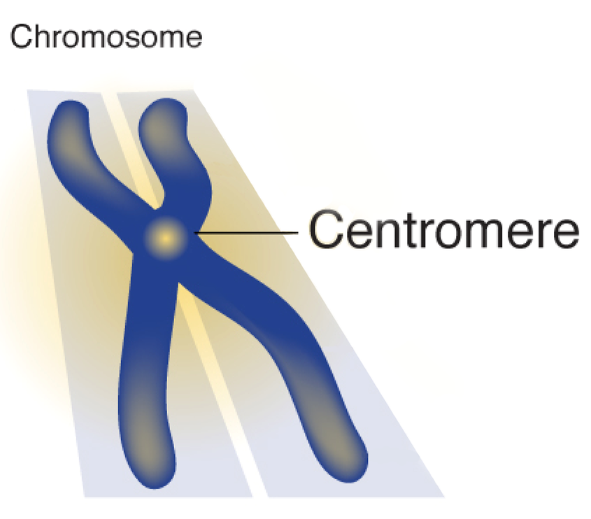
Centromere Localization. On a condensed, duplicated chromosome it is easy to see where the centromere is located . It's the part where the two chromatids are connected and form an X shape. Even on a single condensed chromosome, the centromere forms a constriction that can be seen in the microscope.
What is the function of a centromere?
Centromere Functions. One major function of a centromere is joining the sister chromatids. The two copies of a replicated chromosome are called sister chromatids, and they must stay joined together until it is time for them to be physically pulled into the two future daughter cells.
What is the role of centromeres in eukaryotes?
In eukaryotes, a centromere is a region of DNA that is responsible for the movement of the replicated chromosomes into the two daughter cells during mitosis and meiosis . There is one centromere on each chromosome, and centromeres are responsible for two major functions. You must c C reate an account to continue watching.
Why are centromeres important?
First, they provide one way to help recognize which chromosome is which under the microscope. Also, centromeres are good landmarks for describing the locations of genes along the length of a chromosome. Often, the centromeres are not exactly in the center, so they divide chromosomes into long arms and short arms.
What is the name of the centromere that is located in the center of the chromosome?
They can have various different positions, as shown in this diagram. When the centromere is approximately in the center of a chromosome, it is called metacentric. 'Meta' means middle, so this makes sense. Submetacentric centromeres are closer to one end of the chromosome than the other.
What is the term for centromeres that are close to one end of the chromosome?
Centromeres that are very close to one end of the chromosome are called acrocentric. 'Acro' means top or extremity. Telocentric centromeres are positioned at the very end of a chromosome. 'Telo' means last or end.
What is the segment of DNA that dictates the movement of chromosomes when replicating during cell division?
A centromere is the segment of DNA that dictates the movement of chromosomes when replicating during cell division. Discover the functions, structure, and locations of the centromere in DNA. Updated: 09/16/2021
What Is a Centromere?
A centromere has a highly conserved sequence of 170 bases that are repeated from 5,000 to 15,000 times.
Where is the centromere located in the submetacentric chromosome?
In the submetacentric chromosome, the centromere lies slightly off from the middle
What Are ‘Diffuse’ Centromeres?
Consequently, in the diffuse centromeres of these unique chromosomes, kinetochore bind with microtubules to spread across the chromosome.
What is the difference between a kinetochore and a centromere?
The centromere is often confused with the kinetochore because they share many similarities. However, the centromere is the DNA sequence while the kinetochore is a protein complex connecting to the centromere.
What is the role of centromeric chromatin in the formation of kinetochores?
Centromeric chromatin establishes centromere identity and creates a foundation for the kinetochore.
Why is cancer caused by the inaccurate segregation of chromosomes into daughter cells?
Cancer may be caused by the inaccurate segregation of chromosomes into daughter cells because of defects in the centromere at the DNA and protein level. This inaccuracy results in an imbalance in the number of chromosomes.
What are two highly conserved features that show up at many different phases of cell evolution?
A highly conserved feature shows up at many different phases in cell evolution. Two discovered in cell biology are the centromere and telomere.
What is centromere antibody?
Centromere Antibody is a biomarker for scleroderma and CREST syndrome.
What percentage of Raynaud's patients have anti-centromere antibodies?
Twenty five percent of patients with Raynaud’s Syndrome have anti-centromere antibodies. They are also detected in 12% of patients with primary biliary cirrhosis, half of whom have clinical features of scleroderma.
What percentage of patients have centromere antibodies?
In various reported clinical studies, centromere antibodies occur in 50% to 96% of patients with calcinosis, Raynaud phenomenon, esophageal dysfunction, sclerodactyly, and telangiectasis (CREST) syndrome.
What is the CREST syndrome?
Centromere antibodies occur primarily in patients with the calcinosis, Raynaud phenomenon, esophageal dysfunction, sclerodactyly, and telangiectasis (CREST) syndrome variant of systemic sclerosis (scleroderma). CREST syndrome is characterized by the following clinical features: calcinosis, Raynaud phenomenon, esophageal hypomotility, sclerodactyly, and telangiectasia. (1) Centromere antibodies were originally detected by their distinctive pattern of fine-speckled nuclear staining on cell substrates used in the fluorescent antinuclear antibody test. (2) In subsequent studies, centromere antibodies were found to react with several centromere proteins of 18 kDa, 80 kDa, and 140 kDa named as CENP-A, CENP-B, and CENP-C, respectively. (3) Several putative epitopes associated with these autoantigens have been described. The CENP-B antigen is believed to be the primary autoantigen and is recognized by all sera that contain centromere antibodies.
Overview
The centromere links a pair of sister chromatids together during cell division. This constricted region of chromosome connects the sister chromatids, creating a short arm (p) and a long arm (q) on the chromatids. During mitosis, spindle fibers attach to the centromere via the kinetochore.
The physical role of the centromere is to act as the site of assembly of the kine…
Position
In humans, centromere positions define the chromosomal karyotype, in which each chromosome has two arms, p (the shorter of the two) and q (the longer). The short arm 'p' is reportedly named for the French word "petit" meaning 'small'. The position of the centromere relative to any particular linear chromosome is used to classify chromosomes as metacentric, submetacentric, acrocentric, te…
Centromere types
An acentric chromosome is fragment of a chromosome that lacks a centromere. Since centromeres are the attachment point for spindle fibers in cell division, acentric fragments are not evenly distributed to daughter cells during cell division. As a result, a daughter cell will lack the acentric fragment and deleterious consequences could occur.
Chromosome-breaking events can also generate acentric chromosomes or acentric fragments.
Sequence
There are two types of centromeres. In regional centromeres, DNA sequences contribute to but do not define function. Regional centromeres contain large amounts of DNA and are often packaged into heterochromatin. In most eukaryotes, the centromere's DNA sequence consists of large arrays of repetitive DNA (e.g. satellite DNA) where the sequence within individual repeat elements is similar but not identical. In humans, the primary centromeric repeat unit is called α-satellite (or a…
Inheritance
Since centromeric DNA sequence is not the key determinant of centromeric identity in metazoans, it is thought that epigenetic inheritance plays a major role in specifying the centromere. The daughter chromosomes will assemble centromeres in the same place as the parent chromosome, independent of sequence. It has been proposed that histone H3 variant CENP-A (Centromere Protein A) is the epigenetic mark of the centromere. The question arises whether there must be …
Structure
The centromeric DNA is normally in a heterochromatin state, which is essential for the recruitment of the cohesin complex that mediates sister chromatid cohesion after DNA replication as well as coordinating sister chromatid separation during anaphase. In this chromatin, the normal histone H3 is replaced with a centromere-specific variant, CENP-A in humans. The presence of CENP-A is believed to be important for the assembly of the kinetochore on the centromere. CENP-C has be…
Centromeric aberrations
In rare cases, neocentromeres can form at new sites on a chromosome as a result of a repositioning of the centromere. This phenomenon is most well known from human clinical studies and there are currently over 90 known human neocentromeres identified on 20 different chromosomes. The formation of a neocentromere must be coupled with the inactivation of the previous centromere, since chromosomes with two functional centromeres (Dicentric chromoso…
Dysfunction and disease
It has been known that centromere misregulation contributes to mis-segregation of chromosomes, which is strongly related to cancer and miscarriage. Notably, overexpression of many centromere genes have been linked to cancer malignant phenotypes. Overexpression of these centromere genes can increase genomic instability in cancers. Elevated genomic instability on one hand relates to malignant phenotypes; on the other hand, it makes the tumor cells more …