Kocuria rosea (Micrococcus
Micrococcus
Micrococcus is a genus of bacteria in the Micrococcaceae family. Micrococcus occurs in a wide range of environments, including water, dust, and soil. Micrococci have Gram-positive spherical cells ranging from about 0.5 to 3 micrometers in diameter and typically appear in tetrads. They ar…
What is the genus and species of Kocuria rosea?
Kocuria rosea and Micrococcus luteus (Table 4.1) are members of the family Micrococcaceae (Actinobacteria). Laura R. Jarboe, ... James C. Liao, in Nitric Oxide (Second Edition), 2010 The inhibitory effect of NO on E. coli was first noted in 1965 ( Russell, 1965 ).
Is Kocuria spp a normal flora of the human skin?
Kocuria spp have been reported to be normal flora of human skin and oral cavity and are usually regarded as laboratory contaminants and ignored when isolated in the clinical specimens undermining its pathogenic potential.
Is Kocuria rosea Gram positive or negative?
Kocuria rosea is a gram-positive bacteria that is catalase -positive and oxidase -positive. It has a coccus shape and is a strict aerobe that grows best from 25 to 37 °C. K. rosea has also been found to cause urinary tract infections in people with weakened immune systems.
Is Kocuria aerobic or anaerobic?
They have rigid cell walls and are either aerobic or facultative anaerobic. Kocuria has been found to live on human skin and oral cavity. It is generally considered non-pathogenic but can be found in some infections.
See more

What is the taxonomic status of micrococcus?
Bacteria belonging to the genus Micrococcus are Gram-positive, spherical, 0.5–2.0 μm in diameter, nonsporing, and seldom motile. They occur in pairs, tetrads, or irregular clusters, not in chains.
How many species of micrococcus are there?
Nine species of Micrococcus, Micrococcus agilis, Micrococcus halobius, Micrococcus kristinae, M. luteus, Micrococcus lylae, Micrococcus nishinomiyaensis, Micrococcus roseus, Micrococcus sedentarius, and Micrococcus varians were recognized until 1995. Data on the G+C content of the DNA, fatty and mycolic acid patterns, peptidoglycan type, and 16S rDNA sequences revealed large heterogeneity within this genus. Consequently, strains previously identified as belonging to seven species of Micrococcus were transferred to genera Arthrobacter, Dermacoccus gen. nov., Kocuria gen. nov., Kytococcus gen. nov., and Nesterenkonia gen. nov. Thus, M. agilis, which grows best at 22–25 °C, has been renamed as Arthrobacter agilis; M. nishinomiyaensis as Dermacoccus nishinomiyaensis; M. kristinae, M. roseus, and M. varians as Kocuria kristinae, Kocuria rosea, and Kocuria varians, respectively; M. sedentarius as Kytococcus sedentarius; and M. halobius, which requires 5% NaCl in the culture medium, as Nesterenkonia halobia. Currently, only two species, M. luteus and M. lylae, remain in the genus Micrococcus.
What are micrococcus cells?
After these changes, a new description of the genus Micrococcus (Cohn, 1872; Stackebrandt et al., 1995) was given, as follows: Micrococcus cells are spherical and nonmotile; endospores are nonformed, Gram-positive, and aerobic; chemoorganotrophic; metabolism is strictly respiratory; catalase and oxidase positive; and mesophilic and nonhalophilic. The peptidoglycan contains l -lysine as the diagnostic amino acid. The peptidoglycan variation is either A2, with the interpeptide bridge consisting of a peptide subunit, or A4α. The predominant menaquinones are either MK-8 and MK-8 (H 2) or MK-8 (H 2 ); MK-7 or MK-7 (H 2) and MK-9 (H 2) occur in minor amounts. The cytochromes are cytochromes aa3, b557, b567, and d626; cytochromes c550, c551, b563, b564, and b567 may be present. Mycolic acids and teichoic acids are absent; teichuronic acids may be present. Mannosamine-uronic acid may be present as an amino sugar in the cell wall polysaccharide. Cellular fatty acids are iso- and anteiso-branched fatty acids, with anteiso-C 15:0 and iso-C 15:0 predominating. Polar lipids are phosphatidylglycerol, diphosphatidylglycerol, and unknown ninhydrin-negative phospholipids and glycolipids; phosphatidylinositol may be present. The major aliphatic hydrocarbons (br-Δ-C) are C 27 to C 29 hydrocarbons. The G+C content of the DNA is 69–76 mol% (as determined by the Tm method). The primary habitat is mammalian skin. The type species is M. luteus (Schroeter; Cohn, 1872).
Is micrococcus a cocci?
Micrococcus traditionally has been included in the Micrococcaceae family, together with the genera Staphylococcus, Stomatococcus, and Planococcus of aerobic and facultative anaerobic Gram-positive, catalase-positive cocci. However, a higher 5S rRNA sequence similarity of Micrococcus luteus strains ATCC 9341 and ATCC 4698 with Streptomyces griseus 45-H than with Staphylococcus epidermidis strain ATCC 14990 and Staphylococcus aureus strain Smith was found. According to some authors, Streptomyces and Micrococcus, characterized by a high genomic G+C content, emerged from the Gram-positive bacterial stem at an early time during bacterial evolution, and their unique 5S rRNA secondary structure is closer to the Gram-negative type than to the Gram-positive type.
Systems Approaches to Unraveling Nitric Oxide Response Networks in Prokaryotes
Laura R. Jarboe, ... James C. Liao, in Nitric Oxide (Second Edition), 2010
Biological Warfare of the Spiny Plant
Malka Halpern, ... Simcha Lev-Yadun, in Advances in Applied Microbiology, 2011
Comprehensive insights into microbial keratinases and their implication in various biotechnological and industrial sectors: A review
Mohamed A. Hassan, ... Eman Abbas, in International Journal of Biological Macromolecules, 2020
A global perspective on carotenoids: Metabolism, biotechnology, and benefits for nutrition and health
Manuel Rodriguez-Concepcion, ... Changfu Zhu, in Progress in Lipid Research, 2018
The Importance of Amine-degrading Enzymes on the Biogenic Amine Degradation in Fermented Foods: A review
AOs were firstly mentioned by Yamada et al. [ 58] in Aspergillus niger, they reported that AOs can be formed in fungal mycelium when strains were grown on the medium with monoamine or diamine as a single nitrogen source.
What is the treatment for CAPD peritonitis?
The intraperitoneal administration of amikacin and cefazolin was started for the empirical treatment of CAPD peritonitis. In addition, intravenous injections of vancomycin (1 g) and amikacin were given for 5 days. Improvement of the patient was visible after the initiation of antibiotic treatment, with fever subsiding and a decrease in the C-reactive protein level. Two methods of susceptibility testing showed that PKS1409 was susceptible to vancomycin. The patient responded well to the vancomycin treatment and did not develop any other complications. The patient was symptomless after 14 days of antibiotic therapy and catheter removal and was discharged.
What is the infection of K. marina?
K. marinaand K. rhizophilahave also joined the emerging spectrum of Kocuriaspecies causing human infections.13,14Cases of K. marinaperitonitis in patients undergoing CAPD revealed that dialysis fluid from these patients becoming turbid and straw colored and an increase in white blood cell and platelet counts was noticed;14empirical therapy failed in these cases, and only catheter removal resolved the infection.14The first case of K. rhizophilainfection was reported in a boy with methylmalonic aciduria where the boy suffered multiple episodes of sepsis for more than two years, and the Port-A-Cath was shown to be the focus of infection. Indeed, the Port-A-Cath device might have provided a favorable niche for prolonged survival of the organism and subsequent recurrence.13K. rhizophilahas been widely used as a quality-control strain (American Type Culture Collection-9341) for sterility testing,30and a recent case of K. rhizophilainfection presented as persistent BSI associated with a damaged CVC in a girl with Hirschsprung's disease; advanced molecular methods were used for the identification in this case.23
What is the cause of brain abscess?
A brain abscess caused by Kocuria varianshas been described, affecting the right occipital region, and was successfully treated with surgical excision and antimicrobial therapy.19The potential source of the organism was suspected to be the hematogenous spread to brain parenchyma. The K. variansidentification was based on Vitek-2, which showed a relatively reduced probability level (93%); however, the main drawback in this case was the lack of a 16S rRNA gene sequence analysis for species confirmation. CVC placement was suspected to be a possible risk factor for K. variansperitonitis relapse in patients undergoing dialysis, and the treatment of such infections might be a complicated task, as one study demonstrated that K. variansisolated from the environment was capable of producing biofilms.24,29Interestingly, one investigation used Vitek-2 for the detection of a ventriculoatrial shunt infection by phenotypically variable Staphylococcus epidermidisthat was masquerading as polymicrobial bacteremia caused by CoNS and K. varians.11Therefore, the erroneous identification of CoNS as Kocuriaor vice-versa is possible and should be excluded with certainty only through a genome-based analysis. Clearly, it is now more important than earlier to confirm whether K. variansindeed can cause different types of infection or not.
Can K. rosea cause bacteremia?
K. roseais yet another species capable of causing human infections, and the first report of K. roseainfection describes multiple episodes of febrile neutropenia in a patient with relapsed Hodgkin disease undergoing peripheral blood stem cell transplantation.31Although the isolate was susceptible to vancomycin, its administration did not alter the clinical picture of the infection until the catheter was removed. It was previously reported that K. roseacan cause catheter-related bacteremia and peritonitis; however, no genotypic identification methods were employed to confirm this, thus the etiology is uncertain.16,31,32Only an abstract was available as the reference in two of three cases because the original publication was not in English; thus, precise information could not be obtained. Slightly more information was available for the third case: the bacteremic patient was human immunodeficiency virus positive and was successfully treated with vancomycin and catheter removal.32Such scanty information forced us to rely on a single existing report.16However, the present study conclusively confirms K. roseaas an opportunistic pathogen that is able to cause peritonitis and BSI.
How long does it take for peritoneal dialysis fluid to grow?
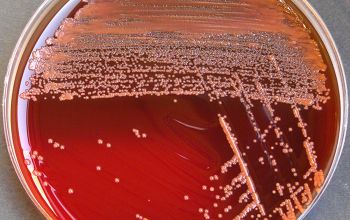
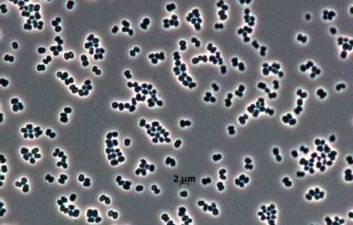
The peritoneal dialysis fluid (7 mL) inoculated into a BacT-alert FA bottle showed growth within 12 h (BacT-alert 240; bioMérieux, France). Direct microscopy of the sample showed gram-positive cocci in pairs and clusters. We performed all culturing procedures according to the 2005 update of the International Society for Peritoneal Dialysis recommendations and guidelines using specimens collected prior to antibiotic treatment.3Subcultures of the peritoneal fluid were performed using sheep blood agar, MacConkey agar and chocolate agar; the plates were incubated at 35 °C for 48 h. After incubation, sheep blood agar yielded the pure luxuriant growth of pale-pink non-hemolytic colonies that were 1–2 mm size (Figure 1A). Subculture on nutrient agar yielded smooth, small, pale-cream to pale-pink colonies (Figure 1B). Gram staining of the culture revealed the cells to be gram-positive cocci in pairs or clusters (Figure 2). The organism was preliminarily identified as Kocuriabased on phenotypic test results, such as positive reactions for catalase, oxidase, nitrate reduction and growth in 5% NaCl and motility test negativity.
Why is it difficult to identify Kocuria?
Furthermore, accurate identification is difficult because commercially available databases do not include all the classified Kocuriaspecies. Even cellular fatty acid profile analyses were not much of use in identification.1,34However, Kocuriaclassification is now typically confirmed by 16S rRNA gene sequencing, and one study even used matrix-assisted laser desorption/ionization time-of-flight mass spectrometry for species identification, which appears to be an efficient method.2,26
Is Kocuria a human disease?
Although not previously known to cause human infections, Kocuriaspecies have now emerged as human path ogens, mostly in compromised hosts with severe underlying disease. Recently, there has been an increasing incidence of different types of Kocuriainfections reported, most likely due to the adoption of better identification methods. Here, we report a case of peritonitis caused by Kocuria roseain a diabetic nephropathy patient who was on continuous ambulatory peritoneal dialysis. Sepsis and peritonitis caused by K. roseain our case yielded two identical Kocuriaisolates from the peritoneal dialysate fluid within a period of three days. The infection was subsequently resolved by antibiotic treatment and catheter removal. In addition to reporting this case, we herein review the literature concerning the emergence of Kocuriaas a significant human pathogen. The majority of cases were device-related, acquired in the hospital or endogenous, and different Kocuriaspecies appear to share a common etiology of peritonitis. The overall disease burden associated with Kocuriaappears to be high, and the treatment guidelines for diseases associated with Kocuriahave not yet been clearly defined.
How long does it take for Kocuria to appear on blood agar?
Appearance of Kocuria spp on blood agar after 24 hours of aerobic incubation
Why is Kocuria spp so elusive?
Identification of Kocuria spp remains elusive because most clinical microbiology laboratories have limited or no access to advanced molecular techniques. Laboratory identification of Kocuriaspp can be made conventionally only after high laboratory suspicion. Properties such as morphological variability between these bacteria and other similar gram-positive cocci, as well as biochemical properties including the antimicrobial susceptibility patterns against selective antibiotics could be used to presumptively identify Kocuriaspp. Infections of Kocuria spp normally involve patients with various debilitated conditions. In the era of drug resistance, and prevalence of multi-drug resistant bacteria, occurrence of Kocuria spp in hospitalized patients should not always be ignored as contaminants. Further studies emphasizing the determination of the virulence, pathogenic potential, predisposing factors and antimicrobial susceptibility patterns of Kocuria spp are warranted.
What is a kocuria?
Kocuriais a Gram-positive cocci arranged in pairs, short chains, tetrads, cubical packets of eight and irregular clusters. Kocuriabelongs to the phylum Actinobacteria, class Actinobacteria, order Actinomycetales, sub order Micrococcinaeand family Micrococcaceae. This bacterium was first identified and described by Miroslav Kosur, a Slovakian microbiologist. Currently, there are more than 18 species of Kocuriaidentified based on the 16S rRNA phylogenetic studies. The species of Kocuriaidentified thus far include Kocuriaassamensis, Kocuriaaegyptia, Kocuriagwangalliensis, Kocuriaatrinae, Kocuriacarniphila, Kocuriaflava, Kocuriapalustris, Kocuriahalotolerans, Kocuriahimachalensis, Kocuriakoreensis, Kocuria kristinae, Kocuriamarina, Kocuriapolaris, Kocuriarhizophila, Kocuriarosea, Kocuriasalsicia, Kocuriasediminis, Kocuriaturfanensis, and Kocuriavarians. Kocuria species (Kocuria spp) inhabit the normal skin and mucous membrane of human and animals [1]. Kocuria was also isolated from various environmental and ecological niches [2]. These are usually considered as non-pathogenic bacteria which are rarely associated with human infections. Recently there has been a rise in the incidence of infections caused by Kocuriaspp causing both superficial infections and deep-seated/invasive infections. The cause of concern is that this bacterium appears to have a broad host range involving both immunocompromised as well as immunocompetent individuals. This review attempts to update the morphology, cultural characteristics, pathophysiological properties, and laboratory diagnosis of Kocuriaspp.
Is Kocuria a pathogen?
The same study has also noted that although Kocuria spp are commensals of humans, animals and are present in the environment, they should be considered as potential pathogens in patients who are immunocompromised, undergoing critical care treatment and neonates. A study which included 12 pediatric age patients suffering from underlying debilitating conditions like premature birth and cancer had noted that more than 50% of patients suffered from invasive infections with Kocuriaspp [28]. Reports of infections caused by Kocuria spp among previously healthy and immunocompetent individuals are showing an increased trend. Kocuriarosea was isolated from a case of descending necrotizing mediastinitis in a 58-year-old woman who was taking medications for gout and hypertension [13]. Another very recent report has observed endocarditis caused by Kocuriarosea in a 10-year-old female patient. Although the patient was healthy before suffering from the infection, a history of surgery to correct congenital heart disease was present [9]. Evaluation of biofilm production by Kocuriaspp isolated from a case of peritonitis showed that the strain was negative for biofilm production [18]. Isolation of K. marina showing tolerance to severe alkaline conditions in a 7-year-old patient receiving epoprostenol therapy should be considered as an alarming signal regarding the potential of Kocuriaspp in causing both opportunistic and nosocomial infections [29-30].
Do Kocuriaspp produce hemolysis?
Kocuriaspp do not produce hemolysis on blood agar, unlike most clinical isolates of Staphylococci. They usually form 2-3 mm whitish, small, round, raised, convex colonies on initial isolation and might develop non-diffusible yellowish pigmentation after prolonged incubation, as shown in Figure Figure11.
Where is Kocuria found?
Kocuria is named after Miroslav Kocur, a Slovakian microbiologist. It has been found in the milk of water deer and reindeer. Cells are coccoid, resembling Staphylococcus and Micrococcus, and can group in pairs, chains, tetrads, cubical arrangements of eight, or irregular clusters.
What is the color of Kocuria?
They grow best in neutral pH environments. Depending on the species, they appear in a range of color such as: orange, pink, red, yellow or cream. They are shown to lack hemolytic ability on a blood agar plate.
Is Kocuria a pathogen?
Kocuria has been found to live on human skin and oral cavity. It is generally considered non-pathogenic but can be found in some infections. Specific infection associated with Kocuria are urinary tract infections, cholecystitis, catheter-associated bacteremia, dacryocystitis, canaliculitis, keratitis, native valve endocarditis, peritonitis, ...