What is the lamina propria?
The lamina propria is one of three layers which make up the mucosa, or mucous membrane. The lamina propria is a large layer of connective tissue which separates the innermost layer of epithelial cells from a layer of smooth muscle tissue called the muscularis mucosa.
Where do lymphatics drain into the lamina propria?
Lymphatics penetrate the mucosa and lie below the basement membrane of the epithelium, from there they drain the lamina propria. The fast rate of cell death and regeneration of the epithelium leaves behind many apoptotic cell bodies. These have been found to go into the lamina propria, most of which are inside its macrophages.
Why is the lamina propria modified in the intestines?
In the intestines, there is less of a need gas exchange. Instead, the lamina propria is modified to allow the passage of a large number of ducts, which connect to the liver, pancreas and salivary glands. These ducts allow the passage of materials needed for digestion.
Why is the lamina propria of the lungs arranged around vessels?
Therefore, the lamina propria of the lungs is arranged around and between these vessels, providing maximum surface area. In the intestines, there is less of a need gas exchange. Instead, the lamina propria is modified to allow the passage of a large number of ducts, which connect to the liver, pancreas and salivary glands.

What is the lamina propria and where is it located?
The lamina propria constitutes the layer of loose connective tissue and interstitial matrix located just below the epithelium. In the stomach, the lamina propria tends to be relatively inconspicuous, filling the interstitial spaces between the tubular gastric glands.
Where in the body is the lamina propria?
mucous membranesThe lamina propria is a thin layer of connective tissue that forms part of the moist linings known as mucous membranes or mucosa, which line various tubes in the body, such as the respiratory tract, the gastrointestinal tract, and the urogenital tract.
What layer contains the lamina propria?
The lamina propria is one of three layers which make up the mucosa, or mucous membrane. The lamina propria is a large layer of connective tissue which separates the innermost layer of epithelial cells from a layer of smooth muscle tissue called the muscularis mucosa.
What is lamina propria and its function?
Lamina propria is loose connective tissue in a mucosa. Lamina propria supports the delicate mucosal epithelium, allows the epithelium to move freely with respect to deeper structures, and provides for immune defense. Compared to other loose connective tissue, lamina propria is relatively cellular.
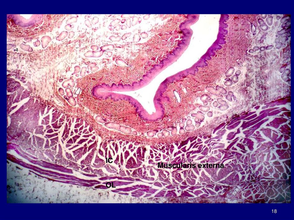
Does the stomach have a lamina propria?
The epithelium of the mucosa of the fundus and body of the stomach forms invaginations called gastric pits. The lamina propria contains gastric glands, which open into the bases of the gastric pits. These glands are responsible for the synthesis and secretion of the gastric juice.
Is there lamina propria in skin?
Although the skin is exposed to the external environment, it does not have lamina propria. Instead, the skin has a specialized layer of connective tissue called the dermis.
In which layer is the lamina propria found quizlet?
The mucosa consists of an inner epithelium, a loose connective tissue called lamina propria, and a thin layer of smooth muscle called muscularis mucosae.
Does Colon have lamina propria?
Abstract. The lamina propria of colonic mucosa normally contains eosinophils, lymphocytes, plasma cells, and a few neutrophils. If the number of such cells is judged to be increased, colonic inflammation is said to be present. However, the number of cells present in normal mucosa has not been clearly established.
Which layer contains the lamina propria quizlet?
The mucosa contains a lamina propria (areolar connective tissue) and a muscularis mucosae (smooth muscle).
What type of tissue is lamina propria?
connective tissueA type of connective tissue found under the thin layer of tissues covering a mucous membrane.
Where is lamina propria found in bladder?
The lamina propria (also called the submucosa) is a thin layer of connective tissue that surrounds the urothelium. It contains blood vessels, nerves and glands. The muscularis propria is the thick, outer muscle layer of the bladder.
What is lamina propria chronic inflammation?
Chronic gastritis is a persistent inflammatory reaction in the gastric mucosa that is characterized by the accumulation of lymphocytes and plasma cells in the lamina propria. Chronic active gastritis implies that ongoing active inflammation is causing damage to epithelial cells.
In which layer is the lamina propria found quizlet?
The mucosa consists of an inner epithelium, a loose connective tissue called lamina propria, and a thin layer of smooth muscle called muscularis mucosae.
What does lamina propria mean?
Listen to pronunciation. (LA-mih-nuh PROH-pree-uh) A type of connective tissue found under the thin layer of tissues covering a mucous membrane.
Which layer contains the lamina propria quizlet?
The mucosa contains a lamina propria (areolar connective tissue) and a muscularis mucosae (smooth muscle).
Why is the lamina propria important?
Because the epithelium is often under external stress and is somewhat delicate, the lamina propria hosts many immune cells. In the intestinal tract the immune system must have tolerance to the normal intestinal flora, yet respond to pathogenic microorganisms. Imbalance of this causes inflammation diseases such as inflammatory bowel disease. The lamina propria’s richness in macrophages and lymphoid cells makes it a key place for immune responses to occur. It forms part of the barrier that protects internal tissues from external pathogenic microorganisms, especially from the gastrointestinal tract.
Where do myofibroblasts live?
It has been suggested that myofibroblasts also reside in the lamina propria of several organs. These cells have characteristics of both smooth muscle and fibroblasts. The lamina propria may also be rich in vascular networks, lymphatic vessels, elastic fibers, and smooth muscle fascicles from the muscularis mucosae.
What is the mucosa?
Thus, the term mucosa or mucous membrane refers to the combination of the epithelium and the lamina propria. The connective tissue of the lamina propria is loose and rich in cells. The cells of the lamina propria are variable and can include fibroblasts, lymphocytes, plasma cells, macrophages, eosinophilic leukocytes, and mast cells.
What is the thin layer of connective tissue?
Thin connective layer forming part of the mucous membranes. Lamina propria. The lamina propria , a thin layer of connective tissue, is part of the mucosa. Here is an example of the mucosa of the mouth. Details.
What is the name of the thin layer of connective tissue that forms part of the moist linings of the?
Mucosa. Identifiers. Latin. lamina propria mucosæ. FMA. 62517. Anatomical terminology. The lamina propria is a thin layer of connective tissue that forms part of the moist linings known as mucous membranes or mucosa, which line various tubes in the body, such as the respiratory tract, the gastrointestinal tract, and the urogenital tract.
How does a tumor get exposed to lymphatics?
As soon as the tumors breach the basement membrane and reach the lamina propria, they are exposed to lymphatics which may increase the rate of metastasis and cancer progression. Deeper invasion into the submucosa will increase the exposure to lymphatics.
Which organs require expansion?
The connective tissue and architecture of the lamina propria is very compressible and elastic, this can be seen in organs that require expansion such as the bladder. The collagen in the lamina propria of elastic organs has been shown to play a major role in mechanical function.
Which proteinase cleaves the N terminal of a helical structure?
Extracellularly, the -C terminal and part of -N terminal of this helical structure are cleaved by -C and -N proteinases, respectively, leading to the formation of tropocollagen – a 5-unit quarter stagger microfibril. The remaining part of the N-terminal is cleaved by procollagen peptidase. In this way, the large collagen molecule is trimmed. The stabilizing of the collagen molecule occurs through the cross-linking of the molecule by oxidation of the lysine and hydroxylysine residues by lysyl oxidase [ Figure 2 ]. [ 10, 11]
Where does collagen come from?
The word “collagen” is derived from Greek word – “kolla” and “gen” ( kolla – glue and gen – producer). They are a group of fibrous proteins that occur in vertebrates as the chief constituent of connective tissue fibrils and in bones. [ 2] .
Is collagen a part of the oral cavity?
As collagen is an integral part of the oral cavity, both in its soft tissue and in its hard tissue, it is very important to know about its structure, function and distribution. Any aberration in its formation and its structure can alter its function which ultimately leads to various pathologies in the body and the oral cavity.
Is collagen made from fibroblasts?
Collagen is not synthesized from fibroblasts alone but by various other cells such as cementoblasts, odontoblasts, chondroblasts, osteoblasts, muscle cells, epithelial cells, endothelial cells and Schwann cells. [ 10, 11] Although these cells secrete collagen in the same manner as that of fibroblasts, the types of collagen they secrete vary.
Is collagen found in dentin?
The amino acid analysis reveals that the collagen of dentin, alveolar bone, and cementum in human teeth is similar. [ 18] . Type III, which is a less cross-linked collagen, is found in high concentrations during development, repair, and regeneration of mineralized tissues like cementum. [ 19]
Do PDL fibroblasts contract?
PDL fibroblasts contract and transmit a contractile force to the extracellular environment; this permits summation of contractile forces. They also exhibit fibronexuses by which such forces can be transmitted to the collagen fiber bundles. Although the in vitro observations conclude the myofibroblastic nature of the fibroblast and the existence of fibronexus, in vivo findings do not support the migratory nature or features of myofibroblast and the existence of fibronexus between fibroblast and fibers. Therefore, these cells would not be able to transmit a tractional force required to pull the tooth in eruption. [ 11]
Overview
- The lamina propria varies in chemical composition from animal to animal, and from organ to organ. In general, the lamina propria is a complex mesh of extracellular proteins and structural molecules. These molecules include collagen, a standard animal structural protein, as well as la…
Structure
Function
Clinical significance
The lamina propria is a thin layer of connective tissue that forms part of the moist linings known as mucous membranes or mucosa, which line various tubes in the body, such as the respiratory tract, the gastrointestinal tract, and the urogenital tract.
The lamina propria is a thin layer of loose (areolar) connective tissue, which lies beneath the epithelium, and together with the epithelium and basement membrane constitutes the mucosa. A…
See also
The lamina propria is a loose connective tissue, hence it is not as fibrous as the underlying connective tissue of the submucosa. The connective tissue and architecture of the lamina propria is very compressible and elastic, this can be seen in organs that require expansion such as the bladder. The collagen in the lamina propria of elastic organs has been shown to play a major role in mechanical function. In the bladder the collagen composition of its lamina propria allows for st…
External links
Because the epithelium is often under external stress and is somewhat delicate, the lamina propria hosts many immune cells. In the intestinal tract the immune system must have tolerance to the normal intestinal flora, yet respond to pathogenic microorganisms. Imbalance of this causes inflammation diseases such as inflammatory bowel disease. The lamina propria’s richness in macrophages and lymphoid cells makes it a key place for immune responses to occur. It forms p…