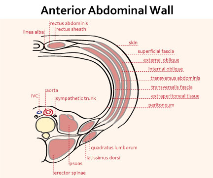
Where is the anterior wall of the abdomen?
The anterior abdominal wall forms the anterior limit of the abdominal viscera and is defined superiorly by the xiphoid process of the sternum and costal cartilages and inferiorly by the iliac crest and pubic bones of the pelvis.
What is US anterior abdominal wall?
The anterior abdominal wall is composed of three layers: skin and adipose tissues; the myofascial layer, consisting of muscles and their fascial envelopes; and a deep layer, consisting of the transversalis fascia, preperitoneal fat, and the parietal peritoneum.
What are the regions of the anterior abdominal wall?
Components of the anterior abdominal wallSkin and subcutaneous tissue.Superficial fascia. ... External oblique muscle.Internal oblique muscle.Transversus abdominis muscle.Deep fascia (transversalis fascia): fuses with the deep fascia of the thigh (fascia lata)Preperitoneal adipose tissue.Parietal peritoneum.
What is the anterior abdominal wall formed by?
The anterior wall is formed by the aponeuroses of the external oblique and half of the internal oblique. The posterior wall is formed by the aponeuroses of half of the internal oblique and transversus abdominis.
What does abdominal wall mean?
The abdominal wall is defined cranially by the xiphoid process of the sternum and the costal margins and caudally by the iliac and pubic bones of the pelvis. It extends to the lumbar spine, which joins the thorax and pelvis and is a point of attachment for some abdominal wall structures [1].
What is abdominal wall pain?
Chronic abdominal wall pain (CAWP) refers to the pain originating from the abdominal wall which is often misdiagnosed as arising from a source inside the abdominal cavity, often resulting in inappropriate diagnostic investigations, unsatisfactory treatment, and considerable costs.
What is anterior abdominal wall hernia?
Anterior abdominal wall hernias, also known as ventral hernias, are a leading cause of abdominal surgery in the United States (,1). These hernias involve the protrusion of part of the peritoneal sac through a defect in the muscle layers of the anterior abdominal wall.
What causes abdominal wall defects?
Causes. No genetic mutations are known to cause an abdominal wall defect. Multiple genetic and environmental factors likely influence the development of this disorder. Omphalocele and gastroschisis are caused by different errors in fetal development.
Why would a doctor order an abdominal ultrasound?
An abdominal ultrasound can help your doctor evaluate the cause of stomach pain or bloating. It can help check for kidney stones, liver disease, tumors and many other conditions. Your doctor may recommend that you have an abdominal ultrasound if you're at risk of an abdominal aortic aneurysm.
What is anterior abdominal wall hernia?
Anterior abdominal wall hernias, also known as ventral hernias, are a leading cause of abdominal surgery in the United States (,1). These hernias involve the protrusion of part of the peritoneal sac through a defect in the muscle layers of the anterior abdominal wall.
Which is a role of the anterior abdominal wall muscles?
The muscles of the anterior abdominal wall are largely involved in protecting the contents of the abdominal cavity, but also function to move the trunk and assist in other bodily functions.
Will an abdominal ultrasound show a hernia?
Abdominal wall hernia Sometimes a hernia cannot be diagnosed through a physical exam alone, and other diagnostic tests are needed. Some examples of these include: Ultrasound.
What is the anterior wall of the abdomen?
The anterior abdominal wall forms the anterior limit of the abdominal viscera and is defined superiorly by the xiphoid process of the sternum and costal cartilages and inferiorly by the iliac crest and pubic bones of the pelvis.
How many layers are there in the abdominal wall?
In general, the anterior abdominal wall has nine layers (from superficial to deep): Scarpa's fascia is deep to the skin and subcutaneous fat in the lower part of the wall and is fused with Colle's fascia in the perineum. The muscle layers include the external oblique muscle, internal oblique muscle, transversus abdominis muscle anterolaterally ...
What vessels drain into the axillary and sternal nodes?
The lymphatic vessels above the umbilicus drain into axillary and sternal nodes. The vessels below the umbilicus drain into superficial inguinal nodes.
Which muscle layer is anterior to the other three muscle layers?
Three muscle layers ( external oblique, internal oblique, transverse abdominis) can be seen anterolaterally in cross section and also the rectus muscle and its sheath can be seen anterior to the other three muscle layers. ventral abdominal wall hernia.
Where does the fascia fuse?
The fascia surrounding the 3 anterolateral muscles fuse anteriorly to attach to the rectus abdominis at the linea semilunaris. The fascia then continues medially surrounding the rectus abdominis as the rectus sheath further fusing in the midline with the contralateral fascia at the linea alba.
What are the quadrants of the abdomen?
The two intersecting planes divide the abdomen into four quadrants, described as right and left upper and lower quadrants. The four-quadrant system is straightforward when used to describe anatomic location. For example, the appendix is located in the lower right quadrant of the abdomen. + +. Figure 7-1.
How many regions does the abdomen have?
The first method partitions the abdomen into four quadrants. The second method partitions the abdomen into nine regions. + + +.
How is the abdomen divided?
The two intersecting planes divide the abdomen into four quadrants, described as right and left upper and lower quadrants. The four-quadrant system is straightforward when used to describe anatomic location. For example, the appendix is located in the lower right quadrant of the abdomen.
Where is the pylorus located?
The pylorus of the stomach, the first part of the duodenum, the fundus of the gallbladder, the neck of the pancreas, the origin of the superior mesenteric artery, the hepatic portal vein, and the splenic vein are all located along the level of the transpyloric plane. + + +.
Which plane is the subcostal plane?
Subcostal (upper horizontal) plane. Transversely courses inferior to the costal margin, through the level of the L3 vertebra. The L3 vertebra serves as an important anatomic landmark in that it indicates the level of the inferior extent of the third part of the duodenum and the origin of the inferior mesenteric artery.
What nerve is used to connect the umbilicus to the thoracic spine?
However, the skin around the umbilicus is supplied by the thoracic spinal nerve T10 (T10 dermatome) A helpful mnemonic is “T10 for belly but-ten”. ... Your MyAccess profile is currently affiliated with ' [InstitutionA]' and is in the process of switching affiliations to ' [InstitutionB]'.
General characteristics of the Anterior abdominal wall (anterolateral)
Musculocutaneous sheet anchored to the skeleton (ribs, lumbar vertebrae, pelvis).
Abdominal wall layers
1st layer= fatty superficial layer AKA Camper’s fascia. This thickness varies a great deal.
Muscles of the abdominal wall
Innervation: anterior rami of T7-T12 spinal nerves, called thoracoabdominal nerves. They pass the costal margin and travel between the 2 nd and 3 rd layers of muscles as thoracoabdominal muscles. L1 may / may not participate.
Appropriate incisions of the anterior abdominal wall based on avoiding damage
Along linea alba at the midline . Problem is that it doesn’t heal well due to low blood supply.
What is the superficial fascia of the anterior abdominal wall?
Superficial fascia of anterior abdominal wall: In the lower part of the anterior abdominal wall (below the line passing through the midpoint between the umbilicus and pubic symphysis), the superficial fascia has two layers:
Where does the lymphatic system drain from the anterior abdominal wall?
Lymphatics from anterior abdominal wall above the level of umbilicus drains into axillary lymph nodes.
What muscles are used to protect the abdominal viscera?
Anterior abdominal wall muscles. Support and protect the abdominal viscera. Help in forced expiration that occurs during coughing, sneezing, vomiting, Increase the intra-abdominal pressure and thereby help in defecation, micturition (urination), and parturition (childbirth).
Which fascia is attached laterally to the conjoint ischiopubic rami?
Medially it passes downwards over the body of the pubis and becomes continuous with the superficial perineal fascia (Colle’s fascia). The superficial perineal fascia is attached laterally to conjoint ischiopubic rami and posteriorly it is fused with the posterior border of perineal membrane.
How many abdominal regions are there?
5 Name the nine abdominal regions and their main contents.
Which layer of the body is devoid of fat?
Superficial fatty layer (Camper’s fascia) Deep membranous layer (Scarpa’s fascia) The superficial fatty layer is continuous with the superficial fascia of the rest of the body, the membranous layer is devoid of fat and has more of elastic fibers.
Why do loops of the small intestine get covered by amnion?
Occurs due to failure of reduction of physiological herniation of midgut loop, herniated loops of small intestine are covered by amnion.
How is the abdomen divided?
The two intersecting planes divide the abdomen into four quadrants, described as right and left upper and lower quadrants. The four-quadrant system is straightforward when used to describe anatomic location. For example, the appendix is located in the lower right quadrant of the abdomen.
What is the lower horizontal plane of the iliac crest?
Transtubercular (lower horizontal) plane. Transversely courses between the two tubercles of the iliac crest, through the level of the L5 vertebra.
Where is the umbilicus located?
Umbilicus. The umbilicus lies at the vertebral level between the L3 and L4 vertebrae. However, the skin around the umbilicus is supplied by the thoracic spinal nerve T10 (T10 dermatome) A helpful mnemonic is “T10 for belly but-ten”.
Which muscle attaches to the iliac crest?
The external oblique muscle is the most superficial of the anterolateral muscles and attaches to the outer surfaces of the lower ribs and iliac crest ( Figure 7-2A ). The external oblique muscle continues anteriorly as the external oblique aponeurosis, which courses anteriorly to the rectus abdominis muscle and inserts into the linea alba. The inferior border of the external oblique aponeurosis, between the anterior superior iliac spine and the pubic tubercle, is called the inguinal ligament.
What is the abdominal wall?
In anatomy, the abdominal wall represents the boundaries of the abdominal cavity. The abdominal wall is split into the anterolateral and posterior walls.
What are the three layers of the abdominal wall?
In medical vernacular, the term 'abdominal wall' most commonly refers to the layers composing the anterior abdominal wall which, in addition to the layers mentioned above, includes the three layers of muscle: the transversus abdominis (transverse abdominal muscle), the internal (obliquus internus) and the external oblique (obliquus externus).
What is the anterior abdominal wall?
The anterior abdominal muscles are part of the musculature that contributes to the anterolateral abdominal wall, along with the lateral abdominal muscles on either side. They collectively form part of the boundaries of the abdominal cavity. The muscles of the anterior abdominal wall are located near ...
Where are the muscles of the anterior abdominal wall located?
The muscles of the anterior abdominal wall are located near the midline between the costal margin superiorly and the pubis inferiorly. There are two pairs of muscles, each located immediately lateral to the linea alba.
What muscles are involved in the anterior abdominal wall?
Key facts about the anterior abdominal muscles. Rectus abdominis muscle. Origin - Pubic symphysis, Pubic crest.
Where is the lateral border of the lateral abdominal muscle?
The lateral borders of the muscle create a semicircular border, the linea semilunaris, which extends from the tip of the 9th costal cartilage to the pubic tubercle. They are enclosed in the rectus sheath, and enveloped by the aponeuroses of the lateral abdominal muscles as they pass to insert onto the linea alba.
Which artery supplies the rectus abdominis muscle?
The inferior epigastric artery is the main artery supplying the rectus abdominis muscle. There are sometimes contributions from the lower intercostal and subcostal arteries, posterior lumbar, and deep circumflex iliac arteries. The venous drainage of the muscles mirrors the arterial supply.
Where does the rectus abdominis originate?
The rectus abdominis has two points of origin. The lateral head originates from the crest of the pubis, between the pubic symphysis and the pubic tubercle. The medial head originates from the pubic symphysis, interlacing with the fibres of the muscle on the contralateral side.
How to diagnose a hernia in the gut?
Diagnosing an umbilical hernia is typically done by physical examination. Ultrasound or CT can also be used to confirm the diagnosis. Umbilical hernias can be dangerous as strangulation of the intestines can occur, leading to a blockage of the gut. The part of the intestines that protrudes through the abdominal wall might also be deprived of adequate blood supply, which can cause necrosis of that section of the gut. Should complications present, surgery may be required to repair the herniation. The surgical procedure is relatively straightforward. It involves a small incision being made near the umbilicus through which the intestines can be safely pushed back into the abdomen followed by closure with stitches. The abdominal wall is usually reinforced by a mesh to prevent further herniation before the incision is closed.
