What is the location of the mandible?
The mandible is the largest bone in the human skull. It holds the lower teeth in place, it assists in mastication and forms the lower jawline. The mandible is composed of the body and the ramus and is located inferior to the maxilla. The body is a horizontally curved portion that creates the lower jawline.
Where is the posterior mandible?
The posterior mandible is usually defined as the part of the mandible posterior to the mental foramen (Fig. 14.1). Hence, the posterior mandible comprises the posterior body of the mandible, the angle, the ascending ramus, the coronoid, and the condylar processes of the mandible.
What is the front of the mandible called?
Introduction to Mandible Bone Anatomy: The anterior portion of the mandible, called the body, is horseshoe-shaped and runs horizontally. At the posterior ends of the body are two vertical or perpendicular extensions called rami (singular; ramus).
Is the mandible on top or bottom?
It forms the lower part of the jaw and part of the mouth. The mandible is the only moveable bone of the skull and is attached to muscles involved in chewing and other mouth movements. It also holds the bottom teeth in place. Also called lower jaw bone.
Which type of bone is mandible?
The mandible is a U-shaped bone. It is the only mobile bone of the facial skeleton, and, since it houses the lower teeth, its motion is essential for mastication. It is formed by intramembranous ossification.
What is the bone under your tongue called?
Torus mandibularis. Other names. Tori mandibulares, mandibular torus, mandibular tori. Mandibular torus in premolar area.
What is the bone above your teeth called?
The maxilla is the bone that forms your upper jaw. The right and left halves of the maxilla are irregularly shaped bones that fuse together in the middle of the skull, below the nose, in an area known as the intermaxillary suture. The maxilla is a major bone of the face.
Why does my mandible bone hurt?
Dental problems A toothache, usually because of a cavity or an abscess. Teeth that are cracked, crowded, or sensitive to temperature or pressure. Gum disease, which can damage your jawbone.
How important is the mandible?
The mandible plays a major role in mastication, because many muscles used for chewing are attached to it, and it holds in place 16 teeth (14 with the third molar extracted) (White et al., 2012). The two most basic parts that assist with mastication are the corpus of the mandible and the ramus (White et al., 2012).
What is another word for mandible?
What is another word for mandible?beakbilljawjawbonemaxillamouthmouthpiecedentarydentary bonejowl9 more rows
How many mandibles do we have?
We have one mandibular body and two rami. The rami provide articulation points by way of two processes – the coronoid and condyloid (or condylar) processes. More will be said about these later on. The ramus of the mandible has two surfaces – lateral and medial.
What is the mandible attached to?
temporal bonesThe mandible sits beneath the maxilla. It is the only movable bone of the skull (discounting the ossicles of the middle ear). It is connected to the temporal bones by the temporomandibular joints.
What is the mandible attached to?
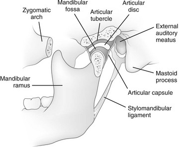
The condylar process has an articular surface (the condyle), via which the mandible articulates with the articular tubercle of the temporal bone to form the temporomandibular joint.
What is mandible fracture?
A mandibular fracture is similar to a bone fracture in any body part. The bone becomes stressed, usually from excessive force, and breaks under that pressure. Beyond accidents, mandible fractures can result from violence.
What is the upper jaw called?
maxillaThe upper part is the maxilla. It doesn't move. The moveable lower part is called the mandible.
In which direction do the mandibles move?
During chewing, the jaw moves in a specific manner as delineated by the two TMJs. The side of the mandible that moves sideways is referred to as either the working or rotating side, while the other side is referred to as either the balancing or orbiting side.
What is the mandible in the face?
Rony Kampalath, MD, is a board-certified diagnostic radiologist specializing in imaging of the abdomen. Also known as the lower jawbone, the mandible is the largest and strongest bone of the face. Tasked with holding the lower set of teeth in place, this bone has a symmetrical, horseshoe shape. Not directly connected to other bones ...
What muscles are involved in the mandible?
Furthermore, other muscles link to the mandible, including: 1 The platysma arises from the collarbone and progresses to the underside of the mandible. 2 Inserting into the side surface of the ramus is the superficial masseter, which is a major muscle of chewing and mouth movement. 3 The deep masseter also inserts into the mandible at the outside surface of the ramus and is involved in chewing motion. 4 The medial angle of the mandibular angle (the outer corner of the mandible) and ramus is the site where the medial pterygoid muscle inserts. This thick, roughly rectangular muscle is also involved in chewing function. 5 At the condyloid process, the inferior head of the lateral pterygoid muscle, which moves the jaw downward and from side to side and is, therefore, another important structure for chewing. 6 The temporalis muscle, a broad, fan-shaped structure along the sides of the head that also work to help with chewing, accesses the coronoid process of the mandible.
What is the ramus on the mandible?
Representing the “wings” of the mandible, the ramus arises on each side of the body, terminating at two ridges separated by the mandibular notch: the one towards the front called the coronoid process and the other towards the back of the head the condylaris process. These bound the temporomandibular joint, which allows the bone to move.
Where does the deep masseter insert?
The deep masseter also inserts into the mandible at the outside surface of the ramus and is involved in chewing motion. The medial angle of the mandibular angle (the outer corner of the mandible) and ramus is the site where the medial pterygoid muscle inserts. This thick, roughly rectangular muscle is also involved in chewing function.
What is the oblique line of the mandible?
The oblique line of the mandible is where the depressor labii inferioris and depressor anguli oris emerge. These are associated with frowning.
Why do they do corrective surgeries on the mandible?
In addition, corrective surgeries may be performed on the mandible to correct misalignment due to improper development of the jaw.
Which joint is responsible for the movement of the jaw?
These bound the temporomandibular joint, which allows the bone to move. The lower surfaces of the ramus define the jawline, and the outer sides are connected to the masseter muscle (for chewing). The inner surfaces contain several openings (fossa) that allow important nerves and arteries to access the mouth region.
What is the Mandible
The mandible is the largest, strongest, and the only skull bone capable of movement. It forms the lower jaw, and thus is also known as the lower jaw bone. It helps with the process of chewing along with the maxilla or upper jaw bone.
Where is the Mandible Bone Located
As stated, the mandible is located in the lower jaw, just inferior to the maxilla or upper jaw. You can easily feel the bone by touching your lower jawline.
Functions
Forms the lower jawline, shaping the face and chin. The teeth in the lower jaw are also rooted to this bone.
Parts and Anatomy of the Mandible
It is a single, horseshoe-shaped bone, consisting of a horizontal body on the anterior side and two vertical rami on the posterior side.
Ossification and Development
Ossification of the mandible starts during the sixth week of intrauterine development. It is the second bone to ossify.
Where is the mandible located?
The mandible is marked by two foramina. The mandibular foramen is located on the internal surface of the ramus of the mandible . It serves as a conduit for the inferior alveolar nerve and inferior alveolar artery. They travel through the mandibular foramen, into the mandibular canal, and exit at the mental foramen.
Which muscle attaches to the mandible?
Mandibular rami – masseter, temporalis, medial pterygoid and lateral pterygoid. The temporalis muscle attaches to the coronoid process, and the masseter attaches to the rami. The lateral pterygoid inserts into the neck of the mandible, and the medial pterygoid inserts into the ramus near the angle of the mandible.
What is the mental protuberance of the chin?
This is a small ridge of bone that represents the fusion of the two halves during development. The symphysis encloses a triangular eminence – the mental protuberance, which forms the shape of the chin. Lateral to the mental protuberance is the mental foramen (below the second premolar tooth on either side).
What is the mandible shaped like?
The body of the mandible is curved, and shaped much like a horseshoe. It has two borders: Alveolar border (superior) – contains 16 sockets to hold the lower teeth. Base (inferior) – site of attachment for the digastric muscle medially. The body is marked in the midline by the mandibular symphysis.
What is the foramen?
A foramen refers to any opening through which neurovascular structures can travel. The mandible is marked by two foramina.
How many rami are there in the mandible?
There are two mandibular rami, which project perpendicularly upwards from the angle of the mandible. Each ramus contains the following bony landmarks: Head - situated posteriorly, and articulates with the temporal bone to form the temporomandibular joint.
Where does the lateral pterygoid insert?
The lateral pterygoid inserts into the neck of the mandible, and the medial pterygoid inserts into the ramus near the angle of the mandible. The mandible articulates with the temporal bone to form the temporomandibular joint which is discussed in more detail here. A mandibular fracture rarely occurs in isolation.
When the mandible is dislocated anteriorly, the coronoid process and anterior border of the?
When the mandible is dislocated anteriorly, the coronoid process and anterior border of the ramus can be palpated easily over the cheek on the affected side (figure 1).
Why are the fingers placed behind the angle of the mandible?
The fingers are placed behind the angle of the mandible to stabilize the grip.
What is anterior dislocation?
Anterior dislocation of the mandible is a clinical scenario that is not infrequently encountered by the ED provider and requires prompt intervention. The classic technique for reduction of the mandible requires the provider to place his/her thumbs or fingers into the patient’s mouth along the lower molars and apply force inferiorly and posteriorly. However, this technique is fraught with difficulties and inefficiencies including the following:
Where is the coronoid process located?
Note that when the mandible is in its resting position and no dislocation is present, the coronoid process and anterior border of the ramus are not palpable as they are located posterior to the body of the zygoma (figure 2).
Who should stand in front of the patient?
The provider should stand in front of the patient.
Is sedation required for anterior mandible surgery?
from the Department of Plastic and Reconstructive Surgery at Chang Gung University in Taiwan describe a simple, effective, and novel technique to reduce the anteriorly displaced mandible. 1 In their experience, no sedation was required for any of their cases.
What force is applied to the anterior mandible?from msdmanuals.com
Simultaneously apply a reciprocal upward force on the anterior mandible (ie, rock the chin upward), which may enhance the condylar distraction.
Where to place thumbs on mandible?from msdmanuals.com
Place your thumbs on the external oblique ridge on either side of the mandible, lateral to the third molar area. Alternatively, wrap your thumbs with layers of gauze and place them as posteriorly as possible on the occlusal surface of the lower molars bilaterally (this increases the risk of being bitten during reduction).
What symptoms relate to a dislocated jaw?from healthdirect.gov.au
A dislocated jaw can interfere with eating and sleeping. It will also feel stiff, swollen and sore. The sooner you see a doctor, the better, since this will reduce the chances of future complications.
What causes a dislocated jaw?from healthdirect.gov.au
Sometimes, it happens just because they open their mouth too wide, for example when they are eating, yawning, vomiting or having a dental procedure.
Why should mandibular dislocations be followed up?from exodontia.info
All patients with reduced mandibular dislocations should be followed-up by an appropriate specialist because of the possibility of jaw instability, ligamentous damage and chronic TMJ pain.
What is the relationship between the mandible and the maxilla?from plasticsurgerykey.com
The relationship of the mandible with the maxilla should be understood, because not all patients have a normal intermaxillary skeletal relationship. In cases with protruding mandible showing class III occlusion, or those with relative underdevelopment of the lower jaw showing class II occlusion, orthognathic surgery may be needed to improve this disorder. If mandibular contouring surgery is to be done without correcting class II or class III skeletal problems, certain characteristics should be considered to avoid aggravating the intermaxillary problems. In cases with prominent mandible showing a skeletal class III relationship, a long jaw line may appear more accentuated if the angle is resected too much during mandible reduction. Therefore, the angle should be conservatively resected and sagittal shaving should be properly performed to minimize aggravation of prognathic appearance. In patients with a retruded mandible showing a class II profile, excessive resection of the mandible angle causes a more obscure cervico-facial line. Therefore, conservative mandible resection and maximal sagittal shaving of the body of the mandible, combined with advancement genioplasty are recommended. In a long face, angle resection should be performed in a limited way to prevent the aggravation of steep mandibular plane.
How long after dislocated jaw can you open your mouth?from healthdirect.gov.au
You should not open your mouth wide for 6 weeks after you have dislocated your jaw. Support your jaw in your hand every time you sneeze or yawn during this time.
