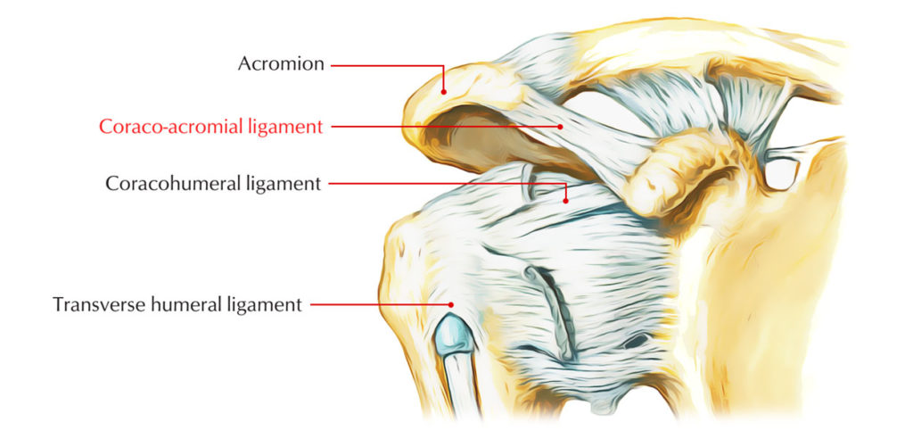
What does the coracoacromial ligament do?
Coracoacromial ligament: one of the ligaments of the shoulder. Ligaments are bands of tough fibrous connective tissue that connect bones or cartilage. The coracoacromial ligament joins two parts of the shoulder blade (scapula), connecting the acromion to the coracoid process.
What structures are found in the coracoacromial arch?
The coracoacromial arch is formed (anteriorly to posteriorly) by the coracoid process, coracoacromial ligament, and acromion. Structures located within the coracoacromial arch include: subacromial-subdeltoid bursa. supraspinatus tendon. long head of biceps tendon.
How is the displacement of the coracoacromial ligament evaluated in overhead athletes?
Evaluating displacement of the coracoacromial ligament in painful shoulders of overhead athletes through dynamic ultrasonographic examination. Arch Phys Med Rehabil. 2010;91:278–282.
Does release of the coracoacromial ligament lead to glenohumeral laxity?
Release of the coracoacromial ligament can lead to glenohumeral laxity: a biomechanical study. J Shoulder Elbow Surg. 2001;10:68–72.

Where is the coracoclavicular ligament located?
The coracoclavicular ligament is a ligament of the shoulder. It connects the clavicle to the coracoid process of the scapula.
Is the coracoacromial ligament part of the shoulder joint?
The coracoacromial ligament is a strong triangular ligament between the coracoid process and the acromion. It protects the head of the humerus. Its acromial attachment may be repositioned to the clavicle during reconstructive surgery of the acromioclavicular joint (shoulder joint).
What ligaments make up the coracoacromial ligament?
The conoid and trapezoid ligaments are continuous inferiorly at the coracoid process attachment but separate at an angle before attaching to the inferior aspect of the clavicle superiorly. [2] These two parts of the coracoclavicular ligament are often separated either by a bursa or by fat.
Where is coracoacromial?
The coracoacromial ligament is a strong triangular band, extending between the coracoid process and the acromion. It is attached, by its apex, to the summit of the acromion just in front of the articular surface for the clavicle; and by its broad base to the whole length of the lateral border of the coracoid process.
What movement does the coracoacromial ligament prevent?
A ligamentous connection between the CAL and the rotator interval capsule has been coined the “coracoacromial veil” and is thought to prevent inferior migration of the glenohumeral joint. The CAL is bordered superiorly by the clavicle and deltoid as well as inferiorly by the subacromial bursa and supraspinatus tendon.
What are the consequences of rupture of coracoclavicular ligament?
Injuries to the acromioclavicular joint and coracoclavicular ligaments are common. Many of these injuries heal with nonoperative management. However, more severe injuries may lead to continued pain and shoulder dysfunction.
How do you palpate the coracoacromial ligament?
0:126:13Shoulder Palpation - YouTubeYouTubeStart of suggested clipEnd of suggested clipAnd go along the clavicle go to the sternoclavicular junction. And basically palpate around thatMoreAnd go along the clavicle go to the sternoclavicular junction. And basically palpate around that Junction just lateral and posterior to that you can find the first rib.
What are the symptoms of a torn ligament in your shoulder?
Symptoms of a Shoulder Ligament Tear Shoulder pain and swelling. Increased pain with arm movement or shrugging your shoulder. Distortion in the normal contour of the shoulder.
How long does it take for a torn ligament in the shoulder to heal?
Usually, mild rotator cuff tears or sprains will heal within four weeks. In other severe cases, the recovery might take 4 to 6 months or even longer based on several factors such as the severity of the tear, age, and other health complications.
How do you test for coracoclavicular ligament?
Assesses integrity of the acromioclavicular and coracoclavicular ligaments. Grasp the proximal forearm with one hand as you place your other hand on the mid-clavicle. Attempt to distract the acromion process from the clavicle by applying a downward force to the arm directed along the longitudinal axis of the humerus.
What is coracoacromial ligament release?
Release of the CA ligament resulted in increased anterior and inferior translation of the internally and externally rotated glenohumeral joint. The CA ligament has previously been implicated only as an important soft tissue structure that contributes to rotator cuff pain.
What are the ligaments of shoulder joint?
Glenohumeral ligaments- Composed of a superior, middle, and inferior ligament, these three ligaments combine to form the glenohumeral joint capsule connecting the glenoid fossa to the humerus.
What is shoulder joint?
The shoulder is one of the largest and most complex joints in the body. The shoulder joint is formed where the humerus (upper arm bone) fits into the scapula (shoulder blade), like a ball and socket. Other important bones in the shoulder include: The acromion is a bony projection off the scapula.
Where is the acromioclavicular joint?
shoulderThe acromioclavicular (AC) joint is formed by the cap of the shoulder (acromion) and the collar bone (clavicle). It is held together by strong ligaments (figure 1). The outer end of the clavicle is held in alignment with the acromion by the acromioclavicular ligaments and the coracoclavicular (CC) ligaments.
Is the Scapulothoracic joint a true joint?
The scapulothoracic joint is not a true synovial joint. Rather, the scapulothoracic articulation is formed by the convex surface of the posterior thoracic cage and the concave surface of the anterior scapula.
How does atmospheric pressure stabilize the shoulder joint?
It was found that the intact shoulder subluxated after percutaneous puncture even without division of the overlying muscles or the capsule. Our findings suggest that negative pressure and muscle tone are the main static stabilisers of the shoulder, rather than the joint capsule.
What is the coracoacromial ligament?
The coracoacromial ligament is a flat triangular band that plays a supportive role for the shoulder joint.
Can decreased space within the coracoacromial arch result in subacromial imping?
It is thought that decreased space within the coracoacromial arch can result in subacromial impingement.
What is the coracoacromial arch?
This ligament forms a part of the coracoacromial arch and provides support to the humeral head. Coracoclavicular and coracohumeral ligaments are other ligaments originating from the coracoid process. Coracoacromial ligament injury is mostly related to trauma or clavicular fracture and isolated injury to the coracoacromial ligament is rare.
Is coracoacromial ligament injury rare?
Coracoacromial ligament injury is mostly related to trauma or clavicular fracture and isolated injury to the coracoacromial ligament is rare. However here I present a case with isolated old tear of this ligament.
What is the magnetic resonance image of the coracoacromial ligament?
Parasagittal magnetic resonance image of the coracoacromial ligament attachment demonstrates the normal coracoacromial ligament, which may mimic an enthesophyte (white arrows). (Reprinted with permission from Rudez et al.50)
How many articles were included in the study of coracoacromial ligament?
In addition, reference lists from all identified articles were reviewed for studies that the search terms may have omitted. Ninety-four articles from 1958 to 2016 were identified and reviewed for possible inclusion. Inclusion criteria were studies with levels 1 through 5 evidence according to the American Academy of Orthopaedic Surgeons Evidence-Based Practice committee and publication in a peer-reviewed journal in the English language. Twenty-eight articles failed to meet our inclusion criteria and were excluded, leaving 66 articles for this analysis. There were 30 cadaveric studies, 25 clinical research articles, and 11 review articles included in this study.
What is the role of the glenohumeral joint?
The CAL is thought to play an important role in shoulder stability via both static restraint and dynamic interactions with other shoulder capsular elements including ligaments, muscles, and osseous structures. Simply by its position anterosuperior to the glenohumeral joint, it passively restricts upward displacement of the humeral head.4,41,47The CAL also acts to transmit loads across the scapula. Serving as a tension band, forces exerted on the coracoid process by the coracobrachialis, pectoralis minor, and biceps (short head) muscles are transmitted to the acromion.18,55Likewise, the acromial distortion due to forces exerted by the deltoid and trapezius muscles is limited by the action of the CAL.48While of uncertain clinical significance, the CAL appears to serve as a dynamic brace within the shoulder girdle.
What is the CAL ligament?
Like all other ligaments, the CAL is a soft collagenous tissue made up of fascicles containing elementary fibrils, reticular fibers, glycoproteins, and intervening fibroblasts. In particular, types I, III, and VI collagens along with proteoglycans, including chondroitin IV sulfate, keratan sulfate, dermatan sulfate, versican, tenascin, and cartilage oligomeric matrix protein are present throughout the CAL.40Parallel collagen fibers are oriented along the ligament’s longitudinal axis, usually within wavy (>2 inflection points per 100 μm) bundles.33Perhaps uniquely, the full length of the CAL is highly fibrocartilagenous, with areas of both uncalcified and calcified fibrocartilage. While often cited as a sign of degenerative change, the presence of fibrocartilage in both young specimens and those devoid of shoulder pathology implies that it is an integral component of the “normal” CAL. Thought to resist shear and compression forces, the fibrocartilage is concentrated most heavily at the entheses and is made up of type II collagen, link protein, and aggrecan, the latter of which is typical of articular cartilage.40Along its posteroinferior surface at the acromial enthesis is a synovial layer coating the ligament’s fibrous core.46,51In bipartite and multipartite ligaments, intervening ligamentous tissue thins progressively, resulting in formation of distinct bands of collagenous tissue.23Concurrent along the midsubstance, there is reduced vascularization, reduced cell number, and some loss of collagen fiber orientation (Figure 3).
What is the CAL in shoulder?
The coracoacromial ligament (CAL) was first described as a pain generator by Dr Charles Neer in the early 1970s. Since that time, considerable controversy regarding CAL management during acromioplasty has persisted. This review aims to better understand the role of the CAL in shoulder physiology and pathology. Sixty-six articles from 1958 to 2016 were identified using an electronic search of PubMed, Cochrane Library, AccessMedicine, and MD Consult for case series as well as cohort and prospective studies. The authors used “coracoacromial ligament” and “coracoacromial veil” as medical subject headings (MeSH). In addition, reference lists from all identified articles were reviewed for studies that the search terms may have omitted. The CAL plays an important role in shoulder biomechanics, joint stability, and proprioception. Morphological variance of the CAL is evident throughout the literature. Age-dependent changes due to chronic stress and cellular degradation cause thickening and stiffening of the CAL that may contribute to a spectrum of shoulder pathology from capsular tightness to rotator cuff tear arthropathy and impingement syndrome. The CAL is an integral component of the coracoacromial arch. CAL release during acromioplasty remains controversial. Future clinical outcomes research should endeavor to advance the understanding of the CAL to refine clinical and intraoperative decision making regarding its management.