Where are the respiratory centers located in brain?
The respiratory centers are also referred to in medial science as the pons. They house the cranial nerves and are located below the mid-brain. The respiratory centers are an important part of the brain stem.
Where is the conscience located in the brain?
This is about the brain’s connection to morality. This means that the Oxford scientists, without apparently realising what they’ve done, have located the conscience. One, it’s called the lateral frontal pole. Two, it’s unique to humans – they ran tests on monkeys in the course of the research at Oxford and, nope, they don’t have it.
Where are cardiac and vasomotor centers in brain?
cardiac center located in the medulla oblongata, and regulates heartbeat respiratory center located in the medulla oblongata, and regulates rate and depth of breathing vasomotor center located in the medulla oblongata, and controls diameter of blood vessels and blood pressure
What are the 4 lobes of the cerebral cortex?
The Four Lobes
- Frontal Lobe. This lobe is located at the front of the brain and is associated with reasoning, motor skills, higher level cognition, and expressive language.
- Parietal Lobe. ...
- Temporal Lobe. ...
- Occipital Lobe. ...

What is the function of cortex in brain?
The cerebral cortex is the largest site of neural integration in the central nervous system. It plays a key role in attention, perception, awareness, thought, memory, language, and consciousness.
Is the cortex the same as the cerebral cortex?
The cerebrum consists of two cerebral hemispheres the outer layer called the cortex (gray matter) and the inner layer (white matter). There are four lobes in the cortex, the frontal lobe, parietal lobe, temporal lobe, occipital lobe. This review article will focus on the functions of the cerebral cortex.
What does the cortex control?
Your cortex is involved in higher processes in the human brain, including memory, thinking, learning, reasoning, problem-solving, emotions, consciousness and functions related to your senses.
What happens if cerebral cortex is damaged?
The cerebral cortex plays a crucial role in nearly all brain functions. Damage to it can cause many cognitive, sensory, and emotional difficulties.
What does cerebral mean?
Definition of cerebral 1a : of or relating to the brain or the intellect. b : of, relating to, affecting, or being the cerebrum cerebral edema cerebral arteries. 2a : appealing to intellectual appreciation cerebral drama. b : primarily intellectual in nature a cerebral society books for cerebral readers.
Which are of the brain is not part of the cerebral cortex?
Which area of the brain is not part of the cerebral cortex? d) Temporal lobe 6.
What is the center of your brain called?
The basal ganglia are a cluster of structures in the center of the brain. The basal ganglia coordinate messages between multiple other brain areas. The cerebellum is at the base and the back of the brain. The cerebellum is responsible for coordination and balance.
What is the parietal lobe?
The parietal lobe is situated between the frontal and occipital lobes, and separated from them by the central and parieto-occipital sulci respectively. It is involved in language and calculation, as well as the perception of various sensations such as touch, pain, and pressure. The lobe consists of two parts called lobules (superior and inferior) separated by an intraparietal sulcus. Other important landmarks include the postcentral sulcus together with the postcentral, angular, and supramarginal gyri. The parietal lobe is supplied by branches of the anterior, middle, and posterior cerebral arteries. The latter originates from the basilar artery.
How many Brodmann areas are there in the brain?
The latter results in Brodmann areas, of which there are 52 in total. Together this information can help us start to form an understanding of the functional areas of the brain.
What are the landmarks of the parietal lobe?
The parietal lobe is supplied by branches of the anterior, middle, and posterior cerebral arteries. The latter originates from the basilar artery.
Which lobe of the cerebrum is involved in processing visual stimuli?
The occipital lobe is the most posterior portion of the cerebrum and it is involved in processing visual stimuli. It rests on the tentorium cerebelli, a fold of dura mater that separates it from the cerebellum. The occipital lobe is separated from the parietal and temporal lobes by the parieto-occipital sulcus and preoccipital notch, respectively. Additional important features and landmarks include the superior and inferior occipital gyri (divided by the lateral occipital sulcus), lingual gyrus and the cuneus. The vascular supply of the occipital lobe stems from the posterior cerebral artery .
What is the role of the sulci and gyri?
Instead, the cerebrum is full of grooves and ridges running in every direction. These are called sulci and gyri, respectively. Their role is to increase surface area, and hence the number of neurons, within the cerebrum. This permits larger processing and cognitive abilities within the cerebral hemispheres.
Which lobe of the cerebrum is the most anterior?
The frontal lobe is the most anterior part of the cerebrum. It is involved in activities like muscle control, higher intellect, personality, mood, social behaviour, and language. Posteriorly, the frontal lobe is separated from the parietal lobe by the central sulcus (of Rolando) and inferiorly from the temporal lobe by the lateral sulcus (of Sylvius). The most significant convolutions of the frontal lobe are the precentral, superior, middle, inferior and orbital gyri. The entire frontal lobe is supplied by the anterior and middle cerebral arteries, which are branches of the internal carotid artery.
Which lobe of the cerebrum is responsible for memory, language, and hearing?
Continuing down the list, we have another lobe of the cerebrum called the temporal lobe. It is responsible for memory, language and hearing. It sits below the previous two lobes, from which it is separated by the lateral sulcus. The temporal lobe consists of the superior, middle, and inferior temporal gyri that are delimited by the superior and inferior sulci. It is supplied by the middle and posterior cerebral arteries.
What are the cortical microcircuits?
These cortical microcircuits are grouped into cortical columns and minicolumns. It has been proposed that the minicolumns are the basic functional units of the cortex. In 1957, Vernon Mountcastle showed that the functional properties of the cortex change abruptly between laterally adjacent points; however, they are continuous in the direction perpendicular to the surface. Later works have provided evidence of the presence of functionally distinct cortical columns in the visual cortex (Hubel and Wiesel, 1959), auditory cortex, and associative cortex.
How thick is the brain?
In the human brain it is between two and three or four millimetres thick, and makes up 40 per cent of the brain's mass. 90 per cent of the cerebral cortex is the six-layered neocortex with the other 10 per cent made up of allocortex.
How is the cerebral cortex folded?
The cerebral cortex is folded in a way that allows a large surface area of neural tissue to fit within the confines of the neurocranium. When unfolded in the human, each hemispheric cortex has a total surface area of about 0.12 square metres (1.3 sq ft). The folding is inward away from the surface of the brain, and is also present on the medial surface of each hemisphere within the longitudinal fissure. Most mammals have a cerebral cortex that is convoluted with the peaks known as gyri and the troughs or grooves known as sulci. Some small mammals including some small rodents have smooth cerebral surfaces without gyrification.
How many layers does the cerebral cortex have?
The cerebral cortex mostly consists of the six-layered neocortex, with just 10% consisting of allocortex. It is separated into two cortices, by the longitudinal fissure that divides the cerebrum into the left and right cerebral hemispheres. The two hemispheres are joined beneath the cortex by the corpus callosum.
What are the two areas of the motor cortex?
Two areas of the cortex are commonly referred to as motor: 1 Primary motor cortex, which executes voluntary movements 2 Supplementary motor areas and premotor cortex, which select voluntary movements.
What is the molecular layer of the cerebral cortex?
Layer I is the molecular layer, and contains few scattered neurons, including GABAergic rosehip neurons. Layer I consists largely of extensions of apical dendritic tufts of pyramidal neurons and horizontally oriented axons, as well as glial cells. During development, Cajal-Retzius cells and subpial granular layer cells are present in this layer. Also, some spiny stellate cells can be found here. Inputs to the apical tufts are thought to be crucial for the feedback interactions in the cerebral cortex involved in associative learning and attention. While it was once thought that the input to layer I came from the cortex itself, it is now realized that layer I across the cerebral cortex mantle receives substantial input from matrix or M-type thalamus cells (in contrast to core or C-type that go to layer IV).
What is the fold in the brain called?
In mammals with a small brain there is no folding and the cortex is smooth. A fold or ridge in the cortex is termed a gyrus ( plural gyri) and a groove is termed a sulcus (plural sulci). These surface convolutions appear during fetal development and continue to mature after birth through the process of gyrification.
How are the hemispheres of the cerebral cortex connected?
The two hemispheres are connected via bundles of nerve fibers called the corpus callosum, to allow both hemispheres of the cerebral cortex to communicate with each other and for further connections ...
What is the outermost layer of the brain?
The cerebral cortex is the outermost layer of the brain that is associated with our highest mental capabilities. The cerebral cortex is primarily constructed of grey matter (neural tissue that is made up of neurons), with between 14 and 16 billion neurons being found here. Although the cerebral cortex is only a few millimeters in thickness, ...
How many lobes are there in the cerebral hemisphere?
Each cerebral hemisphere can be subdivided into four lobes, each associated with different functions. Together the lobes serve many conscious and unconscious functions such as being responsible for movement, processing sensory information from the senses, processing language, intelligence, and personality.
What causes frontal lobe injury?
Frontal lobe injury symptoms can include one or more of the following: memory issues, personality changes, issues with problem-solving, difficulties with working memory, inattentiveness, emotional deficiencies, socially inappropriate behavior, behavioral changes, aphasia, weakness, and paralysis. Common causes of damage to this area of the cortex include traumatic brain injuries or neurogenerative diseases such as dementia. A literature review investigated the frontal lobe’s association with schizophrenia and found that many patients had differences in grey matter volumes and functional activity in their frontal lobes, compared to those without the disorder (Mubarik & Tohid, 2016).
What is the thickness of the cerebral cortex?
Although the cerebral cortex is only a few millimeters in thickness, it consists of approximately half the weight of the total brain mass. The cerebral cortex has a wrinkled appearance, consisting of bulges, also known as gyri, and deep furrows, known as sulci. The many folds and wrinkles of the cerebral cortex allow for a wider surface area ...
Which lobes of the brain are located between the frontal and occipital lobes
Parietal Lobes. The parietal lobes of the cerebral cortex are situated between the frontal and occipital lobes, above the temporal lobes. This region is especially important for integrating the body’s sensory information, so we can build a picture of the world around us.
Where are association areas located?
Association Areas. The association areas are spread throughout the cerebral cortex in the four lobes . These areas act by integrating information from these brain regions, often adding more complexity to their functions.
What is the condition that causes a person to have difficulty walking?
Individuals may have difficulty walking, be unable to dress, or unable to use common objects appropriately. 1 Apraxia is often observed in those with Alzheimer’s disease, Parkinson's disorders, and frontal lobe disorders. 2 . Damage to the cerebral cortex parietal lobe can cause a condition known as agraphia.
Why is the cerebral cortex gray?
It is covered by the meninges and often referred to as gray matter. The cortex is gray because nerves in this area lack the insulation that makes most other parts of the brain appear to be white.
What is the term for the inability to perform certain motor tasks?
A number of disorders result from damage or death to brain cells of the cerebral cortex. The symptoms experienced depend on the area damaged. Apraxia is a group of disorders characterized by the inability to perform certain motor tasks, although there is no damage to the motor or sensory nerve function.
How many lobes are there in the cerebral cortex?
The cerebral cortex is divided into four lobes that each have a specific function. These lobes include the frontal lobes, parietal lobes, temporal lobes, and occipital lobes .
What is the brain's structure that covers the cerebellum?
The cortex also covers the cerebellum . The cortex makes up about two-thirds of the brain's total mass and lies over and around most of the brain's structures. It consists of folded bulges called gyri that create deep furrows or fissures called sulci. The folds in the brain add to its surface area and increase the amount ...
What is the condition where you can't write?
Damage to the cerebral cortex parietal lobe can cause a condition known as agraphia. These individuals have difficulty writing or are unable to write altogether. 3 . Damage to the cerebral cortex may also result in ataxia. These types of disorders are characterized by a lack of coordination and balance.
Which part of the brain is responsible for thinking, perceiving, producing, and understanding language?
The cerebrum is the most highly developed part of the human brain and is responsible for thinking, perceiving, producing, and understanding language. Most information processing occurs in the cerebral cortex. The cerebral cortex is divided into four lobes that each have a specific function. These lobes include the frontal lobes, parietal lobes, ...
What is HSF2 in alcohol?
Upon fetal alcohol exposure, heat shock factor 2 (HSF2) is essential for the triggering of HSF1 activation, which is accompanied by distinctive posttranslational modifications, and HSF2 steers the formation of atypical alcohol-specific HSF1–HSF2 heterocomplexes.
Why are nanofibrous scaffolds important?
Since the ordered and layered cortical structure is important for its function, nanofibrous scaffolds should be carefully designed for controlling cell organization and integration following transplantation into the lesion site, which is critical to the success of stem cell-based therapy after a brain injury or stroke.
What is the gyri?
Gyri: Complex convolutions of brain cortex; hypoechoic on ultrasound (US) Premotor cortex: Within gyrus just anterior to precentral gyrus (motor cortex) Medial surface of parietal lobe is precuneus, in front of parietooccipital sulcus.
What is the brain cortex?
Brain cortex has a very organized layered structure. Stroke and traumatic injury often lead to the loss of nerve tissue and the formation of a lesion cavity that is primarily located to the cortex. Furthermore, the ongoing inflammation at the lesion site and the lack of supportive tissue structure and vasculature within the cavity present a hostile environment that result in low cell survival, as well as poor control over differentiation and engraftment of transplanted stem cells. Radially aligned nanofibers, mimicking some of the physical characteristics of brain cortex, have been implanted into the injured lesion cavity to promote host brain tissue regrowth and regeneration after injury. Aligned, electrospun PLA nanofibers have induced robust and functional vascularization in the fiber orientation, neurogenesis, and integration of the newly generated neurons into a normal brain circuitry (Fig. 26.4 ). 55 However, this nanofibrous scaffold failed to fill the lesion cavity cohesively. Another study has looked at implanting, self-assembled hydrogels with nanofibrous structure to deliver exogenous NSCs for brain tissue regeneration after injuries. The RADA16-IKVAV peptide solution, when injected into the injured brain lesion site, rapidly assembled into nanofibrous hydrogel in situ that filled the lesion cavity. The hydrogel not only created a permissive environment for axons to regenerate at the lesion site but also connected the brain tissue together. 65 The hydrogel further enhanced the survival of encapsulated NSCs and reduced the formation of reactive glial cells. 66 However, few premature neurons survived at the lesion site 6 weeks posttransplantation; and these cells also exhibited a disorganized structure with limited integration with host cortical tissue. Since the ordered and layered cortical structure is important for its function, nanofibrous scaffolds should be carefully designed for controlling cell organization and integration following transplantation into the lesion site, which is critical to the success of stem cell-based therapy after a brain injury or stroke.
How does ethanol affect neurons?
Ethanol decreases the viability of neurons and disrupts their functions in two main ways: (1) inhibits insulin signals, which is required for viability, metabolism, synapse formation, and acetylcholine production; (2) functions as a neurotoxin, causing oxidative stress, DNA damage, and mitochondrial dysfunction.
What is the pathway of phosphoinositide metabolism?
The phosphoinositide signaling pathway is a major mechanism through which neurotransmitters affect intracellular calcium and diacylglycerol levels, thus playing an important role in neuronal function. Activation of phosphoinositide-specific phospholipase C (PI–PLC) through guanine nucleotide-binding (G) proteins is the proven or suspected pathway of transmembrane signaling. Hydrolysis of the phosphoinositides by PI–PLC is promoted by a variety of neurotransmitters in a guanine nucleotide-dependent manner. In conclusion with the exogenous substrate assay for PI–PLC activation, it is possible to get functional data from autopsied human tissue concerning the coupling of neurotransmitter receptors to G proteins and then to PI–PLC. In these assays, the PI in membranes is labeled with [3 H] inositol through the reverse reaction of PI synthase, and this PI is subsequently converted to PI (4) P and PIP 2 by the appropriate kinase activities in the presence of ATP. Finally, the newly made [ 3 H] PIP and [ 3 H] PIP 2 are degraded by PI–PLC when stimulated by GTPγS and carbachol.
What are the causes of stroke in young people?
Other causes of stroke, particularly in the young, are inflammation, infectious and immunological disorders, drug use, and trauma. View chapter Purchase book.
What is the brainstem?
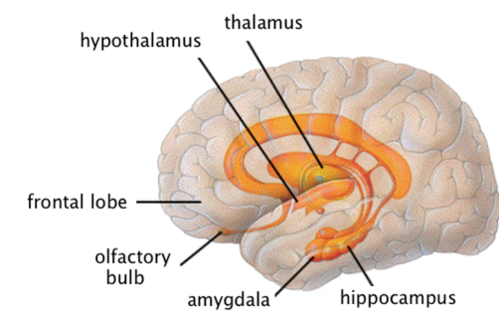
The brainstem (middle of brain) connects the cerebrum with the spinal cord. The brainstem includes the midbrain, the pons and the medulla. Midbrain. The midbrain (or mesencephalon) is a very complex structure with a range of different neuron clusters (nuclei and colliculi), neural pathways and other structures.
How do different signals control different processes?
Different signals control different processes, and your brain interprets each. Some make you feel tired, for example, while others make you feel pain. Some messages are kept within the brain, while others are relayed through the spine and across the body’s vast network of nerves to distant extremities.
What is the difference between gray and white matter?
In the brain, gray matter refers to the darker, outer portion, while white matter describes the lighter, inner section underneath. In the spinal cord, this order is reversed: The white matter is on the outside, and the gray matter sits within.
What organ controls memory, emotion, touch, motor skills, vision, breathing, temperature, hunger, and every other process
The brain is a complex organ that controls thought, memory, emotion, touch, motor skills, vision, breathing, temperature, hunger and every process that regulates our body. Together, the brain and spinal cord that extends from it make up the central nervous system, or CNS.
Where is the spinal cord located?
The spinal cord extends from the bottom of the medulla and through a large opening in the bottom of the skull. Supported by the vertebrae, the spinal cord carries messages to and from the brain and the rest of the body.
What is the name of the outer layer of the cerebrum?
Cortex is Latin for “bark,” and describes the outer gray matter covering of the cerebrum. The cortex has a large surface area due to its folds, and comprises about half of the brain’s weight.
How much fat is in the brain?
Weighing about 3 pounds in the average adult, the brain is about 60% fat. The remaining 40% is a combination of water, protein, carbohydrates and salts. The brain itself is a not a muscle. It contains blood vessels and nerves, including neurons and glial cells.
Overview
The human brain controls and executes various kinds of movements throughout the body.
Summary
The motor cortex is frequently split into two main territories: the primary motor cortex and the nonprimary cortex
Location
The motor cortex is located in the frontal lobe, and it extends across a region of the cortex slightly anterior to the central sulcus, which extends down the side of the cerebral hemispheres.
Structure
The motor cortex is frequently split into two main territories: the primary motor cortex, which is located in a gyrus recognized as the precentral gyrus in front of the central sulcus, and the nonprimary motor cortex, which is located anterior to the primary motor cortex and includes two notable areas that are referred to as the premotor cortex and supplementary motor cortex (Figure 1)..
Function
Employing conscious dogs as their subjects, doctors Gustav Theodor Fritsch and Eduard Hitzig electrically activated the part of the brain presently known as the motor cortex and discovered that the activation led the dogs to act unconsciously.
Complications involving Motor Cortex
Whenever the primary motor cortex is injured, the individual often exhibits poor movement coordination and dexterity. The individual, for instance, cannot frequently conduct fine motor motions.
Conclusion
The motor cortex is divided into three sections of the frontal lobe, all of which are located directly anterior to the central sulcus. The power of a movement is encoded by the primary motor cortex.
Why is the sensory cortex important?
The sensory cortex of the human brain is very important because it enables users to carry out our daily activities with ease. Dysfunctions of the sensory cortex may result in losing some sensing abilities such as hearing, sight or balance.
What are the three areas of the brain?
The cortex of the human brain is categorized into three functionally unique areas namely; associative, sensory and motor (2). The motor cortex is responsible for planning, controlling and executing voluntary movements. Moreover, the associative cortex integrates generated visual, auditory, gustatory and other general sensory signals.
What is the sensory cortex?
The sensory cortex is defined as all cortical areas linked with sensory functions (1). In another definition, the sensory cortex is a section of the cerebral cortex which is responsible for receiving and interpreting sensory information from different parts of the body. Stimuli.
What are stimuli received from?
Stimuli. received from different receptors such as nociceptors and thermoreceptors are. transduced to an action potential which is conveyed along one or more afferent. neuron to a specific section of the brain (3). According to studies, the. sensory cortex comprises the visual cortex, auditory cortex, the primary.
Which part of the sensory cortex is responsible for taste?
It is part of the sensory cortex responsible for tasting. Neurons in the gustatory cortex respond to sourness, sweetness, saltiness, and bitterness. They also code the intenseness of the taste stimulus (1). The gustatory cortex is made up of 2 substructures namely 1) The anterior insula and 2) The frontal operculum.
Which part of the auditory system is responsible for language switching?
The auditory cortex is one part of the auditory system that does common and higher roles in hearing like language switching. Also, the auditory cortex is comprised of sections belongs to the transverse temporal gyri, the superior temporal gyrus that is, planum temporal and the planum polare (2).
Which part of the brain receives sensory input from the thalamus?
The section of the visual cortex that receives sensory input from the thalamus is called the primary visual cortex, also referred to as visual area 1 (V1) or striate cortex (1). Both the right and left hemispheres of the human brain contain the visual cortex. The visual cortex found in the left hemisphere receives radiations from ...
Overview
Structure
The cerebral cortex is the outer covering of the surfaces of the cerebral hemispheres and is folded into peaks called gyri, and grooves called sulci. In the human brain it is between two and three or four millimetres thick, and makes up 40 per cent of the brain's mass. 90 per cent of the cerebral cortex is the six-layered neocortex with the other 10 per cent made up of allocortex. There are between 14 and 16 billion neurons in the cortex, and these are organized radially in cortical colu…
Blood supply and drainage
Blood supply to the cerebral cortex is part of the cerebral circulation. Cerebral arteries supply the blood that perfuses the cerebrum. This arterial blood carries oxygen, glucose, and other nutrients to the cortex. Cerebral veins drain the deoxygenated blood, and metabolic wastes including carbon dioxide, back to the heart.
The main arteries supplying the cortex are the anterior cerebral artery, the middle cerebral artery, …
Development
The prenatal development of the cerebral cortex is a complex and finely tuned process called corticogenesis, influenced by the interplay between genes and the environment.
The cerebral cortex develops from the most anterior part, the forebrain region, of the neural tube. The neural plate folds and closes to form the neural tube. From the cavity inside the neural tube develops the ventricular system, and, from the neuroepithelial cells of its walls, the neurons and glia of …
Evolution
Of all the different brain regions, the cerebral cortex shows the largest evolutionary variation and has evolved most recently. In contrast to the highly conserved circuitry of the medulla oblongata, for example, which serves critical functions such as regulation of heart and respiration rates, many areas of the cerebral cortex are not strictly necessary for survival. Thus, the evolution of the cerebral cortex has seen the advent and modification of new functional areas—particularly asso…
Function
The cerebral cortex is connected to various subcortical structures such as the thalamus and the basal ganglia, sending information to them along efferent connections and receiving information from them via afferent connections. Most sensory information is routed to the cerebral cortex via the thalamus. Olfactory information, however, passes through the olfactory bulb to the olfactory cortex (piriform cortex). The majority of connections are from one area of the cortex to another, …
Clinical significance
Neurodegenerative diseases such as Alzheimer's disease and Lafora disease, show as a marker, an atrophy of the grey matter of the cerebral cortex.
Other diseases of the central nervous system include neurological disorders such as epilepsy, movement disorders, and difficulties in speech (aphasia).
Brain damage from disease or trauma, can involve damage to a specific lobe such as in frontal lo…
History
In 1909, Korbinian Brodmann distinguished different areas of the neocortex based on cytoarchitectural difference and divided the cerebral cortex into 52 regions.
Rafael Lorente de Nó, a student of Santiago Ramon y Cajal identified more than 40 different types of cortical neurons based on the distribution of their dendrites and axons.