What is the decidua basalis?
With continued growth of the embryo, the decidua basalis becomes incorporated into the maternal component of the definitive placenta. The remaining decidua, which consists of the decidualized endometrial tissue on the sides of the uterus not occupied by the embryo, is the decidua parietalis.
What is the decidua?
The decidua is composed of glands, immune cells, blood and lymph vessels , and decidual stromal cells (DSCs) and fetal extravillous trophoblast (Mori et al., 2016).
What is decidualization in mammals?
Decidualization denotes the transformation of the endometrial stroma into the decidual matrix that supports embryo implantation and subsequent placenta formation. This process is foremost characterized by the differentiation of endometrial stromal cells (EnSCs) into secretory decidual cells ( Gellersen and Brosens, 2014 ). Decidualization only occurs in species where the trophoblast breaches the luminal endometrial epithelium and invades maternal tissues. The depth of decidual transformation is determined by the degree of placental trophoblast invasion ( Ramsey et al., 1976 ). In most mammals, decidualization is initiated upon embryo implantation. However, in a handful of species, including humans, Old World monkeys, some bats, elephant shrew, and spiny mouse, decidualization is “spontaneous,” meaning that it is initiated independently of an implanting embryo during the midluteal phase of each cycle ( Emera et al., 2012). Once triggered, the decidual phenotype is strictly dependent on sustained progesterone signaling. In the absence of pregnancy, falling ovarian progesterone production triggers a cascade of inflammatory events in the decidualizing endometrium, which upon recruitment and activation of leucocytes becomes irrevocable and leads to partial tissue destruction, bleeding and menstrual shedding. Hence, spontaneous decidualization is inextricably linked to cyclic menstruation; and the term “decidua,” derived from the Latin verb “decidere” (meaning to fall off, to detach, or to die), aptly captures the nature of the process.
What is ectopic decidua?
Ovarian ectopic decidua is an incidental finding in the majority of cases. Macroscopically, ectopic decidua is seen as a tan or red spot on the ovarian surface. At microscopic examination, the cells of ectopic decidua are similar to the cells of decidualized endometrium.
What is the outermost layer of the uterus?
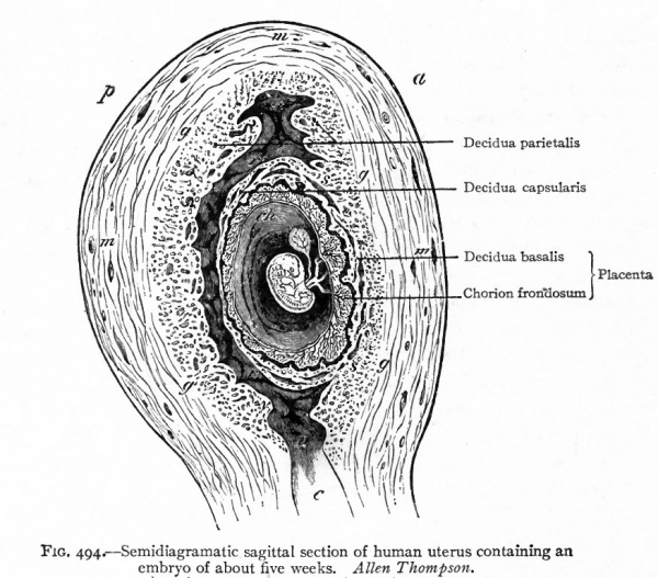
The superficial layer of endometrium becomes circumferentially decidualized throughout the uterus. The decidua capsularis is the portion underlying the chorion laeve and fuses with it to become the outermost component of the peripheral membranes. As the amniotic cavity expands to fill the uterus, the decidua capsularis becomes thinner until the chorioamnion is in direct contact with the decidua parietalis. The decidua basalis is the portion at the base of the placental disk, underneath the chorion frondosum. The chorionic stem villi extend down and anchor to the decidua. As the villi grow throughout pregnancy, they gradually replace most of the decidua basalis. The remaining portion of decidua, unassociated with the placenta, is known as decidua parietalis.
What is the function of the decidua?
The main functions of the decidua are to regulate syncytiotrophoblast invasion, provide nutrition and gas exchange, and produce hormones. The decidualization of the inner layer of endometrium prepares the uterus for pregnancy, with trophoblast invading into it and spiral arterioles sprouting out of it.
What is the function of dendritic cells?
The principal function of dendritic cells is to regulate the adaptive immune response through a specialized capacity to take up, process, and present antigens to T lymphocytes, and to govern the activation response of T cells through powerful immune-regulatory activity. Although fewer in number than macrophages, dendritic cells are ontologically related; a large proportion of dendritic cells in the uterus exhibits characteristics consistent with a myeloid origin. Decidual dendritic cells fall into two broad categories: CD83 + mature dendritic cells, which comprise approximately 1% of decidual stromal cells, 218 and CD83 + immature macrophage/dendritic cells.
What is the decidua basalis?
With continued growth of the embryo, the decidua basalis becomes incorporated into the maternal component of the definitive placenta. The remaining decidua, which consists of the decidualized endometrial tissue on the sides of the uterus not occupied by the embryo, is the decidua parietalis.
What is the decidua?
The decidua is composed of glands, immune cells, blood and lymph vessels, and decidual stromal cells (DSCs) and fetal extravillous trophoblast (Mori et al., 2016 ).
What is decidualization in mammals?
Decidualization denotes the transformation of the endometrial stroma into the decidual matrix that supports embryo implantation and subsequent placenta formation. This process is foremost characterized by the differentiation of endometrial stromal cells (EnSCs) into secretory decidual cells ( Gellersen and Brosens, 2014 ). Decidualization only occurs in species where the trophoblast breaches the luminal endometrial epithelium and invades maternal tissues. The depth of decidual transformation is determined by the degree of placental trophoblast invasion ( Ramsey et al., 1976 ). In most mammals, decidualization is initiated upon embryo implantation. However, in a handful of species, including humans, Old World monkeys, some bats, elephant shrew, and spiny mouse, decidualization is “spontaneous,” meaning that it is initiated independently of an implanting embryo during the midluteal phase of each cycle ( Emera et al., 2012). Once triggered, the decidual phenotype is strictly dependent on sustained progesterone signaling. In the absence of pregnancy, falling ovarian progesterone production triggers a cascade of inflammatory events in the decidualizing endometrium, which upon recruitment and activation of leucocytes becomes irrevocable and leads to partial tissue destruction, bleeding and menstrual shedding. Hence, spontaneous decidualization is inextricably linked to cyclic menstruation; and the term “decidua,” derived from the Latin verb “decidere” (meaning to fall off, to detach, or to die), aptly captures the nature of the process.
Is ectopic decidua a polyp?
Ectopic decidua is common in the cervix in pregnancy .272 It may present as a polyp, as plaquelike deposits on the surface, or within endometriosis. The important points of recognition pertain to the association of a coexisting pregnancy and distinction from mesenchymal neoplasia ( Fig. 15.34 ).
What is ectopic decidua?
Ovarian ectopic decidua is an incidental finding in the majority of cases. Macroscopically, ectopic decidua is seen as a tan or red spot on the ovarian surface. At microscopic examination, the cells of ectopic decidua are similar to the cells of decidualized endometrium.
What is the outermost layer of the uterus?
The superficial layer of endometrium becomes circumferentially decidualized throughout the uterus. The decidua capsularis is the portion underlying the chorion laeve and fuses with it to become the outermost component of the peripheral membranes. As the amniotic cavity expands to fill the uterus, the decidua capsularis becomes thinner until the chorioamnion is in direct contact with the decidua parietalis. The decidua basalis is the portion at the base of the placental disk, underneath the chorion frondosum. The chorionic stem villi extend down and anchor to the decidua. As the villi grow throughout pregnancy, they gradually replace most of the decidua basalis. The remaining portion of decidua, unassociated with the placenta, is known as decidua parietalis.
What are the functions of the decidua?
Function. The main functions of the decidua are to regulate syncytiotrophoblast invasion, provide nutrition and gas exchange, and produce hormones. The decidualization of the inner layer of endometrium prepares the uterus for pregnancy, with trophoblast invading into it and spiral arterioles sprouting out of it.
What is the primary villi?
primary villi - ( primary chorionic villi) Term describing the earliest stage of embryonic placenta development. In humans, the conceptus during week 2 this first stage of chorionic villi development consists of only the trophoblastic shell cells ( syncitiotrophoblasts and cytotrophoblasts) forming finger-like extensions into maternal decidua. ...
Which superfamily is responsible for decidualization of endometrial cells?
Member of the a transforming growth factor beta (TGFbeta) superfamily , contributes to human endometrial stromal cells (HESC) decidualization and has been localized to decidual cells in the human endometrium. (possibly also BMP2 and TGFbeta1)
What is the term for the uterine endometrium?
decidua capsularis - The term given to the uterine endometrium which has been converted to decidua surrounding the conceptus on the smooth chorion side. decidua parietalis - The term given to the remainder of the uterine endometrium, away from the site of implantation, that gradually becomes comverted to decidua.
What is CA in medical terms?
chorioamnionitis - (CA) An intraamniotic puerperal infection described as having 3 forms: histologic, clinical (clinical chorioamnionitis, IAI), and subclinical. Intraamniotic infection is a common (2-4%) event in labor and the systemic inflammatory response can also lead to preterm birth and neonatal complications.
How big is the placenta?
In human, the placenta at term is a discoid shape "flat cake" shape; 20 cm diameter, 3 cm thick and weighs 500-600 gm. Placenta are classified by the number of layers between maternal and fetal blood (Haemochorial, Endotheliochorial and Epitheliochorial) and shape (Discoid, Zonary, Cotyledenary and Diffuse).
How is placental thickness measured?
placental thickness - is measured at its mid-portion from the chorionic plate to the basilar plate, on a longitudinal plane (less than 4 cm at term). Excludes any abnormalities (fibroids, myometrial contractions, or venous lakes). The placental thickness approximates in millimeters to the weeks of gestation.
Why does blood flow increase during pregnancy?
Flow increase is due to the trophoblast cell invasion of the spiral arteries opening them into blood-filled spaces of the placenta.
Where are Mesenchymal stem cells obtained?
Mesenchymal stem cells (MSCs) that meet the International Society for Cellular Therapy (ISCT) criteria are obtained from placental tissue by plastic adherence. Historically, no known single marker was available for isolating placental MSCs (pMSCs) from the decidua basalis. As the decidua basalis is derived from the regenerative endometrium, we hypothesised that SUSD2, an endometrial perivascular MSC marker, would purify maternal perivascular pMSC. Perivascular pMSCs were isolated from the maternal placenta using SUSD2 magnetic bead sorting and assessed for the colony-forming unit-fibroblasts (CFU-F), surface markers, and in vitro differentiation into mesodermal lineages. Multi-colour immunofluorescence was used to colocalise SUSD2 and α-SMA, a perivascular marker in the decidua basalis. Placental stromal cell suspensions comprised 5.1%SUSD2 + cells. SUSD2 magnetic bead sorting of the placental stromal cells increased their purity approximately two-fold. SUSD2 + pMSCs displayed greater CFU-F activity than SUSD2 − stromal fibroblasts (pSFs). However, both SUSD2 + pMSC and SUSD2 − pSF underwent mesodermal differentiation in vitro, and both expressed the ISCT surface markers. Higher percentages of cultured SUSD2 + pMSCs expressed the perivascular markers CD146, CD140b, and SUSD2 than SUSD2 − pSFs. These findings suggest that SUSD2 is a single marker that enriches maternal pMSCs, suggesting they may originate from eMSC. Placental decidua basalis can be used as an alternative source of MSC for clinical translation in situations where there is no access to endometrial tissue.
What is the ethical code for Monash Health?
All procedures performed in studies involving human participants were in accordance with the ethical standards of the National Health and Medical Research Council of Australia Guidelines as approved by Monash Health Human Research Ethics Committee (Ethics Number: 10103B) and with the 1964 Helsinki declaration and its later amendments or comparable ethical standards. All participants gave written informed consent. This article does not contain any studies with animals performed by any of the authors.
Is SUSD2 a marker for pMSC?
Our study aimed to determine the utility of SUSD2 as a marker for enriching human decidua basalis pMSCs by assessing colony-forming ability, surface marker expression, and differentiation potential. For the first time, our findings show that maternal placental SUSD2 + cells with MSC properties are perivascular cells in the decidua basalis of human placenta. These pMSCs display greater CFU-F activity than SUSD2 − eSF, but similar mesodermal differentiation in vitro, and also similarly express ISCT markers. In contrast, higher percentages of cultured SUSD2 + pMSCs express the perivascular markers CD146, CD140b, and as expected, SUSD2. These results indicate that SUSD2 is a suitable single marker for enriching maternal pMSCs from human placenta. The combination of another perivascular marker with SUSD2, such as CD146, might markedly improve the enrichment factor for maternal pMSC.
Introduction

Maternal Decidua
- The maternal uterine endometrium stromal cells (fibroblast-like) are transformed by steroid hormones (progesterone) and embryonic signals into the decidua. The entire maternal decidua is divided into three regions: decidua basalis, decidua capsularis and decidua parietals (decidua vera). These 3 regions are named by their positional relationship to the conceptus. Immunostain…
Maternal Immune
- How does the implanting conceptus avoid immune rejection by the maternal immune system? There are a number of maternal and embryonic mechanisms that are thought to act to prevent immune rejection of the implanting conceptus, though the complete mechanism(s) are unknown. This is particularly relevant to Assisted Reproductive Technologyinvolving donor eggs. Below ar…
Decidualization Factors
- There are a number of known molecular signals involved in the conversion of uterine stromal cells into decimal cells.
References
- Articles
{{#pmid:27219485]] Plaisier M. (2011). Decidualisation and angiogenesis. Best Pract Res Clin Obstet Gynaecol , 25, 259-71. PMID: 21144801 DOI. - Search PubMed
Search Pubmed: Maternal Decidua | Decidualization