What causes an enlarged hilum of the lung?
Some common disorders that affect the hilum include:
- Sarcoidosis. Sarcoidosis is an inflammatory condition that causes atypical cells to form in various organs, like the hilar tissue in your lung.
- Hilar masses or tumors. Lung cancers or lymphomas can cause tumors or masses to form in the hilar tissue.
- Asymmetrical hila. ...
- Infection. ...
- Lymphadenopathy. ...
- Pulmonary artery or venous hypertension. ...
What does right sided hilar mass in lungs indicate?
Treatment for a right hilar mass on the lung can be surgery, radiation, chemotherapy or medications, depending on what is causing the mass, says About.com. The mass can be caused by lung cancer and lymphomas or pulmonary arterial or venous hypertension. A right hilar mass can also be caused by Behcet’s syndrome, according to PubMed.
What is a hilar region?
The hilar region is where the bronchi, arteries, veins, and nerves enter and exit the lungs. This area can be difficult to visualize on a chest X-ray, and further tests such as computerized tomography (CT) scan with contrast are often needed to determine if a problem exists.
Where does lung cancer cause pain?
While chest pain is one of the most common symptoms of lung cancer, other symptoms include:
- Persistent coughing
- Wheezing
- Shortness of breath
- Hoarseness
- Weight loss
- Loss of appetite
- Fatigue or feeling weak
- Respiratory infections (like bronchitis or pneumonia) that don’t go away or keep returning 3

What part of the lung is the hilar?
The hilum of the lung is the wedge-shaped area on the central portion of each lung, located on the medial (middle) aspect of each lung. The hilar region is where the bronchi, arteries, veins, and nerves enter and exit the lungs.
What is hilar cancer?
Hilar cholangiocarcinoma is a type of bile duct cancer that occurs in the bile ducts that lead out of the liver (hepatic ducts) and join with the gallbladder. Hilar cholangiocarcinomas are also known as Klatskin tumors.
What does the medical term hilar mean?
Medical Definition of hilar : of, relating to, affecting, or located near a hilum hilar lymph nodes of the lung.
Can a hilar mass be cured?
Based on the results of early hilar lung carcinoma, we concluded that these lesions are curable if they are properly diagnosed and treated.
What can cause a hilar mass?
The PET-CT scan revealed a high metabolic activity in the hilar tumor mass, which can appear in lung cancer and in several other conditions with active granuloma formation, such as: sarcoidosis, tuberculosis, nontuberculous mycobacterium granuloma, and fungal infections.
Can hilar lymph nodes be removed?
The interlobar lymph nodes and hilar lymph nodes around the vessels should not be removed separately; rather, they should be dissociated to the distal end of the vessel and then removed en bloc with the right upper pulmonary lobe. This is more consistent with the principles of surgical oncology.
What is hilar abnormality?
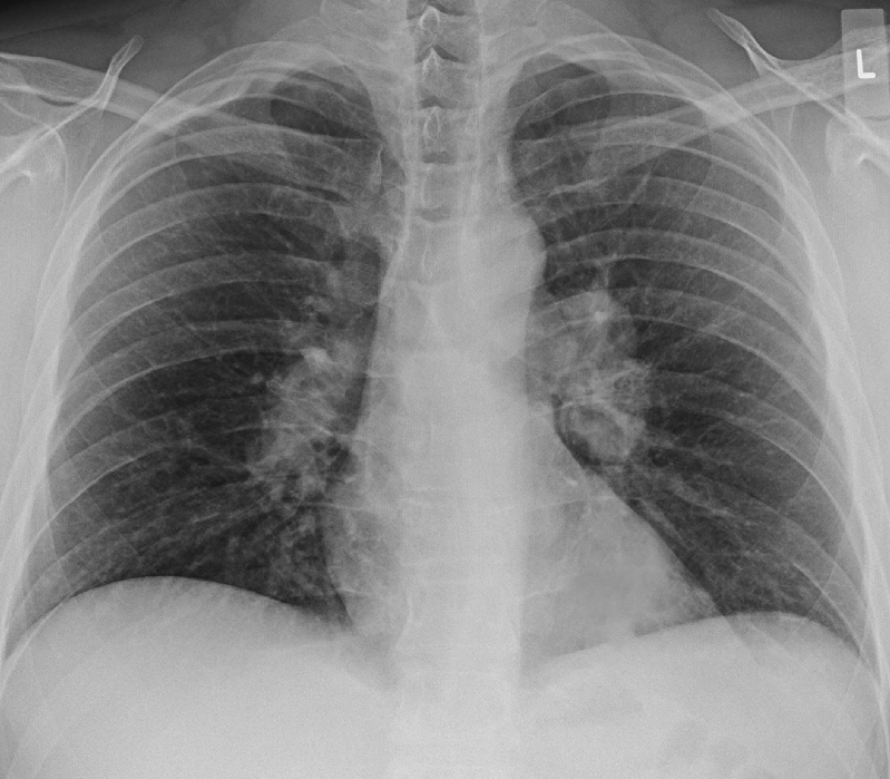
The hila consist of vessels, bronchi and lymph nodes. On a chest X-ray, abnormalities of these structures are represented by a change in position, size and/or density.
What do hilar lymph nodes do?
Hilar lymph nodes collect lymph from the pulmonary nodes, and drain to the tracheobronchial nodes. Synonym: bronchopulmonary lymph node, bronchial gland.
What does a hilar mass mean?
Sarcoidosis is an inflammatory condition that causes atypical cells to form in various organs, like the hilar tissue in your lung. Hilar masses or tumors. Lung cancers or lymphomas can cause tumors or masses to form in the hilar tissue.
Can a CT scan tell if a lung nodule is cancerous?
Can a CT scan tell if a lung nodule is cancerous? The short answer is no. A CT scan usually isn't enough to tell whether a lung nodule is a benign tumor or a cancerous lump. A biopsy is the only way to confirm a lung cancer diagnosis.
At what size should a lung nodule be removed?
Nodules between 6 mm and 10 mm need to be carefully assessed. Nodules greater than 10 mm in diameter should be biopsied or removed due to the 80 percent probability that they are malignant.
What percentage of lung masses are cancerous?
Most lung nodules are benign, or non-cancerous. In fact, only 3 or 4 out of 100 lung nodules end up being cancerous, or less than five percent.
Is Klatskin tumor curable?
As Klatskin tumors are typically resistant to chemotherapy and radiotherapy, surgical resection of the tumor is the only curative treatment but it is not always an option in those patients with widespread metastasis.
Is hilar cholangiocarcinoma curable?
Hilar cholangiocarcinoma is the most common malignant tumor affecting the extrahepatic bile duct. Surgical treatment offers the only possibility of cure, and it requires removal of all tumoral tissues with adequate resection margins.
What is the survival rate for bile duct cancer?
The 5-year survival rate for intrahepatic bile duct cancer is 9%. If the cancer is diagnosed at an early stage, the 5-year survival rate is 25%. If the cancer has spread to the regional lymph nodes, the 5-year survival rate is 8%. If the cancer has spread to a distant part of the body, the 5-year survival rate is 2%.
How rare is hilar cholangiocarcinoma?
Cholangiocarcinoma is rare. On average, it affects fewer than 6 in 100,000 people around the world. In the United States, cholangiocarcinoma affects about 8,000 people per year. In some places, though, cholangiocarcinoma is more common.
Which lung has a hilum?
Both the right and the left lung have a hilum which lies roughly midway down the lungs, and slightly towards the back (closer to the vertebrae than to the front of the chest). Each lung may be visualized as having an apex (the top), a base (the bottom), a root, and a hilum.
Why is the hilum of one or both lungs enlarged?
There are four main reasons why the hilum of one or both lungs may appear enlarged on an X-ray. These include: 1 . Tumors and lymphadenopathy: Cancers such as lung cancers and lymphomas, as well as cancer that has spread to this region from other parts of the body (metastatic cancer) can cause masses in this region.
Why is the hilum enlarged?
Enlargement of the hilum may occur due to tumors (such as lung cancer), pulmonary hypertension, or enlarged hilar lymph nodes due to conditions such as infections (especially tuberculosis and fungal infections), cancer (either local or metastatic), sarcoidosis, and more. 1 . An Overview of Pulmonary Hypertension. Theresa Chiechi / Verywell.
What is the hilum on a chest X-ray?
The hilar region is where the bronchi, arteries, veins, and nerves enter and exit the lungs. This area can be difficult to visualize on a chest X-ray, and further tests such as computerized tomography (CT) scan with contrast are often needed to determine if a problem exists. Enlargement of the hilum may occur due to tumors (such as lung cancer), ...
What causes hilar masses?
Tumors, both primary and metastatic, are a far too common cause of both hilar masses and lymphadenopathy. The most common causes overall include tuberculosis worldwide, and conditions such as histoplasmosis, coccidioidomycosis, and sarcoidosis in the United States. 10 .
Why is my hilum abnormal?
Some apparent abnormalities of the hilum may simply be due to positioning, and further views may rule out problems. If a mass or enlargement is noted, possible causes can vary depending on the appearance:
What is the name of the test that shows abnormalities in the hilar region?
In addition to imaging tests, abnormalities in the hilar region may be identified with tests such as a bronchoscopy, a test in which a tube is inserted through the mouth and down into the major airways (bronchi).
Where are the lungs located?
Log In. The lungs are the organs of respiration. They are located in the thorax, either side of the mediastinum. The function of the lungs is to oxygenate blood. They achieve this by bringing inspired air into close contact with oxygen-poor blood in the pulmonary capillaries.
Which lung has inferior and superior lobes?
The left lung contains superior and inferior lobes, which are separated by a similar oblique fissure.
How are the medial surfaces of the lungs suspended?
They are suspended from the mediastinum by the lung root – a collection of structures entering and leaving the lungs. The medial surfaces of both lungs lie in close proximity to several mediastinal structures:
Which bronchioles are terminal?
The segmental bronchi give rise to many conducting bronchioles, which eventually lead into terminal bronchioles. Each terminal bronchiole gives off respiratory bronchioles, which feature thin walled outpocketings that extend from their lumens. These are the alveoli – the site of gaseous exchange.
How many lobes does the right lung have?
The right and left lungs do not have an identical lobular structure. The right lung has three lobes; superior, middle and inferior. The lobes are divided from each other by two fissures: Oblique fissure – Runs from the inferior border of the lung in a superoposterior direction, until it meets the posterior lung border.
What is the role of the lungs in pulmonary embolism?
The lungs are the organs of respiration. They are located in the thorax, either side of the mediastinum. The function of the lungs is to oxygenate blood. They achieve this by bringing inspired air into close contact with oxygen-poor blood in the pulmonary capillaries.
Which bronchus has a higher incidence of foreign body inhalation?
Note: The right bronchus has a higher incidence of foreign body inhalation due to its wider shape and more vertical course.
What is a hilar adenopathy?
Definition: What is Hilar adenopathy? Hilar adenopathy can be defined as the enlargement of the lymph nodes, occurring at the level of the pulmonary hilum. This condition does not appear on its own, always signifying the existence of an underlying pathology.
What are the causes of symmetrical hilar adenopathy?
The following conditions lead to the appearance of unilateral or bilateral, symmetrical hilar adenopathy: primary tuberculosis, fungal infection, atypical mycobacterial infection, viral infection, tularemia, anthrax, bronchogenic carcinoma, lymphoma, sarcoidosis and silicosis. On the other hand, the bilateral symmetrical adenopathy can only be caused by: sarcoidosis ( most common), viral infections (adenovirus or mononucleosis).
What is the difference between enlarged lymph nodes and enlarged pulmonary arteries?
While the enlarged lymph nodes have a lumpy or bumpy appearance (characteristic opacity on the X-ray), the enlarged pulmonary arteries appear to be smooth (in reference to their contour).
Can a chest CT confirm hilar adenopathy?
Chest CT can also be used for the confirmation of hilar adenopathy. Depending on the results provided by the imaging studies, the enlargement of the lymph nodes can be classified as unilateral or bilateral. In the situation that the hilar adenopathy is bilateral, it can be further classified into symmetrical or asymmetrical.
Can a pulmonary X-ray show hilar adenopathy?
Diagnosis. One of the most investigations used for the diagnosis of the hilar adenopathy is the plain pulmonary X-ray. However, has to take into consideration that the pulmonary arteries go through the same area. In the situation that these vessels are enlarged, they might be mistaken for hilar adenopathy.
What is Hilum of Lung?
The hilum is the triangular depressed area that allows the entry of bronchus, blood vessels, and nerves. Through the hilum, the root of the lung enters and exits the lung. It situates in the centre of the medial surface. Each lung has a hilum (plural – hila). Hence, our body has two hila. Both hila are similar in size, with the left hilum usually found slightly higher in the chest than the right hilum.
How many hilums are in each lung?
Each lung has one hilum and one root of the lung.
What is the root of the lung?
The root of the lung is the structures that enter and exit at the hilum area. Therefore, each lung has its own root in the lung. Bronchus, pulmonary artery, pulmonary veins, lymphatics and nerves together make the root of each lung. Furthermore, there is a difference between right and left roots of the lungs.
What is the difference between a hilum and a root of the lung?
The key difference between Hilum and Root of Lung is that the Hilum of the lung is the large depressed area that lies near the centre of the medial surface while the Root of Lung is the all structures entering or leaving the lung at the hilum, forming a pedicle. Lungs are the respiratory organs of our body.
What are the components of the lungs?
There are four main components of each lung. They are apex, base, root and hilum. The root of the lung situates in the hilum of each lung.
What connects the medial surface of the lung with the mediastinum?
The root of lung connects the medial surface of the lung with the mediastinum. Surrounding the root of the lung in each lung, there is a tubular sheath derived from mediastinal pleura.
How many hila are there in the body?
Hence, our body has two hila. Both hila are similar in size, with the left hilum usually found slightly higher in the chest than the right hilum. Tumors can occur in the area of hilum. Furthermore, the enlargement of hilar lymph nodes, as well as abnormalities of the pulmonary arteries or veins can happen.
Right Hilar Lymph Node Location
The location of the lymph nodes may be at various points in the human body, for instance, around the neck, etc. However, the hilar lymph nodes, either right or left hilar lymph nodes, are present in the human respiratory system. Now, the question that arises; what is the location of hilar lymph nodes in a human body?
Right Hilar Lymph Node Metastasis
Metastasis refers to the state of the body when the cancerous cells begin to spread from the place where they get formed first to some other part of the body. The cancer cells tear apart from the original tumor and travel through the lymph system or blood during metastasis. As a result of which, new tumors are formed; in other organs or tissues.
Right Hilar Lymph Node Calcification
Calcification refers to the accumulation of calcium salts in body tissue. It usually occurs in bone formation; however, it sometimes deposits in the soft tissue abnormally, causing it to harden. Calcifications occur in nearly all cells. Similarly, the calcifications are also associated with the lymph nodes.