What is the lateral canthus and medial canthus?
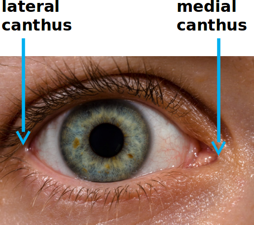
The lateral canthus is the angle or corner of the eye that is located laterally, or away from the face's midplane. It is sometimes referred to as the "outside corner" of the eye. The medial canthus is the opposite angle, located medially, or toward the middle of the face. It is sometimes referred to as the "inside corner" of the eye.
What is the angulus canthus?
the angle formed by the junction of the lateral parts of the upper and lower eyelids. Synonym(s): angulus oculi lateralis[TA], angulus oculi temporalis, external canthus, lateral canthus Farlex Partner Medical Dictionary © Farlex 2012 Want to thank TFD for its existence?
What is lateral Canthoplasty?
Canthoplasty surgery, also known as lateral canthoplasty or cat eye surgery, is a type of eyelid surgery that alters the lateral canthus (the outer corner of the eye where the upper and lower lid meet) by tightening and elongating the eye horizontally. It’s an oculoplastic procedure that creates an almond-shaped or “cat” eye.
What are the 3 pins on the lateral canthus?
The lateral canthus, posterior end of the tragus and commissure were marked as reference points with 3 pins with colored heads and the center of each ligament over the skin were also marked with 3 pins. True Retaining Ligaments of Face as Surgical Landmarks/Los Verdaderos Ligamentos de Retencion de la Cara como Marcadores Quirurgicos
How to treat lateral scleral show?
What is the most prominent portion of the zygomaticomax?
Where is the canthusa drawn?
Which canthusligament was cut to access the caudal aspect of orbit?
About this website
Where is your canthus located?
The canthus (pl. canthi, palpebral commissures) is either corner of the eye where the upper and lower eyelids meet. More specifically, the inner and outer canthi are, respectively, the medial and lateral ends/angles of the palpebral fissure.
What is lateral canthal area?
The outer corner of the eyelids where the upper and lower eyelids meet are rounded instead of joining in a discrete angle. This area is called the outer canthus or the lateral canthus.
Is lateral canthus upper or lower eyelid?
The lateral canthus is the point where the outer edge of the upper and lower eyelids meet. These two structures join to form an angle. Rounding or blunting of this angle may occur after previous eyelid surgery or with aging.
What is the lateral side of the eye?
The lateral rectus muscle is a muscle on the lateral side of the eye in the orbit. It is one of six extraocular muscles that control the movements of the eye. The lateral rectus muscle is responsible for lateral movement of the eyeball, specifically abduction....Lateral rectus muscle.Lateral rectusFMA49038Anatomical terms of muscle11 more rows
What is the function of the lateral canthus?
The lateral canthal tendon is attached to the inner aspect of the frontozygomatic process on the orbital osseous tubercle and is essential to the structural fixation of the lateral canthus as well as a check on the mobility of the lateral canthal angle of the eye itself.
Why does my canthus hurt?
The inner canthus is the area where your top eyelid meets your bottom. If you have accidentally scratched this area, it could be painful, particularly when pressure is placed on the area by your nose pads. Another explanation could be a tiny spot in the area irritating your eye or eyelid.
What is the corner of eye near nose called?
The nasolacrimal duct is known to you and me as the tear duct. It's the area found in the corner of your eyes, closest to the nose.
What is the area between your eyelid and eyebrow called?
This space is also called the palpebral fissure. Typically the palpebral fissure measures between 28 to 30 mm wide and around 9 to 10 mm in height.
What is lower eyelid called?
Similarly, the skin fold in the lower eyelid is called the inferior palpebral sulcus. This lower skin fold is often more prominent in children and can become less prominent as one gets older. Anatomically, the inferior skin crease is seen around 3 to 5 mm below the outer aspect of the lid margin.
Is lateral outside or inside?
A lateral orientation is a position away from the midline of the body. For instance, the arms are lateral to the chest, and the ears are lateral to the head. A medial orientation is a position toward the midline of the body. An example of medial orientation is the eyes, which are medial to the ears on the head.
Is lateral left or right?
(Note: They all apply to a standing human body.) Medial and lateral: Medial refers to being toward the midline of the body or the median plane, which splits the body, head-to-toe, into two halves, the left and right. Lateral is the side of the body or part of the body that is away from the middle.
What is the outside of the eye called?
The sclera is the tough outer layer of the eyeball (the white of the eye). The slight bulge in the sclera at the front of the eye is a clear, thin, dome-shaped tissue called the cornea. The cornea directs light rays into the eye and helps focus them on the retina.
What is canthus of the eye?
plural canthi ˈkan-ˌthī -ˌthē : either of the angles formed by the meeting of an eye's upper and lower eyelids.
What is the canthal?
What Is a Canthal Tilt? The canthal tilt may be described as the angle between the medial canthus (inner corner of the eye) and the lateral canthus (outer corner of the eye). It is an integral concept in periorbital aesthetics and forms the foundation of several eyelid procedures.
What is the medial canthal area?
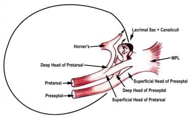
INTRODUCTION. The eyelid and canthal areas are common locations for malignant skin tumors. The medial canthal region includes the medial canthal tendon, lacrimal apparatus, and neurovascular structures from the deep orbit [1].
Where is the medial canthal area?
The medial palpebral ligament (medial canthal tendon) is a ligament of the face. It attaches to the frontal process of the maxilla, the lacrimal groove, and the tarsus of each eyelid. It has a superficial (anterior) and a deep (posterior) layer, with many surrounding attachments.
Where is the lateral canthus of the eye located?
The lateral angle, often known as the corner of the eye, is the point at where the upper and lower eyelids meet. The canthus of the eye is divided...
What is the inside of the upper eyelid called?
The upper and lower eyelids come together at two locations. The one on the inside is known as the medial canthus, while the one on the outside is k...
Is the bridge of the nose lateral to the left eye?
The eye is located lateral to the nose. The nose is located between the ears on the medial side. The ear is shaped like a drum; it is hollow and ha...
What is located lateral to the eyes?
The nose is MEDIAL (or LATERAL) in relation to the eyes. 2. The ears are in the MEDIAL (or LATERAL) position in relation to the eyes. 3. The mouth...
Where are the eyes located on the face?
At actuality, your eyes are about in the center of your head when measured vertically. Most individuals, though, draw them definitely above the cen...
How to treat lateral scleral show?
To treat lateral scleral show, a small amount of filler can be injected below the lateral canthusto provide lift.
What is the most prominent portion of the zygomaticomax?
Various specialties including plastic surgery, otolaryngology and maxillofacial surgery deal with this kind of fracture and differ in their approach towards reduction and fixation of the processes of the zygomatic bone.12 The malar eminence is the most prominent portion of the zygomaticomaxillary complex (ZMC) and is located approximately 2 cm inferior to the lateral canthus.
Where is the canthusa drawn?
Starting at the lateral canthusa line is drawn with a semicircular pattern towards the lateral eyebrow.
Which canthusligament was cut to access the caudal aspect of orbit?
The medial and lateral canthusligaments were cut to access the caudal aspect of orbit.
How to tell if lateral canthal tendon is intact?
Identifying the inferior crus of the lateral canthal tendon can sometimes be tricky. Use scissors to “strum” the area to feel for the inferior crus. If you feel tension (like a plucked string), the tendon is still intact and should be cut.
What is orbital compartment syndrome?
Orbital compartment syndrome is an ophthalmic emergency that can lead to rapid blindness if not promptly diagnosed and managed. Lateral canthotomy and cantholysis is the treatment of choice and involves surgically exposing the lateral canthal tendon and its inferior crus to relieve intraorbital pressure. Illustration by Lauren Chen.
Why do you cut the superior crus?
In some situations, the superior crus may also be cut to further relieve intraorbital pressure.
What nerve is the temporal branch?
The temporal branch is a single nerve with no anastomosis. Lift the lateral portion of the inferior eyelid to expose the lateral canthal tendon. To perform cantholysis, identify and cut the inferior crus of the lateral canthal ligament. Ensure that your scissors are pointing away from the globe during the incision.
Which ligament keeps the eye in the orbit?
Anatomy. The medial canthal ligament and lateral canthal ligament both keep the eye within the orbit. The lateral canthal tendon, which is the focus of this guide, has a superior crus (branch) and an inferior crus.
How long does it take for a hemostat to crush tissue?
Using a needle driver/hemostat, approximate the path of your incision from the lateral canthus to the rim of the orbit and lock the hemostat in place for about 20 seconds to 1 minute to crush the tissue, which helps with hemostasis.
Can a canthotomy be performed on an open globe?
Lateral canthotomy should not be performed in patients with suspected open globe, which may present with a corneal laceration, positive Seidel sign, hyphema, irregularly shaped pupil, herniated uveal tissue, and/or shallow anterior chamber.
How to prevent canthal and lid complications?
Intraoperative prevention of canthal and lid complications of lower blepharoplasty, in addition to lid tightening, depends heavily on minimizing vertical and horizontal forces of wound contracture. Avoiding overresection of skin during lower blepharoplasty is of utmost importance. There is, however, no unerring method for determining skin excess in the lower eyelid. Preoperatively, the pinch technique provides an estimate, but it may lead to overresection unless the lower lid is in its anatomical position as the test is performed (▶ Fig. 23.15). This method does not consider risk factors such as patient age, orbicularis atrophy, ligamentous atrophy, negative vector, or dry-eye symptoms, all of which influence the degree of skin removal. Intraoperative pitfalls that are inherent include overstretching of skin during redraping and unrecognized inferior lid margin displacement during skin resection (▶ Fig. 23.16). The latter may lead to significant skin overresection. Both are avoided by maintaining the eyelid’s anatomical position by traction suture during measurement and resection (▶ Fig. 23.17). Further, draping the skin superiorly without medial or horizontal traction further minimizes risk of overresection. Abnormal projection of the globe beyond the orbital rim (e.g., high myopia, exophthalmos, malar hypoplasia) significantly increases the risk of scleral show. Thus, skin resection is conservative in this setting and follows the preceding guidance. Excessive horizontal eyelid tightening may exacerbate or create scleral show in such cases. In the presence of dry-eye symptoms or advanced age, conservative or no skin resection is advisable. Finally, consideration of adjunctive techniques that reduce the need for skin resection, such as fat repositioning, midfacelift, and orbicularis suspension is recommended during preoperative assessment.12,13,14,15 These measures tend to minimize the foregoing complications while achieving more natural outcomes.
What is the importance of lid laxity?
Preoperative identification of risk factors for lower lid and lateral canthal complications, lid laxity in particular, is essential to achieving refined outcomes in aesthetic lower eyelid surgery. Lower lid tone is evaluated preoperatively and intraoperatively to assess the need for tightening and by what method (tarsoperiosteal fixation with or without tarsal resection or orbicularis suspension). Correct management of lateral canthal components essential to restoration of lid tone is straightforward. Joining the upper and the lower tarsus by suture before performing periosteal attachment minimizes common complications that occur with other methods. Positioning the lateral eyelid attachment at the lateral orbital tubercle maintains anatomical contact between the eyelids and the globe.
What causes laxity in the lower eyelid?
Age-related attenuation of the canthal constituents, particularly the tarsoligamentous, imparts laxity to the lower eyelid. Unrecognized or untreated lower lid laxity may contribute to well-recognized deformities after aesthetic eyelid surgery, such as the round-eye syndrome, canthal malposition, and scleral show.
What is the shape of the lower eyelid?
Lower eyelid shape and appearance at the canthal angle are important indicators of orbicularis oculi muscle tone and tarsotendinous status. A crescent-shaped lower lid contour or lower lid descent at the lateral limbic line implies anterior lamella laxity, possible orbicularis atrophy, and tendon dehiscence (▶ Fig. 23.3). Frank ectropion (▶ Fig. 23.4a) or failure of the lid to return to the globe after downward displacement (without blinking) establishes advanced laxity of both muscular and eyelid ligamentous elements (▶ Fig. 23.4b). Horizontal fissure effacement (i.e., phimosis) and widening of the lateral canthal angle indicate tarsotendinous separation at the lateral orbital rim.
What is tarsal strip?
The tarsal strip technique re-creates a neocanthal tendon from the terminal lower tarsus.10 It is effective and avoids Bick’s disadvantages but may misalign the upper and lower eyelids. Alternatively, direct reapproximation of both the terminal upper and lower tarsi to the periosteum at Whitnall’s tubercle restores tension while preserving the eyelid’s lateral anatomical relationships (see later). It also permits vertical modification of canthal position relative to the medial canthus.
What is the lateral canthus?
The lateral canthus is an important aesthetic facial landmark. It is formed by fusion of the upper and lower tarsal plates and is supported by muscular and fibrous lateral orbital attachments (▶ Fig. 23.1). The posterior limb of the canthal tendon (lateral palpebral ligament) anchors the tarsi to the internal zygoma at the lateral orbital tubercle ...
Where is the tarsal suture anchored?
In this case, the tarsi are anchored by drilling two holes in the lateral orbital wall that converge to Whitnall’s tubercle. The tarsal sutures are retrieved with a 3–0 wire snare and tied at the lateral rim (▶ Fig. 23.10b).
How Much Does Cat Eye Surgery Cost?
According to a 2020 survey by the American Society of Plastic Surgeons (ASPS), the average cost for cat eye surgery is about $4,120.2
Canthoplasty Procedure: What to Expect
Cat eye surgery is an outpatient procedure, meaning you'll go home immediately after surgery. It's primarily performed using local anesthesia and intravenous (IV) sedation to help you relax. 3
How Long Does it Take to Recover From Canthoplasty?
For most people, recovery after cat eye surgery may take about two to three weeks. Full recovery may take several more weeks.
Canthoplasty vs. Other Treatments
Other eyelid treatments exist and may be confused with canthoplasty. These include blepharoplasty and canthopexy.
What should you expect during canthoplasty recovery?
Canthoplasty recovery time is minimal, but you’ll need to take some time before getting back to your routine. You may have to take anywhere from two to four weeks off work, depending on how your surgery was done and how your body heals.
What is canthoplasty surgery?
Canthoplasty surgery, also known as lateral canthoplasty or cat eye surgery, is a type of eyelid surgery that alters the lateral canthus (the outer corner of the eye where the upper and lower lid meet) by tightening and elongating the eye horizontally. It’s an oculoplastic procedure that creates an almond-shaped or “cat” eye.
What are the pros and cons of canthoplasty?
Canthoplasty surgery can make your eyes appear larger and brighter, with a “cat eye” shape that’s widely considered attractive.
How much does canthoplasty cost?
Health insurance will not cover cosmetic canthoplasty, but your surgery may be covered if your canthoplasty is done to correct a medical condition.
Is eyelid lift surgery painful?
As you'll be under some form of anesthesia , you won't feel any pain during the actual surgery. Afterward, you can expect mild to moderate pain for a day or two.
What is the difference between canthopexy and canthoplasty?
While a canthoplasty lifts the lateral canthus, a canthopexy strengthens and stabilizes the lateral canthal tendon and surrounding internal structures of the lower eyelid, without cutting or detaching the tendon and muscles.
What is the surgery to open up the inner corner of the eye?
In addition to lateral canthoplasty, there's also the epicanthoplasty surgery . While the former targets and tightens the outer corner of the eye, an epicanthoplasty is performed on the inner corner of the eye to enlarge and open up its appearance.
Introduction
Most of us have experienced sore outer or inner corner of the eye, either mild or severe, at least once in a while.
Common Causes and Treatment of Pain in Corner of Eye (Outer or Inner)
Most of the factors responsible for eye pain in the outer and inner corners of the eye are similar, while few of them are creating trouble only to the eye corner near to the nose. The potential causes of pain in the corner of your eye are mentioned below with their treatment options.
Pain in Inner Corner of Eye
The corner of the eye near the nose is more susceptible to infection, itchiness, and redness due to the presence of tear ducts, and other eye structures such as caruncle and puncta, as compared to the lateral or outer corner.
Pain in Outer Corner of Eye: Sore Outer Corner of Eye
Similar to pain in the inner corner of the eye, the sore outer corner of the eye is also the consequence of the above-mentioned causative factors.
Home Remedies
Some of the causative factors that are responsible for pain localized in the corner of the eye can be treated at home, while others require medical treatment.
How to treat lateral scleral show?
To treat lateral scleral show, a small amount of filler can be injected below the lateral canthusto provide lift.
What is the most prominent portion of the zygomaticomax?
Various specialties including plastic surgery, otolaryngology and maxillofacial surgery deal with this kind of fracture and differ in their approach towards reduction and fixation of the processes of the zygomatic bone.12 The malar eminence is the most prominent portion of the zygomaticomaxillary complex (ZMC) and is located approximately 2 cm inferior to the lateral canthus.
Where is the canthusa drawn?
Starting at the lateral canthusa line is drawn with a semicircular pattern towards the lateral eyebrow.
Which canthusligament was cut to access the caudal aspect of orbit?
The medial and lateral canthusligaments were cut to access the caudal aspect of orbit.