The palatine bone
Palatine bone
The palatine bones are two irregular bones of the facial skeleton in many animal species.
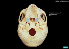
Sphenoid bone
The sphenoid bone is an unpaired bone of the neurocranium. It is situated in the middle of the skull towards the front, in front of the temporal bone and the basilar part of the occipital bone. The sphenoid bone is one of the seven bones that articulate to form the orbit. Its shape somewhat resembles that of a butterfly or bat with its wings extended.
Ethmoid bone
The ethmoid bone is an unpaired bone in the skull that separates the nasal cavity from the brain. It is located at the roof of the nose, between the two orbits. The cubical bone is lightweight due to a spongy construction. The ethmoid bone is one of the bones that make up the orbit of the eye.
Can you feel your palatine bone?
Horizontal Plate of the Palatine Bone The horizontal plate can be felt in the back of the hard palate of the oral cavity. It also forms part of the bottom of the nasal cavity.
What does the palatine bone look like?
The palatine bone has a horizontal and vertical plate as well as a pyramidal process (or pyramid-shaped portion). The horizontal plate makes up the roof of the mouth, and the rear portion of the oral cavity, just behind the nasal cavity; its front end is serrated and its back end is smoother.
Are there 2 palatine bones?
The two palatine bones (L., palatum “palate”) form portions of the hard palate, lateral walls of the nasal cavity, and floors of the orbits. These small, L-shaped, facial bones are located between the palatine processes of the maxilla bones and the pterygoid processes of the sphenoid bones.
Is palatine bone a facial bone?
In anatomy, the palatine bones (/ˈpælətaɪn/) are two irregular bones of the facial skeleton in many animal species, located above the uvula in the throat. Together with the maxillae, they comprise the hard palate.
What bone is on the roof of your mouth?
The hard palate is located at the front of the roof of the mouth, and is comprised of two bones: the palatine bone and the maxilla, each of which are covered by soft tissue. Cancers affecting the hard palate will commonly also involve the upper alveolar ridge, due to its close proximity.
What is the definition of palatine?
Definition of palatine (Entry 1 of 4) 1a : possessing royal privileges. b : of or relating to a palatine or a palatinate. 2a : of or relating to a palace especially of a Roman or Holy Roman emperor.
What is the palatine bone process?
The pyramidal process of the palatine bone projects backward and lateralward from the junction of the horizontal and vertical parts, and is received into the angular interval between the lower extremities of the pterygoid plates. Medial wall of left orbit.
Is the palatine bone single or paired?
The palatine bone is a paired bone located between the maxillae and the pterygoid process of the sphenoid bone. It participates in building the three cavities within the skull; the oral cavity, nasal cavity and the orbits.
Are there 2 maxilla bones?
The maxilla is the bone that forms your upper jaw. The right and left halves of the maxilla are irregularly shaped bones that fuse together in the middle of the skull, below the nose, in an area known as the intermaxillary suture. The maxilla is a major bone of the face.
Are there 2 zygomatic bones?
In the human skull, the zygomatic bone (from Ancient Greek: ζῠγόν, romanized: zugón, lit. 'yoke'), also called cheekbone or malar bone, is a paired irregular bone which articulates with the maxilla, the temporal bone, the sphenoid bone and the frontal bone.
What are the two bones of the mouth?
The mandible sits beneath the maxilla. It is the only movable bone of the skull (discounting the ossicles of the middle ear).
Where is the palatine bone located?
The location of the palatine bone is best understood through its borders and articulations. Its horizontal plate is just behind the maxilla bone of the upper jaw, while lying in front of the soft palate (the soft tissue at the roof of the mouth).
What is the palatine bone?
Treatment. Making up a portion of the nasal cavity and palate, the palatine bone is a paired, L-shaped facial bone. It forms a part of the underside of the skull, and lies between the maxilla bone (the fixed, upper bone of the jaw) and the sphenoid bone (whose wings help form the base of the eye sockets and base of the skull).
What is the most common anatomical variation in the palatine bone?
The most commonly seen anatomical variation in the palatine bone has to do with the positioning of the greater palatine foramen, an opening towards the rear side that allows the descending and greater palatine nerves to pass through.
What percentage of the time is the palatine bone in between the second and third molars?
It also noted a positioning opposite the second molar about 7% of the time, and in between the second and third molar roughly 16% of the time. 1 . While subtle, variations of the palatine bone have significant clinical implications, especially for dentists or dental specialists looking at molar or premolar tooth extraction.
What is the function of the palatine bone?
Primarily, the palatine bone serves a structural function, with its shape helping carve out important structures within the head and defining the lower wall of the inside of cranium. This bone helps form the nasal and oral cavities, the roof of the mouth, and the lower portion of the eye sockets (orbits).
Where is the palatine canal?
Here, the palatine canal, which runs between the sidewall of the palatine bone and the adjacent maxilla bone, is also observed. This portion also includes a sphenopalatine notch on the upper border that connects with the sphenoid bone. Finally, the pyramidal process arises at the juncture between the horizontal and perpendicular plates.
How long does it take to heal torus palatinus?
Typically, this involves an incision in the middle of the palate to allow surgeons to get at the problem. In recovery, which usually takes three to four weeks, 4 pain and inflammation are managed with prescription drugs.
What is the horizontal plate of the palatine bone?
Markings of the Palatine Bone: Horizontal Plate: A horizontal projection that articulates with the palatine process of the Maxilla; forms the posterior portion of the hard palate (or roof of the mouth / floor of the nasal cavity). [ Sagittal view / Inferior view]
What are the two bones that make up the hard palate?
The two palatine bones ( L., palatum “palate”) form portions of the hard palate, lateral walls of the nasal cavity, and floors of the orbits.
What is the lesser palatine foramina?
Lesser palatine foramina: one or more small openings located posterior to the greater palatine foramen; passageways for branches of the lesser palatine nerve. [ Inferior view]
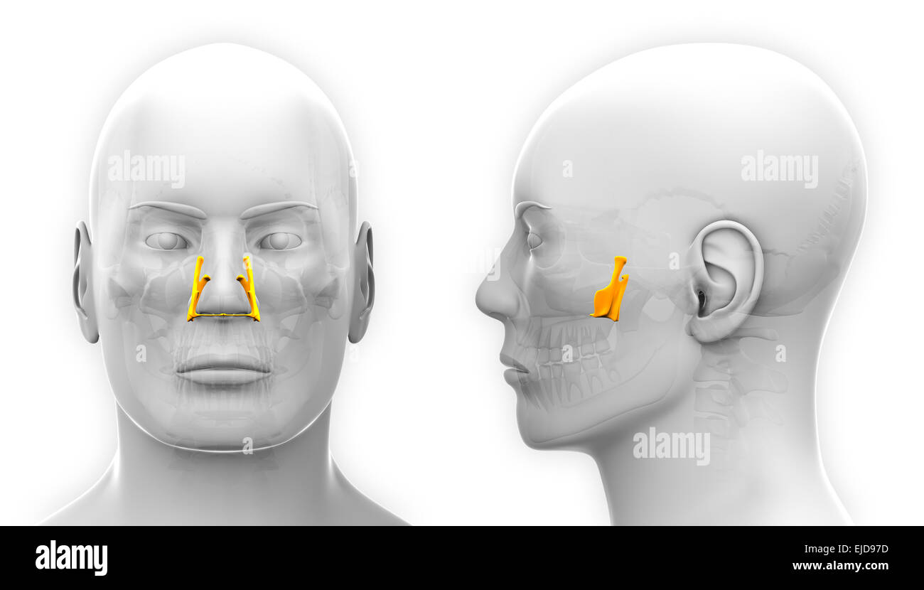
What is the Perpendicular Plate?
Perpendicular plate (vertical plate): A superior projection that forms the posterior portion of the lateral wall of the nasal cavity. [ Sagittal view] Greater palatine foramen (posterior palatine foramen): The largest opening along the posterior part of the horizontal plate; passageway for the greater palatine nerve and vessels. [ Inferior view] ...
Which suture adheres the palatine process of the maxillary bone to the palatine bone?
The transverse palatine suture adheres the palatine process of the maxillary bone to the palatine bone.
What is the median palatine suture?
The median palatine suture connects the horizontal plates of the palatines. It is the posterior continuation of the intermaxillary suture. The transverse palatine suture adheres the palatine process of the maxillary bone to the palatine bone.
What is the groove between the occipital bone and the mastoid process of the temporal bone?
The occipitomastoid suture is the groove between the occipital bone and the mastoid process of the temporal bone. The temporozygomatic suture is the adherence of the temporal bone and the zygomatic bone. The coronal, lambdoid and frontozygomatic sutures are also visible from this angle.
Where is the metopic suture found?
Metopic suture - found in children; on the midline of the frontal bone. Posterior aspect of skull. Sagittal suture - between two parietal bones. Lambdoid suture - between the parietal bone and occipital bone. Lambda - convergence of the sagital and lambdoid suture (resembles to a greek letter 'lambda')
Which suture joins the frontal bone and the nasal bones?
The frontonasalsuture joins the frontal bone and the nasal bones. The frontozygomatic suture articulates the frontal bone and the zygomatic bone. The zygomaticomaxillary suture links the zygoma and the maxilla. The two maxillary bones are anteriorly connected by the intermaxillary joint.
Which suture joins the two parietals?
The sagittal suture joins the two parietals.
What is the name of the joint that allows movement in the fetal skull?
These joints are fixed, immovable, and they have no cavity. They are also referred to as the synarthroses. In fetal skull the sutures are wide and allow slight movement during birth, but later they become rigid and fixed just like in the adults. Key facts. Anterior aspect of skull.
Which bone forms the upper lateral side of the skull?
The parietal bone forms most of the upper lateral side of the skull (see Figure 3). These are paired bones, with the right and left parietal bones joining together at the top of the skull. Each parietal bone is also bounded anteriorly by the frontal bone, inferiorly by the temporal bone, and posteriorly by the occipital bone.
What are the parts of the skull?
Parts of the Skull. The skull consists of the rounded brain case that houses the brain and the facial bones that form the upper and lower jaws, nose, orbits, and other facial structures. Watch this video to view a rotating and exploded skull, with color-coded bones.
How many cranial fossae are there?
Inside the skull, the floor of the cranial cavity is subdivided into three cranial fossae (spaces), which increase in depth from anterior to posterior (see Figure 4, Figure 6b, and Figure 9). Since the brain occupies these areas, the shape of each conforms to the shape of the brain regions that it contains. Each cranial fossa has anterior and posterior boundaries and is divided at the midline into right and left areas by a significant bony structure or opening.
Where are the paranasal sinuses located?
The paranasal sinuses are named for the skull bone that each occupies. The frontal sinus is located just above the eyebrows, within the frontal bone (see Figure 15). This irregular space may be divided at the midline into bilateral spaces, or these may be fused into a single sinus space. The frontal sinus is the most anterior of the paranasal sinuses. The largest sinus is the maxillary sinus. These are paired and located within the right and left maxillary bones, where they occupy the area just below the orbits. The maxillary sinuses are most commonly involved during sinus infections. Because their connection to the nasal cavity is located high on their medial wall, they are difficult to drain. The sphenoid sinus is a single, midline sinus. It is located within the body of the sphenoid bone, just anterior and inferior to the sella turcica, thus making it the most posterior of the paranasal sinuses. The lateral aspects of the ethmoid bone contain multiple small spaces separated by very thin bony walls. Each of these spaces is called an ethmoid air cell. These are located on both sides of the ethmoid bone, between the upper nasal cavity and medial orbit, just behind the superior nasal conchae.
Where is the internal acoustic meatus located?
Located on the medial wall of the petrous ridge in the posterior cranial fossa is the internal acoustic meatus (see Figure 9). This opening provides for passage of the nerve from the hearing and equilibrium organs of the inner ear, and the nerve that supplies the muscles of the face. Located at the anterior-lateral margin of the foramen magnum is the hypoglossal canal. These emerge on the inferior aspect of the skull at the base of the occipital condyle and provide passage for an important nerve to the tongue.
Where is the superior nasal concha?
The superior nasal concha is located just lateral to the perpendicular plate, in the upper nasal cavity.
What is the anterior skull?
The anterior skull consists of the facial bones and provides the bony support for the eyes and structures of the face. This view of the skull is dominated by the openings of the orbits and the nasal cavity. Also seen are the upper and lower jaws, with their respective teeth (Figure 2).