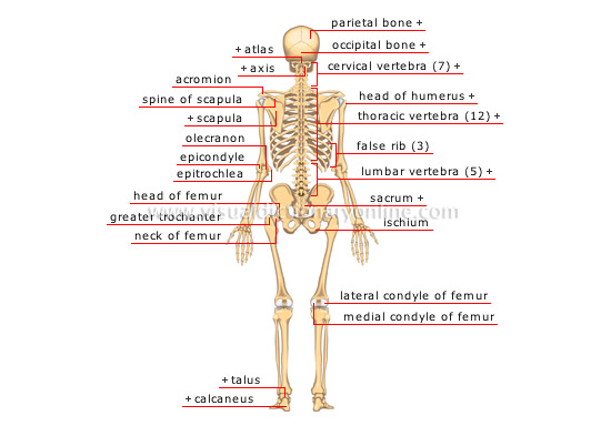
What is the parietal bone function?
The parietal bone helps to shape and protect the brain as a component of the neurocranium. The centre of the skull is covered by the parietal bone, also known as the os parietale, a flat, paired cranial bone. Each bone protects the right and left parietal lobes of the brain.
Where are the parietal and temporal bones located?
the skullThe temporal bones are two major bones in the skull, or cranium. They help form the sides and base of the skull, where they protect the temporal lobe of the brain and surround the ear canal. The other major bones in the skull are: the two parietal bones that make up the top of the skull.
What type of bone is the parietal bone?
quadrilateral skull boneThe parietal bone is a paired, irregular, quadrilateral skull bone that forms the sides and roof of the cranium.
What is the parietal area of the skull?
The parietal bones (/pəˈraɪ. ɪtəl/) are two bones in the skull which, when joined at a fibrous joint, form the sides and roof of the cranium. In humans, each bone is roughly quadrilateral in form, and has two surfaces, four borders, and four angles.
What is the bone above the ear called?
The temporal bone: the base of the skull Forming the lateral-inferior region of the skull, the temporal bone is an even and symmetrical bone. Because of its position, this bone protects the temporal lobe of the brain and the ear. Also, it contributes to the development of the temporomandibular joint.
What happens to the parietal lobe if it is damaged?
Parietal Lobe, Right - Damage to this area can cause visuo-spatial deficits (e.g., the patient may have difficulty finding their way around new, or even familiar, places). Parietal Lobe, Left - Damage to this area may disrupt a person's ability to understand spoken and/or written language.
What does parietal mean in anatomy?
(pəˈraɪɪtəl ) adjective. anatomy, biology. of, relating to, or forming the walls or part of the walls of a bodily cavity or similar structure. the parietal bones of the skull.
How strong is the parietal bone?
The average ultimate tensile strength, loaded in the direction of the long axis, of the compact bone of 15 specimens taken from the parietal bone of adult human embalmed cadavers was 10,230 lb/in. (6,030–15,800).
Which area is part of the temporal bone?
The temporal bones are situated at the sides and base of the skull, and lateral to the temporal lobes of the cerebral cortex. The temporal bones are overlaid by the sides of the head known as the temples, and house the structures of the ears.
Where is the temporal lobe located?
The temporal lobes sit behind the ears and are the second largest lobe. They are most commonly associated with processing auditory information and with the encoding of memory.
On which bone are the temporal lines located?
The parietal bone has an external and internal surface. The external surface is smooth and convex. It has several important features: Superior temporal line which forms an arch that travels between the frontal and occipital borders of the parietal bone.
What are the two temporal bones called?
This part of the temporal bone is usually split into two: the petrous part and the mastoid part. The mastoid part is the most posterior part of the temporal bone. Its outer surface is roughened by muscular attachments. There is a downward conical projection called the mastoid process from the mastoid part.
What is the parietal bone in the head?
The parietal bone makes up a significant part of the skull. It creates the back two-thirds of the top of the skull and curves over the sides to pro...
What type of bone is parietal bone?
The parietal bone has layers of different types of bone. The top and bottom layer are hard, compact bone. Between the two is a layer of spongy bone.
What is the function of parietal bone?
There are two main functions of the parietal bone. The first is to provide structure. The second is to protect the delicate brain tissue.
What is the parietal bone?
Definition. The parietal bone or os parietale is a paired, flat cranial bone that covers the mid portion of the skull. Both bones cover the left and right parietal lobes of the brain respectively. As part of the neurocranium, the parietal bone helps to form the shape of the head and protect the brain. More specifically, both bones form part of the ...
Where is the parietal foramen located?
The parietal foramen is found near the lambda – a midline skull landmark where the lambdoid and sagittal sutures meet. Parietal foramina. Not everybody has these holes – they are “inconsistent” foramina. Alternatively, they can become smaller over time or be overly large, meeting to form one large hole.
How many sutures are there in the skull?
Once fused, the bones of the skull are said to be sutured. The parietal bones have five sutures: Sagittal: joins the left parietal bone to the right parietal bone. Lambdoidal: joins the posterior parietal with the top of the occipital bone.
What is cancellous bone?
Cancellous bone is honeycomb-like with a network of spaces that contain red bone marrow. Red bone marrow produces blood cells; much smaller quantities are produced in the skull than in the long bones.
What are the articulations of the sphenoid, temporal, frontal, and o?
Parietal bone articulations with the sphenoid, temporal, frontal, and occipital bones are called sutures.
Which bone joins the posterior parietal with the top of the occipital bone?
Lambdoidal: joins the posterior parietal with the top of the occipital bone
Which bone has sutures?
Each bone has sutures with the: Greater wing of the sphenoid bone. Temporal bone. Frontal bone. Occipital bone. This means it is very easy to picture the exact location of the parietal bone. Different views of the parietal bone. The parietal bones make up the majority of the top of the head.
What is the parietal bone?
Parietal bone, cranial boneforming part of the side and top of the head. In front each parietal bone adjoins the frontal bone; in back, the occipitalbone; and below, the temporal and sphenoid bones. The parietal bones are marked internally by meningeal blood vessels and externally by the temporal muscles.
Where do parietal bones meet?
The parietal bones are marked internally by meningeal blood vessels and externally by the temporal muscles. They meet at the top of the head (sagittal suture) and form a roof for the cranium. The parietal bone forms in membrane (i.e.,without a cartilaginous precursor); the sagittal suture closes between ages 22 and 31.
What are the bones of the human skeleton?
human skeleton: Development of cranial bones. Each parietal bone has a generally four-sided outline. Together they form a large portion of the side walls of the cranium. Each adjoins the frontal, the sphenoid, the temporal, and the occipital bones and its fellow of the opposite side. They are almost exclusively cranial bones,….
Which bones are marked internally by meningeal blood vessels?
In front each parietal bone adjoins the frontal bone; in back, the occipital bone; and below, the temporal and sphenoid bones. The parietal bones are marked internally by meningeal blood vessels and externally by the . Parietal bone, cranial bone forming part of the side and top of the head. In front each parietal bone adjoins ...
What are the bones that make up the cranial floor?
The parietal and temporal bones form the sides and uppermost portion of the dome of the cranium, and the frontal bone forms the forehead; the cranial floor consists of the sphenoid and ethmoid bones.
Where is the parietal bone?
The parietal bone is usually present in the posterior end of the skull and is near the midline. This bone is part of the skull roof, which is a set of bones that cover the brain, eyes and nostrils. The parietal bones make contact with several other bones in the skull.
What is the definition of parietal bones?
Anatomical terms of bone. The parietal bones ( / pəˈraɪ.ɪtəl /) are two bones in the skull which, when joined together at a fibrous joint, form the sides and roof of the cranium. In humans, each bone is roughly quadrilateral in form, and has two surfaces, four borders, and four angles.
How many parts does the parietal bone have?
Occasionally the parietal bone is divided into two parts, upper and lower, by an antero-posterior suture.
What is the channel of the superior sagittal sinus?
Along the upper margin is a shallow groove, which, together with that on the opposite parietal, forms a channel, the sagittal sulcus, for the superior sagittal sinus; the edges of the sulcus afford attachment to the falx cerebri .
What is the name of the bones that form the sides and roof of the cranium?
Parietal bone. The parietal bones ( / pəˈraɪ.ɪtəl /) are two bones in the skull which, when joined together at a fibrous joint, form the sides and roof of the cranium. In humans, each bone is roughly quadrilateral in form, and has two surfaces, four borders, and four angles. It is named from the Latin paries ( -ietis ), wall.
Which bone articulates with the mastoid?
The mastoid angle is truncated; it articulates with the occipital bone and with the mastoid portion of the temporal, and presents on its inner surface a broad, shallow groove which lodges part of the transverse sinus. The point of meeting of this angle with the occipital and the mastoid part of the temporal is named the asterion.
Which suture separates the parietal bones?
Sagittal suture separates left and right parietal bone. Coronal suture. It separates the parietal bones and the frontal bone . Squamosal suture. It separates the parietal bones and the temporal bone . Lambdoid suture. It separates the parietal bones and the occipital bone .

Overview
In other animals
In non-human vertebrates, the parietal bones typically form the rear or central part of the skull roof, lying behind the frontal bones. In many non-mammalian tetrapods, they are bordered to the rear by a pair of postparietal bones that may be solely in the roof of the skull, or slope downwards to contribute to the back of the skull, depending on the species. In the living tuatara, and many fossil species, a small opening, the parietal foramen, lies between the two parietal bones. This openin…
Surfaces
The external surface [Fig. 1] is convex, smooth, and marked near the center by an eminence, the parietal eminence (tuber parietale), which indicates the point where ossification commenced.
Crossing the middle of the bone in an arched direction are two curved lines, the superior and inferior temporal lines; the former gives attachment to the tempor…
Borders
• The sagittal border, the longest and thickest, is dentated (has toothlike projections) and articulates with its fellow of the opposite side, forming the sagittal suture.
• The frontal border is deeply serrated, and bevelled at the expense of the outer surface above and of the inner below; it articulates with the frontal bone, forming half of the coronal suture. The point where the coronal su…
Angles
• The frontal angle is practically a right angle, and corresponds with the point of meeting of the sagittal and coronal sutures; this point is named the bregma; in the fetal skull and for about a year and a half after birth this region is membranous, and is called the anterior fontanelle.
• The sphenoidal angle, thin and acute, is received into the interval between the frontal bone and the great wing of the sphenoid. Its inner surface is marked by a deep groove, sometimes a canal, fo…
Ossification
The parietal bone is ossified in membrane from a single center, which appears at the parietal eminence about the eighth week of fetal life.
Ossification gradually extends in a radial manner from the center toward the margins of the bone; the angles are consequently the parts last formed, and it is here that the fontanelles exist.
Occasionally the parietal bone is divided into two parts, upper and lower, by an antero-posterior …
Additional images
• Position of parietal bone (shown in green). Animation.
• Parietal bone
• Trajectory of the missile through President Kennedy's skull. The bullet struck posterior part of his right parietal bone from behind.
See also
• Bone terminology
• Terms for anatomical location
• Parietal lobe