Where would you find pleura membrane?
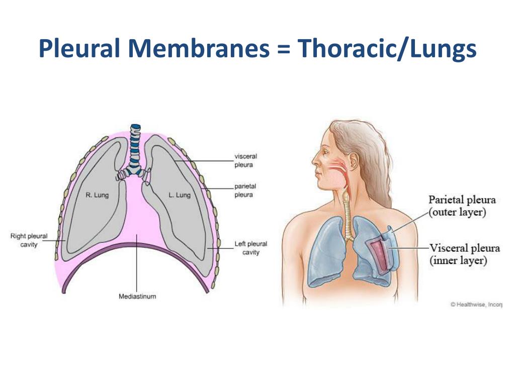
lungsThere are two layers; the outer pleura (parietal pleura) is attached to the chest wall and the inner pleura (visceral pleura) covers the lungs and adjoining structures, via blood vessels, bronchi and nerves.
Where is the pleura in the lungs?
The chest cavity is lined by a thin shiny membrane called the pleura, which covers the inside surface of the rib cage and spreads over the lungs as well. Normally, the pleura produces a small amount of fluid which serves as a lubricant to the lungs as they move back and forth against the chest wall during respiration.
What is pleural membrane and its function?
The visceral pleura surrounds the outside of the lung. The parietal pleura lines the inside of the chest wall and extends over the diaphragm. These membranes secrete a lubricating fluid, which allows for free movement of the lungs against the chest wall when we breathe.
Which organs are surrounded by the pleural membrane?
The pleural membranes are two layers of serous membrane which enclose and protect the lung. The superficial layer is called parietal pleura and lines the wall of the thoracic cavity. The deep layer is called visceral pleura and covers the lungs themselves.
What does pleural mean?
Medical Definition of pleural : of or relating to the pleura or the sides of the thorax.
What is the pleural membrane made of?
The pleura consists of a visceral and parietal layer that is composed of a continuous surface epithelium of mesothelial cells and underlying connective tissue. The visceral pleura covers the lungs and interlobar fissures, whereas the parietal pleura lines the ribs, diaphragm, and mediastinum.
What happens if the pleural membrane is punctured?
If the chest wall, and thus the pleural space, is punctured, blood, air or both can enter the pleural space. Air and/or blood rushes into the space in order to equalise the pressure with that of the atmosphere. As a result, the fluid is disrupted and the two membranes no longer adhere to each other.
Where does pleural fluid come from?
Pleural effusion occurs when fluid builds up in the space between the lung and the chest wall. This can happen for many different reasons, including pneumonia or complications from heart, liver, or kidney disease. Another reason could be as a side effect from cancer.
What happens if lungs are not covered by pleural membrane?
This abolishes the negative intrapleural pressure and the lung in the affected area will collapse. Gas exchange will be seriously impaired because movement of the chest wall will no longer expand the lung.
How does a person get pleurisy?
What causes pleurisy? Most cases are the result of a viral infection (such as the flu) or a bacterial infection (such as pneumonia). In rarer cases, pleurisy can be caused by conditions such as a blood clot blocking the flow of blood into the lungs (pulmonary embolism) or lung cancer.
What causes pleural effusion?
The most common causes of pleural effusion are congestive heart failure, cancer, pneumonia, and pulmonary embolism. Pleural fluid puncture (pleural tap) enables the differentiation of a transudate from an exudate, which remains, at present, the foundation of the further diagnostic work-up.
What causes pleural effusion in the lungs?
Pleural effusion occurs when fluid builds up in the space between the lung and the chest wall. This can happen for many different reasons, including pneumonia or complications from heart, liver, or kidney disease. Another reason could be as a side effect from cancer.
Where is the pleural cavity located?
The pleural cavity is a fluid filled space that surrounds the lungs. It is found in the thorax, separating the lungs from its surrounding structures such as the thoracic cage and intercostal spaces, the mediastinum and the diaphragm. The pleural cavity is bounded by a double layered serous membrane called pleura.
What is the pleural space?
Pleural space. The pleural cavity surrounds the lungs in the thoracic cavity. There are two pleural cavities, one for each lung on the right and left sides of the mediastinum. Each pleural cavity and it’s enclosed lung are lined by a serous membrane called pleura.
What is the parietal pleura?
The parietal pleura is the layer of pleura associated with the walls of the pleural cavity. It lines the internal aspect of the thoracic wall, the thoracic surface of the diaphragm and separates the pleural cavity from the mediastinum.
What is the space between the parietal and visceral pleura?
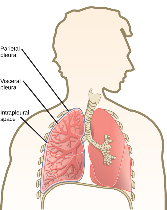
The space between the parietal and visceral pleura is the pleural cavity. The lung itself is not located within the pleural cavity , rather it is surrounded by it. The function of the pleura is to allow optimal expansion and contraction of the lungs during breathing.
What are the two layers of the pleura?
The pleurae are two layers of serous membrane that form the boundaries of the pleural cavity. There are two types of pleura; parietal and visceral. The parietal pleura is the thicker and more durable outer layer that lines the inner aspect of the thoracic cavity and the mediastinum. The visceral pleura is the more delicate inner layer of pleura that lines the outer surface of the lung itself. The parietal and visceral layers are not entirely separate, rather they are continuous with each other at the hilum of the lung. Each layer consists of a single layer of mesothelial cells and supporting connective tissue including collagen, elastin, blood vessels and lymphatics. The pleural cavity containing a small amount of pleural fluid is contained between the parietal and visceral layers of pleura.
How to describe the location of the parietal and visceral pleura?
A common way of describing the location of the parietal and visceral pleura relative to one another is by thinking of pushing your fist into an underinflated balloon, a useful analogy for the developing lung. Your fist represents the developing lung and the balloon, the pleural cavity.
Why is the right lung on the same side?
The build-up of fluid in the pleural space causes the lung on the same side to be pushed upwards. Because the right and left pleural cavities are separate from each other, the opposite lung will not be affected unless its surrounding pleural cavity is also compromised.
Parietal Pleura
The parietal pleura is 30 to 40 micrometers thick and is in contact with the inside of the ribcage, the mediastinum and the upper surface of the diaphragm. The parietal pleura receives its blood supply from the intercostal arteries (arteries between the ribs). It is sensitive to pain and is highly involved in the formation of parietal fluid.
Visceral Pleura
The visceral pleura covers the surface of the lungs. It receives its blood supply from the bronchial (lung) circulation. The thickness of the visceral pleura varies over the surface of the lung, ranging from 20 to 80 micrometers. Due to its lack of sensory innervation, the visceral pleura is not sensitive to pain.
Functions of the Pleural Membrane
The main function of the pleura is to provide mechanical protection and a smooth, lubricating elastic surface for the lungs to move during breathing.