What is the anatomy of the pterygoids?
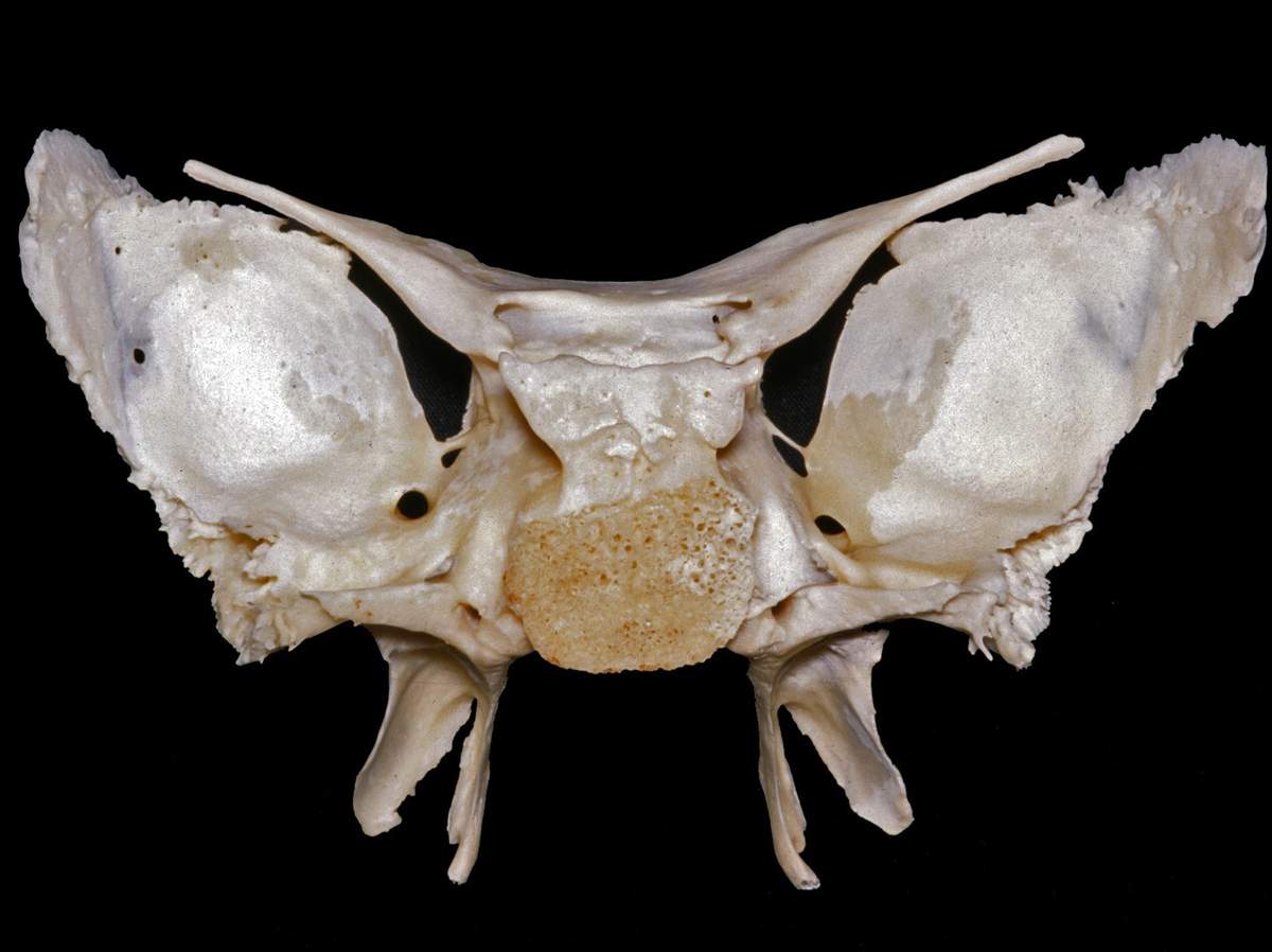
Gross anatomy. Each pterygoid process projects inferiorly from the junction of the body and greater wing of the sphenoid bone and bifurcates into a medial pterygoid plate and a lateral pterygoid plate. At the inferior tip of the medial pterygoid plate is the small hook-shaped process, the pterygoid hamulus.
What is the medial pterygoid plate?
The medial pterygoid plate (or medial pterygoid lamina) of the sphenoid bone is a horse-shoe shaped process that arises from its underside.
What are the pterygoid processes of the sphenoid?
The pterygoid processes of the sphenoid (from Greek pteryx, pterygos, "wing"), one on either side, descend perpendicularly from the regions where the body and the greater wings of the sphenoid bone unite.
Where do the lateral and medial pterygoid muscles originate?
The lateral and medial pterygoid muscles which form some of the muscles of mastication originate from the lateral pterygoid plate of the sphenoid bone. The sphenoid is an unpaired bone. It sits anteriorly in the cranium, and contributes to the middle cranial fossa, the lateral wall of the skull, and the floor and sides of both orbits.
What are the pterygoid processes?
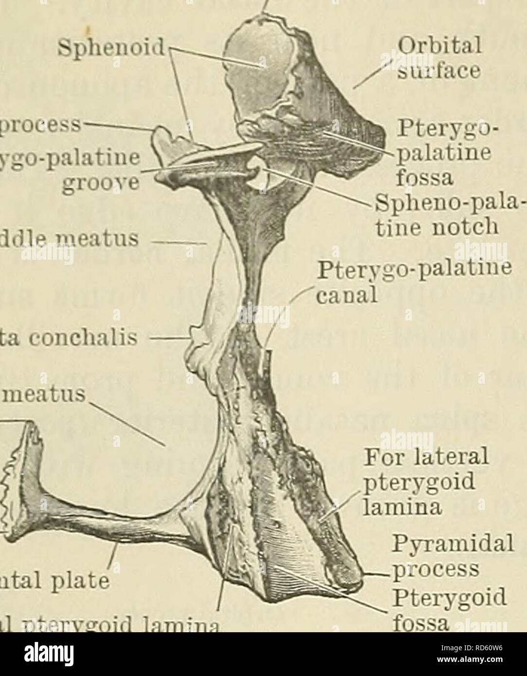
The pterygoid processes, one on either side, descend perpendicularly from the regions where the body and great wings unite. Each process consists of a medial and a lateral plate, the upper parts of which are fused anteriorly; a vertical sulcus, the pterygopalatine groove, descends on the front of the line of fusion.
Where is the pterygoid process of sphenoid bone?
The pterygoid processes of the sphenoid (from Greek pteryx, pterygos, "wing"), one on either side, descend perpendicularly from the regions where the body and the greater wings of the sphenoid bone unite. Sphenoid bone, upper surface....Lateral pterygoid plateFMA54682Anatomical terms of bone6 more rows
Where are the pterygoid processes located relative to the mandible?
The medial pterygoid muscle, a major elevator of the jaw is a square-shaped masticatory muscle, located on the medial aspect of the lower jaw bilaterally. It is also known as internal pterygoid muscle. This muscle lies medial to the lateral pterygoid muscle.
Which bones has pterygoid processes?
The pterygoid processes or pterygoid plates are paired posteroinferior projections of the sphenoid bone.
What is the Pterygoid bone?
Definition of pterygoid bone : a horizontally placed often more or less rodlike bone or group of bones of the upper jaw or roof of the mouth in most lower vertebrates connecting the palatine in front and the quadrate behind and forming part of the palatoquadrate arch.
What does pterygoid mean?
Definition of pterygoid (Entry 1 of 2) : of, relating to, or lying in the region of the inferior part of the sphenoid bone of the vertebrate skull.
What is the origin of the pterygoid?
Medial pterygoid muscleOriginSuperficial part: Tuberosity of maxilla, Pyramidal process of palatine bone; Deep part: Medial surface of lateral pterygoid plate of sphenoid boneInsertionMedial surface of ramus and angle of mandible3 more rows•May 11, 2020
What is the function of the medial pterygoid?
The function of medial pterygoid, while contracting bilaterally, it produces elevation and protrusion of the mandible, while unilaterally contracted, it produces contralateral excursion (Neumann, 2010; Okeson, 2013). It pulls the ramus of the mandible medially and shifting the mandible toward to the contralateral side.
What muscle opens the jaw?
lateral pterygoidMasseter. The masseter muscle is one of four muscles of mastication and has the primary role of closing the jaw in conjunction with two other jaw closing muscles, the temporalis and medial pterygoid muscles. The fourth masticatory muscle, the lateral pterygoid, causes jaw protrusion and jaw opening when activated.
What are the processes of palatine bone?
The palatine bone is composed of two plates, the horizontal and perpendicular, which are connected and form a characteristic L-shape bone. The bone features three processes; pyramidal, orbital and sphenoidal.
What passes through the sphenoid bone?
The blood vessels pass through the foramina present in the wings of the sphenoid bone. The ophthalmic artery passes through the optic canal to enter the orbit. The ophthalmic veins pass out of the orbit through the superior orbital fissure.
Where is the lateral pterygoid muscle located?
Lateral pterygoid is located deep to the temporalis and masseter muscles, spanning between the sphenoid bone and temporomandibular joint. Its muscle belly is separated by a small horizontal fissure into two heads; superior (upper) and inferior (lower).
Where is the lateral pterygoid muscle located?
Lateral pterygoid is located deep to the temporalis and masseter muscles, spanning between the sphenoid bone and temporomandibular joint. Its muscle belly is separated by a small horizontal fissure into two heads; superior (upper) and inferior (lower).
Where does the pterygoid plexus drain into?
Maxillary veinThe function of the pterygoid venous plexus is to collect the blood from the palate, nasal cavity, paranasal sinuses, nasopharynx, and auditory tube....Pterygoid venous plexus.Drains fromVenules of the infratemporal fossaDrains toMaxillary vein2 more rows
Where does the medial pterygoid muscle insertion?
Medial pterygoid muscleOriginSuperficial part: Tuberosity of maxilla, Pyramidal process of palatine bone; Deep part: Medial surface of lateral pterygoid plate of sphenoid boneInsertionMedial surface of ramus and angle of mandible3 more rows•May 11, 2020
Where does the lateral pterygoid insertion?
Structure. The lateral pterygoid muscle has an upper head and a lower head. The upper head originates on the infratemporal surface and infratemporal crest of the greater wing of the sphenoid bone. It inserts onto the articular disc and fibrous capsule of the temporomandibular joint.
What is the pterygoid process?
54682. Anatomical terms of bone. The pterygoid processes of the sphenoid ( from Greek pteryx, pterygos, "wing"), one on either side, descend perpendicularly from the regions where the body and the greater wings of the sphenoid bone unite. Each process consists of a medial pterygoid plate and a lateral pterygoid plate, ...
Which part of the pterygoid process is fused anteriorly?
The superior portion of the pterygoid processes are fused anteriorly; a vertical groove, the pterygopalatine fossa, descends on the front of the line of fusion. The plates are separated below by an angular cleft, the pterygoid notch, the margins of which are rough for articulation with the pyramidal process of the palatine bone .
What is the angular prominence between the posterior margin of the vaginal process and the medial border of the?
The angular prominence between the posterior margin of the vaginal process and the medial border of the scaphoid fossa is named the pterygoid tubercle, and immediately above this is the posterior opening of the pterygoid canal .
What is the medial pterygoid plate?
The medial pterygoid plate (or medial pterygoid lamina) of the sphenoid bone is a horse-shoe shaped process that arises from its underside.
Which muscle is attached to the medial surface of the lateral pterygoid muscle?
Its lateral surface forms part of the medial wall of the infratemporal fossa, and gives attachment to the lateral pterygoid muscle; its medial surface forms part of the pterygoid fossa, and gives attachment to the medial pterygoid muscle. Posterior edge is sharp, and often has sharp projection - pterygospinous process (Civinini process).
Which pterygoid allows the jaw to move in a vertical direction?
Each process consists of a medial pterygoid plate and a lateral pterygoid plate, the latter of which serve as the origins of the medial and lateral pterygoid muscles. The medial pterygoid, along with the masseter allows the jaw to move in a vertical direction as it contracts and relaxes. The lateral pterygoid allows the jaw to move in a horizontal direction during mastication. Fracture of either plate are used in clinical medicine to distinguish the Le Fort fracture classification for high impact injuries to the sphenoid and maxillary bones .
What bone articulates with the posterior border of the vertical part of the palatine bone?
In many animals it is a separate bone called the pterygoid bone .
What is the pterygoid process?from en.wikipedia.org
54682. Anatomical terms of bone. The pterygoid processes of the sphenoid ( from Greek pteryx, pterygos, "wing"), one on either side, descend perpendicularly from the regions where the body and the greater wings of the sphenoid bone unite. Each process consists of a medial pterygoid plate and a lateral pterygoid plate, ...
What is the medial pterygoid plate?from en.wikipedia.org
The medial pterygoid plate (or medial pterygoid lamina) of the sphenoid bone is a horse-shoe shaped process that arises from its underside.
What is the angular prominence between the posterior margin of the vaginal process and the medial border of the?from en.wikipedia.org
The angular prominence between the posterior margin of the vaginal process and the medial border of the scaphoid fossa is named the pterygoid tubercle, and immediately above this is the posterior opening of the pterygoid canal .
Why are pterygoid plates important?from radiopaedia.org
Pterygoid plates are important in diagnosing midface fractures. In Le Fort classification of midface fracture, the involvement of pterygoid plates is necessary to confirm the diagnosis.
Which muscle is attached to the medial surface of the lateral pterygoid muscle?from en.wikipedia.org
Its lateral surface forms part of the medial wall of the infratemporal fossa, and gives attachment to the lateral pterygoid muscle; its medial surface forms part of the pterygoid fossa, and gives attachment to the medial pterygoid muscle. Posterior edge is sharp, and often has sharp projection - pterygospinous process (Civinini process).
Which pterygoid allows the jaw to move in a vertical direction?from en.wikipedia.org
Each process consists of a medial pterygoid plate and a lateral pterygoid plate, the latter of which serve as the origins of the medial and lateral pterygoid muscles. The medial pterygoid, along with the masseter allows the jaw to move in a vertical direction as it contracts and relaxes. The lateral pterygoid allows the jaw to move in a horizontal direction during mastication. Fracture of either plate are used in clinical medicine to distinguish the Le Fort fracture classification for high impact injuries to the sphenoid and maxillary bones .
What bone articulates with the posterior border of the vertical part of the palatine bone?from en.wikipedia.org
In many animals it is a separate bone called the pterygoid bone .
Where is the lateral pterygoid located?
Lateral pterygoid muscle (Musculus pterygoideus lateralis) The lateral pterygoid is a short, two-headed muscle, located in the infratemporal fossa of the skull. The smaller superior head arises from the infratemporal crest of the greater wing of sphenoid bone , while the larger inferior head arises from ...
What is the medial pterygoid muscle?
The medial pterygoid muscle is a thick quadrilateral muscle that arises by two heads, a superficial and a deep head . The smaller superficial head originates from the maxillary tuberosity and pyramidal process of palatine bone . The larger deep head originates from the medial surface of the lateral pterygoid plate and the pyramidal process of sphenoid bone . From their origin, the muscle fibers run posteroinferiorly and laterally, surrounding the lower fibers of the lateral pterygoid muscle. Together, the two heads of the medial pterygoid muscle insert onto the triangular impression located on the medial surface of the mandible.
What is the function of the lateral pterygoid muscle?
The function of the lateral pterygoid muscle is to protrude and depress the mandible when contraction is bilateral. In unilateral contraction, the lateral pterygoid muscle moves the mandible medially.
Where does the deep head come from?
The larger deep head originates from the medial surface of the lateral pterygoid plate and the pyramidal process of sphenoid bone . From their origin, the muscle fibers run posteroinferiorly and laterally, surrounding the lower fibers of the lateral pterygoid muscle.
Which muscle is innervated by the medial pterygoid branch of the mandib?
The medial pterygoid muscle is innervated by the medial pterygoid branch of the mandibular division of the trigeminal nerve (CN V3). It receives blood supply from the pterygoid branches of the maxillary artery. The bilateral contraction of this muscle elevates the mandible and closes the mouth.
Which nerve innervates the pterygoid muscle?
Both muscles are innervated by branches of the mandibular division of the trigeminal nerve (CN V3) , and receive their blood supply from branches of the maxillary artery . This article will introduce you to the anatomy and function of the pterygoid muscles. Muscles of mastication that produce movements of the jaw.
Where do the fibres of the superior head insert?
The fibres of both muscle heads merge and insert on the pterygoid fovea on the neck of the mandible . Some fibres from the superior head insert on the capsule of the temporomandibular joint and its articular disc. The lateral pterygoid muscle is innervated by the nerve to the lateral pterygoid muscle, which is a branch ...
Where does the pterygoid process protrude?
Pterygoid process protrudes down from the junction in the middle of the body as well as greater wing of sphenoid posterior towards the last molar tooth. It splits within medial along with lateral pterygoid plates; the pterygoid fossa divides them from each other. A free posterior border is found in each plate. Pterygoid process.
What is the medial pterygoid plate?
The medial pterygoid plate a.k.a. medial pterygoid lamina of the sphenoid bone is a horse-shoe shaped process which emerges from its bottom.
What is the hamulus of a pterygoid?
The pterygoid hamulus is a hook-like projection from which each medial plate of the pterygoid process inferiorly terminates with. Each medial plate of pterygoid process divides superiorly in order to create the small, shallow scaphoid fossa.
What nerve is in the pterygoid canal?
Nerve of pterygoid canal a.k.a. Vidian’s nerve.
Which muscle is attached to the lateral pterygoid plate?
Lateral pterygoid muscle has it’s the lower part connect to the lateral part of the lateral pterygoid plate. Medial pterygoid muscle is attached towards medial part of the lateral pterygoid plate. Superior pharyngeal constrictor is attached towards inferior end of the medial pterygoid plate.
Where is the pterygoid canal located?
The opening of the pterygoid canal, which travels forward near the anterior margin of the foramen lacerum, is located at the root of the medial plate of the pterygoid process, immediately superior towards the scaphoid fossa.
Which wall of the pterygopalatine fossa is created via the root of the p?
The posterior wall of the pterygopalatine fossa created via the root of the pterygoid process.
Which process is unites rostrally to the palatine bone and to the pterygoi?
On each side of the body of the basisphenoid bone, the pterygoid process (Processus pterygoideus) comes off. It unites rostrally to the palatine bone and to the pterygoid bone.
Which bone forms the pterygoid fossa?
At its junction with the pterygoid bone abd the perpendicular plate of the palatine bone, the pterygoid process also contributes to form the pterygoid fossa.
What is the pterygoid process?from en.wikipedia.org
54682. Anatomical terms of bone. The pterygoid processes of the sphenoid ( from Greek pteryx, pterygos, "wing"), one on either side, descend perpendicularly from the regions where the body and the greater wings of the sphenoid bone unite. Each process consists of a medial pterygoid plate and a lateral pterygoid plate, ...
What is the medial pterygoid plate?from en.wikipedia.org
The medial pterygoid plate (or medial pterygoid lamina) of the sphenoid bone is a horse-shoe shaped process that arises from its underside.
What is the angular prominence between the posterior margin of the vaginal process and the medial border of the?from en.wikipedia.org
The angular prominence between the posterior margin of the vaginal process and the medial border of the scaphoid fossa is named the pterygoid tubercle, and immediately above this is the posterior opening of the pterygoid canal .
Why are pterygoid plates important?from radiopaedia.org
Pterygoid plates are important in diagnosing midface fractures. In Le Fort classification of midface fracture, the involvement of pterygoid plates is necessary to confirm the diagnosis.
Which muscle is attached to the medial surface of the lateral pterygoid muscle?from en.wikipedia.org
Its lateral surface forms part of the medial wall of the infratemporal fossa, and gives attachment to the lateral pterygoid muscle; its medial surface forms part of the pterygoid fossa, and gives attachment to the medial pterygoid muscle. Posterior edge is sharp, and often has sharp projection - pterygospinous process (Civinini process).
Which pterygoid allows the jaw to move in a vertical direction?from en.wikipedia.org
Each process consists of a medial pterygoid plate and a lateral pterygoid plate, the latter of which serve as the origins of the medial and lateral pterygoid muscles. The medial pterygoid, along with the masseter allows the jaw to move in a vertical direction as it contracts and relaxes. The lateral pterygoid allows the jaw to move in a horizontal direction during mastication. Fracture of either plate are used in clinical medicine to distinguish the Le Fort fracture classification for high impact injuries to the sphenoid and maxillary bones .
What bone articulates with the posterior border of the vertical part of the palatine bone?from en.wikipedia.org
In many animals it is a separate bone called the pterygoid bone .
Where does the pterygoid process descend?
The pterygoid process descends inferiorly from the point of junction between the sphenoid body and the greater wing. It consists of two parts:
Where do the lateral and medial pterygoid muscles originate?
The lateral and medial pterygoid muscles which form some of the muscles of mastication originate from the lateral pterygoid plate of the sphenoid bone.
What is the butterfly shape of the sphenoid bone?
The sphenoid bone is said to be ' butterfly-shaped '. It consists of a body, paired greater wings and lesser wings, and two pterygoid processes. Body. The body lies at the centre of the sphenoid bone, and is almost completely cubical in shape. It contains the sphenoidal sinuses, which are separated by a septum - meaning ...
What is the butterfly bone?
The sphenoid bone is said to be ‘ butterfly-shaped ‘. It consists of a body, paired greater wings and lesser wings, and two pterygoid processes.
What is the sphenoid bone?
Log In. The sphenoid bone is one of the eight bones that make up the cranium - the superior aspect of the skull that encloses and protects the brain. Its name is derived from the Greek ‘sphenoeides’, to mean wedge-shaped. In this article, we shall look at the anatomy of the sphenoid bone - its location, structure, and clinical significance.
Which tuberculum forms the anterior wall of the sella turcica?
Tuberculum sellae - forms the anterior wall of the sella turcica, and the posterior aspect of the chiasmatic groove.
What is the medial border of the optic canal?
The medial border of the optic canal is formed by the body of the sphenoid. There is a ‘slit-like’ gap between the lesser and greater wings of the sphenoid – the superior orbital fissure. Numerous structures pass through here to reach the bony orbit.