What causes partially empty sella?
empty sella syndrome may occur as a primary disorder, for which the cause is unknown (idiopathic), or as a secondary disorder, in which it occurs due to an underlying condition or disorder such as a treated pituitary tumor, head trauma, or a condition known as idiopathic intracranial hypertension (also called pseudotumor cerebri) during which …
Which gland is located in the Sella Turcia?
- Gillian Leiberman; Imaging The Turkish Saddle; Retrieve from: http://eradiology.bidmc.harvard.edu/LearningLab/central/Goodman.pdf
- ALBERT L. ...
- What is the Sella turnica; Retrieve from: http://www.wisegeek.com/what-is-the-sella-turcica.htm
- Tekiner H, Acer N, Kelestimur F. ...
What does nearly empty sella Turcia mean?
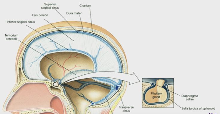
The sella turcica is an indentation in the sphenoid bone at the base of your skull that holds the pituitary gland. If you have empty sella syndrome, your sella turcica is not actually empty. In fact, it means your sella turcica is either partially or totally filled with cerebrospinal fluid (CSF).
What causes empty sella syndrome?
Possible causes of secondary empty sella syndrome:
- Increase in intracranial pressure, for example, because of a cerebral tumor or hydrocephalus
- Idiopathic intracranial hypertension
- Surgery for pituitary tumor
- Sheehan‘s syndrome (postpartum pituitary gland necrosis)
- Sequela of craniocerebral trauma
- Sequela of cerebral radiotherapy

Where is the sella turcica located in the skull?
sphenoid boneStructure. The sella turcica is located in the sphenoid bone behind the chiasmatic groove and the tuberculum sellae. It belongs to the middle cranial fossa. The sella turcica's most inferior portion is known as the hypophyseal fossa (the "seat of the saddle"), and contains the pituitary gland (hypophysis).
What is the sella turcica where is located Why is important?
Within your skull, there's a small, bony nook at the base of your brain that holds and protects your pituitary gland (which controls how hormones work in your body). This tiny structure is called the sella turcica.
Why is the sella turcica important?
During embryological development, the sella turcica area is the key point for the migration of the neural crest cells to the frontonasal and maxillary developmental fields. The neural crest cells are involved in the formation and development of sella turcica and teeth.
In which bone is the sella turcica?
The sphenoid boneIntroduction: The sphenoid bone has a superior depression called the sella turcica, Latin for "Turkish saddle," where the pituitary gland is found.
Can empty sella cause vision problems?
The disorder can also include worsened visual acuity, blurred vision, diplopia, defect in the oculomotor nerve, tunnel vision, quadrantanopia, optical neuritis and CSF rhinorrhea. Endocrine findings include growth hormone deficiency, hypopituitarism, and hyperprolactinemia.
What does empty sella mean on MRI?
When the pituitary gland shrinks or becomes flattened, it cannot be seen on an MRI scan. This makes the area of the pituitary gland look like an "empty sella." But the sella is not actually empty. It is often filled with cerebrospinal fluid (CSF). CSF is fluid that surrounds the brain and spinal cord.
What is the treatment for empty sella?
Treatment. For primary empty sella syndrome: There is no treatment if pituitary function is normal. Medicines may be prescribed to treat any abnormal hormone levels.
Can empty sella cause dizziness?
Background. Primary empty sella is a herniation of the sellar diaphragm into the pituitary space. It is an incidental finding and patients may manifest neurological, ophthalmological and/or endocrine disorders. Episodes of vertigo, dizziness, and hearing loss, have been reported.
Is empty sella syndrome life threatening?
It is not a life-threatening condition. You may not have any symptoms. If symptoms occur, they may include impotence, less desire for sex, and irregular menstrual periods. You may not need treatment if you do not have symptoms, and if your pituitary gland is not enlarged.
What causes partially empty sella turcica?
Secondary empty sella syndrome is caused by a variety of different conditions including injury or trauma to the head, treated pituitary tumors, infection, radiation therapy, surgery on the pituitary region, or rare disorders such as Sheehan syndrome.
Can empty sella cause depression?
Primary empty sella syndrome (PES) occurs due to a defective or weakened sella diaphragm. Reports of neuropsychiatric manifestations of PES are available, with most cases describing depression.
What does it mean when you have a partially empty sella?
Partial empty sella syndrome means your sella is less than half full of CSF, and your pituitary gland is 3 to 7 millimeters (mm) thick. Total empty sella syndrome means more than half of your sella is filled with CSF, and your pituitary gland is 2 mm thick or less.
What is Sellar region?
The sellar and parasellar regions constitute an anatomically complex area comprising various important neurovascular structures within a small space. The sellar region includes the sella turcica and the pituitary gland, together with the ventral adenohypophysis and dorsal neurohypophysis.
What does it mean to have an empty sella?
Empty sella syndrome is a condition in which the pituitary gland shrinks or becomes flattened. The pituitary is a gland attached to the base of the brain. The pituitary secretes hormones that regulate the body's balance of many hormones controlling growth, development, and metabolism of the body.
What is function of pituitary gland?
The main function of your pituitary gland is to produce and release several hormones that help carry out important bodily functions, including: Growth. Metabolism (how your body transforms and manages the energy from the food you eat). Reproduction.
What is a sella turcica mass?
Brain and Skull Base Tumors The sella turcica is a depression in the skull base where the pituitary gland resides. The hypothalamus, optic chiasm, and carotid arteries are all located nearby. Parasellar tumors are a diverse group of brain tumors that share only one common trait: They arise near the sella turcica.
What is the empty space in the sella turcica?
The excessive amount of this fluid provides pressure to the pituitary gland and become shrink in size. This appears as empty space present in the sella turcica and termed as empty sella turcica.
What causes an empty sella?
An empty sella is often an subsidiary anatomical shift and sporadically it consequences in anomalous pituitary function. Despite of the volume, the sella is partially or completely packed with cerebrospinal fluid. It can arise at any age and is not a gender specific condition, though the incidence rate is more in women with increasing age. The different causes associated with Empty sella are as follows: 1 Congenital nonappearance of the diaphragma sellae 2 Incomplete formation of diaphragma sellae 3 Expansion of the suprasellar subarachnoid space.
What is the function of the diaphragma sellae?
Clinical pathology states that due to the weakness of the diaphragma sellae, which is the covering of the pituitary gland and acts to provide barrier of flow to cerebrospinal fluid (CSF) into the sella turcica, and resultant effect shows leakage of the CSF into the sella turcica.
Why does the pituitary fossa turn into empty?
The secondary empty sella syndrome develops, when the pituitary fossa turn into empty due to the surgical removal of the pituitary gland or radiation therapy causes damage in the pituitary gland. Presence of pituitary tumor can cause tissue necrosis or pituitary gland becomes shrink due to internal hemorrhage.
What is the name of the gland that produces thyroid hormone?
Any anomaly or pathological condition in the pituitary gland can apparent from a distorted form of sella turcica, to a disorder in the malfunctioning of the secretion of the hormones secreted by the pituitary, which includes growth hormones (GH), thyroid stimulating hormone (TSH), prolactin, follicular stimulating hormone (FSH), and others. (6)
Is empty sella syndrome common in obese women?
The primary empty sella syndrome is most common in obese female and does not provide any symptom. Usually it is detected, when any other associated condition is diagnosed. Usually affecting individual complain chronic headache due to intracranial pressure.
What is the sella turcica?
ANATOMY. The sella turcica is a bony depression in the sphenoid bone. The sella is bordered laterally by the cavernous sinuses, superiorly by the diaphragma sella (dural fold), anteroinferiorly by the sphenoid sinus and posteriorly by the pontine cistern. The pituitary gland normally sits within the sella.
Where does the sella turcica come from?
The sella turcica derives its name from the Latin words for Turkish saddle. The name reflects the anatomic shape of the saddle-like prominence on the upper surface of the sphenoid bone in the middle cranial fossa, above which sits the pituitary gland.
What is an empty sella?
The empty sella turcica was first described in 1949 as a condition where the sella turcica is only partially filled by the pituitary gland, which appears flattened against the sellar floor ( Fig. 19.1 ). 1 Autopsy studies confirm the high disease prevalence reported to be 5.5% to 20% of the general population. 2 Not surprisingly, many patients who undergo brain imaging will have a partially empty sella. Recent studies report an overall incidence on imaging of 12%. 3 Most patients with an empty sella on imaging are asymptomatic. However, some may develop a constellation of symptoms including hypopituitarism, inferior displacement of the optic tracts with associated visual disturbance, rhinorrhea, and other symptoms pertaining to elevated intracranial pressure (ICP). This has been referred to as the empty sella syndrome ( Figs. 19.2–19.4 ). Other patients may develop an empty sella secondarily in response to pituitary surgery or radiotherapy for adenomas ( Figs. 19.5 and 19.6 ), medical therapy for macroadenomas, spontaneous pituitary apoplexy, trauma, infection, autoimmune disease, and Sheehan syndrome. 4
What is the posterior sella?
Posterior to the sella are the posterior clinoid processes, dorsum sellae, and interpeduncular cistern containing the basilar apex and cranial nerves III and IV. Inferiorly, the sella turcica has a thin floor of cortical bone, below which lies the sphenoid sinus. Adjacent to the posteroinferior aspect of the cavernous sinus lies Meckel’s cave, ...
What is the suprasellar cistern?
The suprasellar cistern contains the supraclinoid internal carotid arteries, pituitary stalk, and the optic nerves, chiasm, and tracts. Lateral to the sella turcica are the cavernous sinuses containing the carotid arteries and cranial nerves III, IV, V1 (ophthalmic division of trigeminal nerve), and VI. Anteriorly, the sella turcica is bound by the ...
What is the parasellar region?
Parasellar Region Anatomy. The sella turcica is a midline depression in the sphenoid bone which contains the pituitary gland and distal portion of the pituitary stalk. The sella is covered by a dural reflection (i.e., diaphragma sellae) above which lies the suprasellar cistern. The suprasellar cistern contains the supraclinoid internal carotid ...
Which process is the sella turica bound by?
Anteriorly, the sella turcica is bound by the tuberculum sellae and anterolaterally by the anterior clinoid processes. Anteroinferiorly, the foramen rotundum conducts V2 (maxillary division of trigeminal nerve). Posterior to the sella are the posterior clinoid processes, dorsum sellae, and interpeduncular cistern containing ...
Why is the sella turcica enlarged?
Enlargement of the sella turcica may arise because of a slow-rising intrasellar bulk or an extension of the meningeal space inside the pituitary fossa via the diaphragm sellae or it can be a reaction to pituitary hyperplasia.
Where is the anterior wall of the sella located?
The anterior wall of the sella is situated perpendicularly at the posterior margin of the chiasmatic sulcus along with its superior point noticeable as a slight elevation referred to as the tuberculum sellae.
What are the lateral projections from the corners of the tuberculum sellae?
Lateral projections from the corners of the tuberculum sellae are prominent sometimes which are also known as the middle clinoid processes.
What is the tumor that spreads from Rathke's pouch?
Craniopharyngioma is an embryonal tumor arising from squamous epithelial residue of Rathke’s pouch causing sellar swelling as well as destruction of variable amount, frequently spreads inside the pituitary fossa.
Which diaphragm splits the sella turcica and the suprasellar subarachnoid space?
The diaphragma sellae splits the sella turcica and the suprasellar subarachnoid space.
Where is the anterior third ventricular recess located?
The hypothalamus and anterior third ventricular recess is located just over the infundibular stalk.
Can sella turcica be destroyed?
Erosion or destruction of the sella turcica can arise with any of numerous intracranial tumors, including
What is the volume of the sella turcica?
The volume is the product of one-half length × width × height. An area greater than 130 mm 2, and a volume greater than 1092 mm 3, have been reported to be abnormal [2]. These techniques are limited because they do not necessarily reflect true pituitary size.
How to determine the size of the sella turcica?
Assessment of size and configuration of the sella Turcica is possible by a single lateral skull roentgenogram. In the past, it was customary to obtain multiple plain skull films in different planes. However, little useful information can be gathered from plain examinations other than lateral projection. On plain films, important bony elements are identified, the size and configuration of the sella Turcica is assessed, calcifications in or around the sella are detected, and pneumatization of the sphenoid sinus can be determined. In obtaining the correct lateral film, it is essential that a true lateral projection be got hold as indicated by the superimposition of the anterior clinoid processes. Rotated films may cause the erroneous impression of a false double floor of the sella.6 Intrasellar masses may cause enlargement of the sella with preservation of cortical bone. There is uniform enlargement with deepening of the floor, thinning, and posterior displacement of the dorsum sellae. This displacement results in an increase in the distance between the tuberculum sellae and the posterior clinoid processes. The cortical bone lining the floor is destroyed with increasing intrasellar pressure. The anterior clinoid processes are often elevated and eroded on the undersurface. Uniform enlargement of the sella is seen in pituitary tumors and empty sella syndrome. It is seen less commonly in intrasellar aneurysms and craniopharyngiomas. 3,7–9 Hypothyroidism, hypogonadism, neurofibromatosis, and oxycephaly are some of the rare pathologies that may cause homogeneous enlargement of the sella. 3
What is the normal diameter of a sella?
According to Taveras and Wood [1], 17 mm is the upper limit of normal for the maximum anteroposterior diameter of the sella. The depth measured perpendicular to the sella floor, from a line drawn between dorsum and tuberculum, should not exceed 13 mm in most cases. The normal width varies between 10 and 15 mm. These are only guidelines and sella turcica enlargement can only be used as a suggestion of pituitary abnormality and is certainly not sufficient for diagnosis.
Is sella turcica normal?
A wide range of normal exists, and this has been expanded with information gained from CT and MR. For instance, visualization of an “enlarged” empty sella in an asymptomatic patient indicates that sella turcica size alone is not a valid determinant of pituitary disease. A small sella turcica may be associated with pituitary insufficiency, but the correlation is poor [1] and most small sellas are of no significance.
What is the sella turcica?
The sella turcica (also called the hypophyseal fossa or pituitary fossa) is a midline saddle-shaped depression in the sphenoid bone that is line d by the dura mater. Although it is a relatively small area, it is an extremely valuable piece of real estate in the brain because it forms the bony seat for the pituitary gland which it houses ...
What is the condition of the Sella Turcica?
Pathological Conditions of the Sella Turcica. This is a rare disorder in which an enlarged or malformed sella turcica is partially filled with CSF and contains a tiny pituitary gland (partially empty sella) or the pituitary is not visualized (completely empty sella).
What is the pituitary fossa?
In addition to the pituitary gland, the pituitary fossa contains the pituitary vessels, the anterior and posterior intercavernous sinuses, and cerebrospinal fluid. Surrounding anatomical structures include the sphenoid sinus, clivus, brainstem, basilar artery, infundibulum, optic chiasm, hypothalamus, and cavernous sinus. In normal individuals, the sella turcica is less than 15 mm long and less than 12 mm deep.
What is the fat in the suprasellar region?
Fat or calcifications in the intrasellar, suprasellar or parasellar region may be indicative of germ cell tumors or craniopharyngioma
What is skull PA axis?
Skull PA Axial (Haas View): This is an occipito-frontal projection that is angled 25 degrees cephalad to the orbitomeatal line (OML). The patient sits or stands facing an upright Bucky with the forehead and nose touching the IR. The neck is flexed to bring the OML perpendicular to the IR. This projection can also be done with the patient prone. The midsagittal plane is aligned perpendicular to the Bucky and the rays are angled at 25 degrees cephalad to the OML. The patient’s head should not be rotated or tilted. This view demonstrates the dorsum sellae in the shadow of the foramen magnum.
What is lateral supine skull?
Skull Lateral Supine: A right or left non-angled lateral view of the skull demonstrates details of the cranium on the side closest to the IR. The sella turcica and dorsum sellae are visualized in profile on this projection. In a properly positioned lateral skull X-ray, the line extending from the outer canthus of the eye to the external auditory meatus is perpendicular to the table and the anterior and posterior skull is visualized.
Is the sella turcica bony?
The anterior, posterior, and inferior walls of the sella turcica are bony while the lateral walls and roof are made of dura that slings between the anterior and posterior clinoid processes. The dural roof of the pituitary fossa has fenestrations for the infundibulum. The terms sella turcica and pituitary fossa are often used synonymously, ...