What is the plexus of the submucosa?
The plexus of the submucosa from the rabbit. X 50. The submucosal plexus ( Meissner's plexus, plexus of the submucosa, plexus submucosus) lies in the submucosa of the intestinal wall.
Where is the myenteric plexus located?
The submucosal (Meissner’s) plexus, located in the submucosa The myenteric plexus is mainly organized as a longitudinal chains of neurons. When stimulated, this plexus increases the tone of the gut as well as the velocity and intensity of its contractions.
What are the two plexuses of the esophageal nerve?
The ENS consists of two plexuses, the submucosal and the myenteric. The myenteric plexus increases the tone of the gut and the velocity and intensity of contractions. The submucosal plexus is involved with local conditions and controls local secretion, absorption, and muscle movements.
Where is Meissner’s plexus located in the stomach?
It contains Meissner’s plexus, an enteric nervous plexus, situated on the inner surface of the muscularis externa. In the gastrointestinal tract, the submucosa is the layer of dense irregular connective tissue or loose connective tissue that supports the mucosa.
What is the submucosal plexus?
The submucosal plexus (Meissner's plexus, plexus of the submucosa, plexus submucosus) lies in the submucosa of the intestinal wall. The nerves of this plexus are derived from the myenteric plexus which itself is derived from the plexuses of parasympathetic nerves around the superior mesenteric artery.
Where is myenteric plexus located?
The myenteric plexus lies in between the outer longitudinal and inner circular smooth muscle layers of the intestines. By stimulating these muscles, it controls motility along the gastrointestinal tract.
What does the submucosal plexus stimulate?
…of neurons is called the Meissner, or submucosal, plexus. This plexus regulates the configuration of the luminal surface, controls glandular secretions, alters electrolyte and water transport, and regulates local blood flow.
What is the function of the submucosal plexus of the digestive tract?
Submucosal plexus (Meissner plexus)LocationSubmucosa of gastrointestinal tractStructureSubmucosal ganglia and motor nerve fibersFunctionRegulation of fluid secretion and absorption Modulation of blood flow Response to stimuli from epithelium and lumen to support bowel function.7 hours ago
What is the difference between myenteric and submucosal plexus?
The myenteric plexus increases the tone of the gut and the velocity and intensity of contractions. The submucosal plexus is involved with local conditions and controls local secretion, absorption, and muscle movements.
What is the function of the myenteric plexus and the submucous plexus?
The myenteric plexus (or Auerbach's plexus) provides motor innervation to both layers of the muscular layer of the gut, having both parasympathetic and sympathetic input (although present ganglion cell bodies belong to parasympathetic innervation, fibers from sympathetic innervation also reach the plexus), whereas the ...
What does submucosal plexus release?
The myenteric plexus controls mainly the gastrointestinal movements, and the submucosal plexus controls mainly gastrointestinal secretion and local blood flow.
Is the submucosal plexus in the esophagus?
The myenteric plexus, also known as Auerbach's plexus, is located between the longitudinal and circular muscle layers of the esophagus, stomach, and small and large intestine. The submucosal plexus, also known as Meissner's plexus, is situated in the submucosal region between the circular muscle and mucosa.
What structures are found on the submucosal plexus?
From outermost to innermost these layers are the serosa, longitudinal muscle layer, circular muscle layer, submucosa, and mucosa. Within the mucosa is (outermost to innermost) the muscularis mucosae, lamina propria, and epithelium.
Is the myenteric plexus located Muscularis?
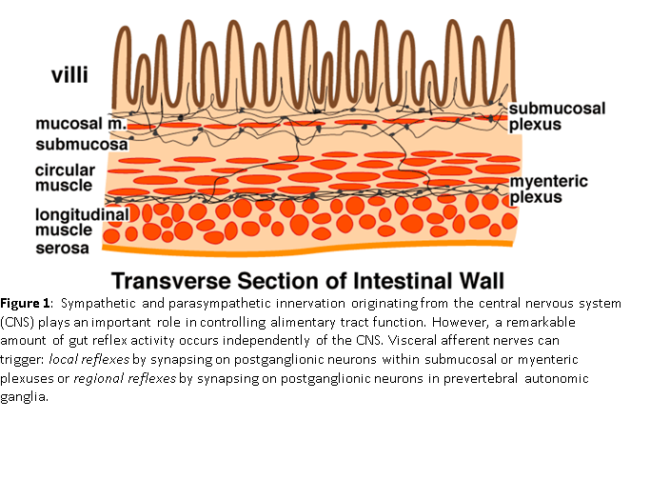
2.1. 2 Enteric Nervous System: Myenteric Plexus. The myenteric plexus of the enteric nervous system lies between the circular and longitudinal layers of the muscularis externa and is the main neuronal regulator of intestinal motor function.
Where is the myenteric plexus quizlet?
The myenteric plexus lies between the circular muscle and the longtidutional muscle. Preganglionic fibers of the sympathetic nervous system are relatively short and synapse in ganglia outside the GI tract.
What is the function of the myenteric plexus quizlet?
The myenteric plexus is located between two muscle layers and it controls the motility of the intestinal tract.
Where is the plexus of the submucosa?
The plexus of the submucosa from the rabbit. X 50. The submucosal plexus ( Meissner's plexus, plexus of the submucosa, plexus submucosus) lies in the submucosa of the intestinal wall. The nerves of this plexus are derived from the myenteric plexus which itself is derived from the plexuses of parasympathetic nerves around ...
Where is the plexus located?
Stomach. Intestines. v. t. e. The submucosal plexus ( Meissner's plexus, plexus of the submucosa, plexus submucosus) lies in the submucosa of the intestinal wall. The nerves of this plexus are derived from the myenteric plexus which itself is derived from the plexuses of parasympathetic nerves around the superior mesenteric artery.
Which plexus is associated with the longitudinal muscle layer?
A tertiary plexus is associated with the longitudinal muscle layer and contains axons of motor neurons that innervate it.
Which plexus contains secretomotor neurons?
Neurons in the submucous plexus of the intestine tend to be smaller than those in the myenteric plexus and contain fewer functional cell types. Nevertheless, the anatomical arrangement of the plexus is complicated with conflicting naming conventions (see Furness and Costa, 1987; Wedel et al., 1999 ). The ganglia include multiple populations of secretomotor neurons projecting to the gut mucosa, as well as vasodilator neurons innervating the local microcirculation ( Vanner and Surprenant, 1996; Hens et al., 2001 ). Submucous ganglia also contain IPANs with receptive endings projecting to the mucosa, and with outputs to other submucous neurons or to the myenteric plexus. About two-thirds of submucosal neurons are likely to be cholinergic, whilst both secretomotor and vasodilator neurons contain some combination of VIP and NOS ( Bosshard et al., 1989; Crowe et al., 1992; Porter et al., 1996, 1999 ). Other submucous neurons contain 5-HT, substance P, or somatostatin ( Crowe et al., 1992; Hens et al., 2001 ). In the colon, some neurons in the submucous plexus project to the deeper layers of the circular muscle. These neurons are located closer to the circular muscle layer; some contain NOS and VIP and are probably inhibitory ( Domoto et al., 1990; Porter et al., 1999 ). Functions of other submucosal neurons projecting to the muscle are unknown.
Where is SPLI found?
SP is found in association with nerves intrinsic to the gastrointestinal tract arising from the submucous and myenteric plexuses. In guinea pigs, substance P-containing nerves have been found in the intestinal mucosa ( Keast et al., 1984 ), frequently near the mucosal walls and beneath the villous epithelium. SPLI is present in the submucous nerve cells and mucosal terminals. To determine whether these nerves were of extrinsic or intrinsic origin, the extrinsic nerves supplying the intestinal mucosa were lesioned. Since these lesions did not result in decreased SPLI density and distribution, the SPLI-containing nerve fibers in the mucosal layers of the intestine must come from local submucosal nerve cell bodies.
Where are the neurons in the colon located?
In the colon, some neurons in the submucous plexus project to the deeper layers of the circular muscle. These neurons are located closer to the circular muscle layer; some contain NOS and VIP and are probably inhibitory ( Domoto et al., 1990; Porter et al., 1999 ). Functions of other submucosal neurons projecting to the muscle are unknown.
What are the cell bodies of enteric neurons?
The cell bodies of most enteric neurons are localized in two extensive networks: the MP (sandwiched between the circular and longitudinal muscle layers of the muscularis externa) and the submucous plexus which lies within the submucosal connective tissue. In the human colon, the MP consists of irregularly spaced stellate ganglia joined by thick interganglionic connectives. Each ganglion contains, on average, 70–80 nerve cell bodies and glial cells.98 In both myenteric and submucous plexuses, human ganglia contained more glia per neuron than in the intensively studied laboratory animal, the guinea pig. Together, the myenteric ganglia and connectives comprise the primary plexus ( Fig. 23.4 ). A secondary plexus (nonganglionated) consists of nerve trunks aligned with circular muscle bundles that innervate the muscle layer and penetrate through it, en route to the submucosa. A tertiary plexus is associated with the longitudinal muscle layer and contains axons of motor neurons that innervate it.
Which type of neuron is found in the submucosal plexus?
Tiny parasympathetic ganglia are scattered around to form the submucosal plexus (or Meissner’s plexus) where preganglionic parasympathetic neurons create synapses with postganglionic nerve fibers that supply the muscularis mucosae.
Which connective tissue is the submucosa?
In the gastrointestinal tract, the submucosa is the layer of dense, irregular connective tissue or loose connective tissue that supports the mucosa, as well as joins the mucosa to the bulk of underlying smooth muscle (fibers that run circularly within a layer of longitudinal muscle).
What vessels run through the mucosa?
Blood vessels, lymphatic vessels , and nerves (all supplying the mucosa) will run through here. Tiny parasympathetic ganglia are scattered around forming the submucosal plexus (or Meissner’s plexus) where preganglionic parasympathetic neurons create synapses with the postganglionic nerve fibers that supply the muscularis mucosae.
What is the submucosa of the stomach?
The submucosa lies under the mucosa and consists of fibrous connective tissue, separating the mucosa from the next layer, the muscularis externa. Layers of stomach lining: Stomach. The serosa is labeled at far right, and is colored yellow.
What are the layers of the GI tract?
The GI tract is composed of four layers. Each layer has different tissues and functions. From the inside out they are called: 1 Mucosa 2 Submucosa 3 Muscularis 4 Serosa
What is the structure of the gut wall?
General structure of the gut wall: The general structure of the gut wall is illustrated. The submucosa consists of a dense irregular layer of connective tissue with large blood vessels, lymphatics, and nerves that branch into the mucosa and muscularis externa.
What is a bundle of neurons with their connective tissue sheaths, blood vessels, and lymphatics?
nerve: A bundle of neurons with their connective tissue sheaths, blood vessels, and lymphatics.
What is the function of myenteric plexus?
Lying within intestinal smooth muscle, the myenteric plexus focuses on muscle control. Upon stimulation, the plexus causes an increase in gut wall tone and in intensity of rhythmical contractions. While mostly associated with excitatory muscle activity, there is also an inhibitory function of the myenteric plexus.
Where are lewy bodies found?
Lewy bodies have been found in enteric neurons, in the Auerbach and Meissner plexuses along the entire GI tract, including the esophagus, stomach, small intestine, and colon, particularly in neurons of the Auerbach plexus in the lower esophagus (Wakabayashi et al., 1988).
Which type of cell is responsible for the transmission of slow waves to smooth muscle?
Interstitial cells of Cajal, a cell type unique to the alimentary tract, are present medial to the inner smooth muscle layer. These specialized cells interact with myenteric neurons, and are thought to exhibit independent electrical activity, generating and transmitting slow waves to smooth muscle, functioning as pacemakers for colonic motility. The ENS is capable and does functions independent of the central nervous system (CNS), with reflex activity, in response to luminal stimuli, including muscle contraction and coordination – i.e., motility, blood flow, and glandular secretion. Modulation of the ENS is via the sympathetic and parasympathetic nervous system.
Where does sympathetic outflow occur?
Sympathetic outflow to the gastrointestinal tract, which arises from preganglionic neurons at the T1–L1 segments of the spinal cord and relays, via the splanchnic nerves, in the celiac and mesenteric ganglia, is involved in reflexes that decrease gut motility.
Where are syn aggregates found?
α-Syn aggregates have been found in post-ganglionic ENS terminals. The large majority of neurons receiving vagal fibers are located in the myenteric Auerbach's plexus and the submucosal Meissner's plexus, whereas α-staining in PD biopsies has been mostly confined to mucosal nerve fibers or to the submucosal ganglionic cells where the fibers originate. Furthermore, α-Syn aggregates has been found in the sigmoid colon where there is no vagal innervation. Also, not all PD patients had α-Syn aggregates in their GI tract. Indeed, immunohistochemical methods to detect α-Syn aggregatesin GI mucosal biopsy is not adequate to be used as biomarkers to predict PD due to the lack of sensibility and specificity (Corbillé et al., 2016; Ruffmann and Parkkinen, 2016 ).
What is the function of the myenteric plexus?
When stimulated, this plexus increases the tone of the gut as well as the velocity and intensity of its contractions . This plexus is concerned with motility throughout the whole gut. Inhibition of the myenteric system helps to relax the sphincters —the muscular rings that control the flow of digested food or food waste.
Which plexus controls the tone of the gut?
The myenteric plexus increases the tone of the gut and the velocity and intensity of contractions. The submucosal plexus is involved with local conditions and controls local secretion, absorption, and muscle movements. While described as a second brain, the enteric nervous system normally communicates with the central nervous system (CNS) ...
Which system is embedded in the lining of the gastrointestinal system?
The enteric nervous system ( ENS), which is embedded in the lining of the gastrointestinal system, can operate independently of the brain and the spinal cord. The ENS consists of two plexuses, the submucosal and the myenteric. The myenteric plexus increases the tone of the gut and the velocity and intensity of contractions.
