What is the thoracic inlet?
What is the structure of the thoracic inlet?
Which muscle is superior to the thoracic inlet?
Where is the brachial plexus?
Where is the oesophagus located?
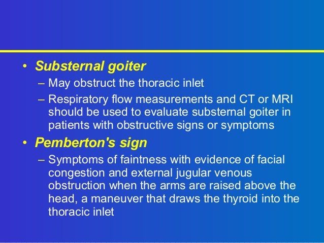
Can a thyroid gland extend inferiorly?
See 3 more
About this website

What do you mean by thoracic inlet?
Abstract. The thoracic inlet is the junctional region between the structures of the root of the neck and the contents of the thoracic cavity. The transverse plane through the thoracic inlet parallels the first rib and is tilted so that it is higher posteriorly than it is anteriorly (Fig. 4.28 a).
What goes through the thoracic inlet?
The right and left subclavian arteries and veins pass through the superior thoracic aperture. These vessels may be compromised by pathologic conditions of the lower cervical or upper thoracic viscera.
How do I open thoracic inlet?
Thoracic inlet release (necklace technique)Contact the thoracic inlet by placing palms over the trapezius muscle and fingers over the medial clavicle and proximal sternum.Determine restricted barriers. ... Place thoracic inlet into the restrictive barriers and apply lateral traction until a release is felt.
Is thoracic inlet the same as thoracic outlet?
It is also clinically referred to as the thoracic outlet, in the case of thoracic outlet syndrome; this refers to the superior thoracic aperture, and not to the lower, larger opening, the inferior thoracic aperture.
Which spinal nerve is affected in thoracic inlet syndrome?
Neurogenic (neurologic) thoracic outlet syndrome. This most common type of thoracic outlet syndrome is characterized by compression of the brachial plexus. The brachial plexus is a network of nerves that come from your spinal cord and control muscle movements and sensation in your shoulder, arm and hand.
Which of the following is are signs of arterial insufficiency in thoracic outlet syndrome?
Arterial thoracic outlet syndrome causes symptoms that affect your fingers, hands or entire arm. The most common sign is a dull ache or numbness in one arm. Symptoms are worse when you use your arm and better when you rest. Usually, people with ATOS don't have any symptoms in their neck or shoulder.
Do you need surgery for thoracic outlet syndrome?
Surgery: Patients with arterial thoracic outlet syndrome often require surgical treatment. Surgery may be performed to remove the first rib and make more room for the vessels and nerves. Surgery may also be performed to repair any structural problems of the artery.
How do you sleep with thoracic outlet syndrome?
Thoracic outlet syndrome sleeping position It is important to avoid sleeping positions that increase the pressure on the outlet. Generally, we recommend sleeping on your back or with two pillows on the affected side to prevent compression of the outlet.
Can thoracic outlet syndrome affect vision?
These symptoms may worsen when the thoracic outlet is narrowed in certain body positions like when the arm is raised overhead. TOS can also lead to eye problems including vision loss because of vertebral artery compression.
Thoracic Inlet or Thoracic Outlet: Which One is Which in ... - ResearchGate
Request PDF | On Mar 1, 2014, Satheesha B Nayak published Thoracic Inlet or Thoracic Outlet: Which One is Which in Anatomical and Clinical Literature? | Find, read and cite all the research you ...
The thoracic inlet: normal anatomy - PubMed
The thoracic inlet is the junction between the neck and the chest. A number of neural structures traverse this region. A knowledge of the location of these various neural structures and their relationship to one another is important when interpreting cross-sectional images of this region. This artic …
Inferior thoracic aperture | Radiology Reference Article | Radiopaedia.org
Gross anatomy. The inferior thoracic aperture is irregular in shape and is more oblique and much larger than the superior thoracic aperture.The diaphragm occupies and closes the inferior thoracic aperture, thereby separating the thoracic and abdominal cavities. For structures to pass between the two cavities, they either pass behind the diaphragm or pass through various diaphragmatic apertures.
Thoracic Inlet Anatomy - Structures Passing Through ... | GrepMed
Thoracic Inlet Anatomy - Structures Passing Through Thoracic Inlet • Muscles: Sternohyoid, Sternothyroid, Longus Cervicis/[ongus Colli • Arteries: Right & Left Internal Thoracic Arteries, Brachiocephalic Trunk/Artery, Left Common Carotid Artery, Left Subclavian Artery, Right And Left Superior Intercostal Arteries • Nerves: Right And Left Vagus Nerves, Left Recurrent Laryngeal Nerve ...
Thoracic inlet or thoracic outlet: which one is which in anatomical and ...
Thoracic inlet or thoracic outlet: which one is which in anatomical and clinical literature?
What is the thoracic outlet?
Thoracic outlet. Thoracic outlet. The thoracic outlet is the space between your collarbone (clavicle) and your first rib. This narrow passageway is crowded with blood vessels, nerves and muscles. Thoracic outlet syndrome is a group of disorders that occur when blood vessels or nerves in the space between your collarbone and your first rib ...
How to treat thoracic outlet syndrome?
Treatment for thoracic outlet syndrome usually involves physical therapy and pain relief measures. Most people improve with these approaches. In some cases, however, your doctor may recommend surgery.
What age is thoracic outlet syndrome most common?
Age. Thoracic outlet syndrome is more common in young adults, between 20 and 40 years old.
What is the most common type of thoracic outlet syndrome?
There are a number of types of thoracic outlet syndrome, including: Neurogenic (neurological) thoracic outlet syndrome. This most common type of thoracic outlet syndrome is characterized by compression of the brachial plexus. The brachial plexus is a network of nerves that come from your spinal cord and control muscle movements ...
What causes a compression in the thoracic outlet?
Poor posture. Drooping your shoulders or holding your head in a forward position can cause compression in the thoracic outlet area. Trauma. A traumatic event, such as a car accident, can cause internal changes that then compress the nerves in the thoracic outlet.
Can thoracic outlet syndrome be caused by repetitive movements?
Athletes, such as baseball pitchers and swimmers, also can develop thoracic outlet syndrome from years of repetitive movements. Pressure on your joints. Obesity can put an undue amount of stress on your joints, as can carrying around an oversized bag or backpack. Pregnancy.
Is thoracic outlet syndrome a common disorder?
Some doctors don't believe it exists, while others say it's a common disorder. People with nonspecific-type thoracic outlet syndrome have chronic pain in the area of the thoracic outlet that worsens with activity, but a specific cause of the pain can't be determined.
What is the thoracic inlet?
The thoracic inlet allows unobstructed passage of the neurovascular bundle (nerves, arteries and veins) from the root of the neck to the axilla (from the neck to the arm pit).
What are the symptoms of thoracic inlet syndrome?
There are many factors that may be involved, including tight neck muscles or abnormal muscular attachment onto the ribs, cervical ribs (either calcific or fibrous), or postural causes.
How to treat neck and back pain?
Physiotherapy treatment will address faulty neck/back postures and joint mobility with mobilising of joints and posture correction exercises. Tightness of neck musculature, in particular the scalenes, can be decreased through stretching and retraining of shoulder girdle muscles. The stabilising muscles of the neck and thoracic spine (such as longus colli, lower trapezius and serratus anterior) are also retrained and strengthened. Modifying household activities and exercise routines is important for positive long-term outcomes. Initial manual physiotherapy can speed recovery and is best combined with a prescribed home exercise programme. Results from conservative treatment can be very successful with 70-90% resolution of symptoms.
Where are the lymph nodes located in the thorax?
Thoracic lymph nodes are separated into two types: parietal lymph nodes located in the thoracic wall, and visceral lymph nodes, which are associated with the internal organs. Due to their location, abnormalities of the lymph nodes in the thorax, or chest, are not easily detected. However, any changes in the size or amount of these lymph nodes could be indicative of several types of extrapulmonary or pulmonary diseases. For diagnostic purposes, lymph nodes of the thorax can be further divided into sub-categories. The lung lymph nodes can be found along the bronchi. The paratracheal and tracheobronchial groups of lymph nodes are located in the neck and also in the junction where the trachea meets the bronchi, respectively. These accept drainage from the heart, lungs, bronchi, and thoracic trachea as well as other lymph nodes. The posterior mediastinal group of lymph nodes, located near the thoracic aorta, is closely linked to the tracheobronchial group and primarily drains into the thoracic duct. The chest wall thoracic lymph nodes receive drainage from the breasts, arms, pectoral muscles, and other muscles and skin located in the upper section of the chest.
Where are the paratracheal and tracheobronchial lymph nodes located?
The paratracheal and tracheobronchial groups of lymph nodes are located in the neck and also in the junction where the trachea meets the bronchi, respectively. These accept drainage from the heart, lungs, bronchi, and thoracic trachea as well as other lymph nodes.
Where is the posterior mediastinal lymph node located?
The posterior mediastinal group of lymph nodes, located near the thoracic aorta, is closely linked to the tracheobronchial group and primarily drains into the thoracic duct.
Which thoracic opening is located in the neck?
The thorax has two major openings: the superior thoracic aperture found superiorly and the inferior thoracic aperture located inferiorly. The superior thoracic aperture opens towards the neck. It is bounded by the bones of the upper thorax; manubrium of sternum, the first pair of ribs, and the body of the vertebra T1.
What is the thoracic wall?
Thoracic wall. The first step in understanding thorax anatomy is to find out its boundaries. The thoracic, or chest wall, consists of a skeletal framework, fascia, muscles, and neurovasculature – all connected together to form a strong and protective yet flexible cage.
What are the arteries that supply breasts?
Anatomy of the female breast (lateral view) They are supplied by several arteries of the thoracic wall, namely branches of the internal thoracic, axillary, lateral thoracic, thoracoacromial, and posterior intercostal arteries.
What is the chest?
The chest, properly called the thorax, is the superior part of the trunk located between the neck and abdomen. It consists of several components: 1 Thoracic wall 2 Several cavities 3 Neurovasculature and lymphatics 4 Internal organs 5 Breasts
How does the thoracic cavity communicate with the neck?
The thoracic cavity communicates with the neck via the superior thoracic aperture and with the abdominal cavity via the inferior thoracic aperture through anatomical spaces piercing the diaphragm. Cavities of the body Explore study unit.
What is the inferior thoracic aperture?
The inferior thoracic aperture is almost completely covered by the diaphragm, separating it from the abdominal cavity. Moving forward with the skeletal scaffold of the thorax, we have the thoracic skeleton. It is made up of the sternum, twelve pairs of ribs, twelve thoracic vertebrae, and interconnecting joints.
What muscles support the thorax?
In addition, all of the thoracic muscles provide further support and strength for the thorax. If you want to learn more about the muscles of the thoracic wall and get one step closer to mastering chest anatomy, take a look at our muscle anatomy charts!
What is the thoracic inlet?
The thoracic inlet, also known as the superior thoracic aperture, refers to the opening at the top of the thoracic cavity. It is also clinically referred to as the thoracic outlet, in the case of thoracic outlet syndrome; this refers to the superior thoracic aperture, and not to the lower, larger opening, the inferior thoracic aperture .
What is the structure of the thoracic inlet?
Structure. The thoracic inlet is essentially a hole surrounded by a bony ring, through which several vital structures pass. The thoracic inlet is bounded by: the first thoracic vertebra (T1) posteriorly; the first pair of ribs laterally, forming lateral C-shaped curves posterior to anterior; and the costal cartilage of the first rib and ...
Which muscle is superior to the thoracic inlet?
Superior to the thoracic inlet is the root of the neck, and the superior mediastinum is inferiorly related. The brachial plexus is a superolateral relation of the thoracic inlet. The brachial plexus emerges between the anterior and middle scalene muscles, superior to the first rib, and passes obliquely and inferiorly, underneath the clavicle, into the shoulder and then the arm. Impingement of the plexus in the region of the scalenes, ribs, and clavicles is responsible for thoracic outlet syndrome .
Where is the brachial plexus?
The brachial plexus emerges between the anterior and middle scalene muscles, superior to the first rib, and passes obliquely and inferiorly, underneath the clavicle, into the shoulder and then the arm.
Where is the oesophagus located?
The oesophagus lies against the body of the T1 vertebra, separated from it by the prevertebral fascia, and the trachea lies in front of the oesophagus, in the midline, and may touch the manubrium. The apices of the lungs lie to either side of the oesophagus and trachea, and is separated from them by the other vessels and nerves listed above. Furthermore, they extend slightly superior past the level of the inlet (e.g. the horizontal plane of the first rib).
Can a thyroid gland extend inferiorly?
There are several other minor, but important, vessels and nerves passing through, and an abnormally large thyroid gland may extend inferiorly through the thoracic inlet into the superior mediastinum.
Overview
- Thoracic outlet syndrome (TOS) is a group of disorders that occur when blood vessels or nerves in the space between your collarbone and your first rib (thoracic outlet) are compressed. This can cause shoulder and neck pain and numbness in your fingers. Common causes of thoracic outlet syndrome include physical trauma from a car accident, repetitive...
Symptoms
- There are three general types of thoracic outlet syndrome: 1. Neurogenic (neurologic) thoracic outlet syndrome.This most common type of thoracic outlet syndrome is characterized by compression of the brachial plexus. The brachial plexus is a network of nerves that come from your spinal cord and control muscle movements and sensation in your shoulder, arm and hand. …
Causes
- Thoracic outlet syndrome is usually caused by compression of the nerves or blood vessels in the thoracic outlet, just under your collarbone (clavicle). The cause of the compression varies and can include: 1. Anatomical defects.Inherited defects that are present at birth (congenital) may include an extra rib located above the first rib (cervical rib) or an abnormally tight fibrous band connecti…
Risk Factors
- There are several factors that seem to increase the risk of thoracic outlet syndrome, including: 1. Sex.Females are greater than three times more likely to be diagnosed with thoracic outlet syndrome than are males. 2. Age.Thoracic outlet syndrome may occur at any age but is most commonly diagnosed in adults between the ages of 20 and 50.
Complications
- Complications from this condition stem from the type of presentation (neurogenic, venous or arterial). For patients with venous or arterial TOS, it is important to seek urgent medical attention to make the correct diagnosis and implement appropriate treatment. For neurogenic TOS, it is important to seek medical attention with appropriate evaluation and testing.
Prevention
- If you're at risk for thoracic outlet compression, avoid repetitive movements and lifting heavy objects. If you're overweight, losing weight may help you prevent or relieve symptoms of thoracic outlet syndrome. Even if you don't have symptoms of thoracic outlet syndrome, avoid carrying heavy bags over your shoulder, because this can increase pressure on the thoracic outlet. Stretc…