What are the bones of the orbit?
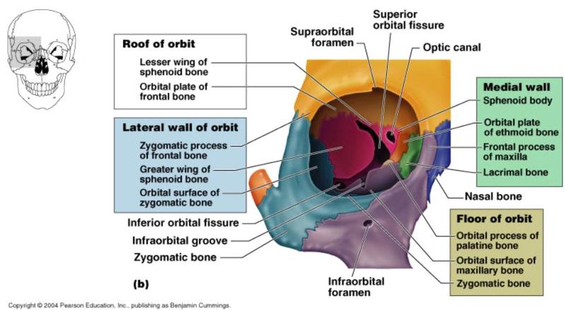
Anatomy The orbit appears as a quadrangular pyramidal cavern in the upper face. It is made up of four facial bones and three cranial bones: maxilla, zygomatic bone, lacrimal bone, palatine bone, frontal bone, ethmoid bone, and sphenoid bone.
What bone forms the posterior wall of the orbital process?
The sphenoid bone forms the entire posterior wall of the bony orbit. A tiny slither of the vertical plate of the palatine bone, known as the orbital process can be seen between the maxilla, the ethmoid bone and the sphenoid bone. Articulations of the Orbit.
What is the structure of the orbit?
The structure of the orbit is made up of several orbital bones that provide a strong base for the eye so that it can perform its functions properly. There are seven orbital bones that make up this structure: the frontal, sphenoid, zygomatic, ethmoid, lacrimal, palatine and maxilla bones. Each of these plays a role in keeping the eyeball protected.
What bone separates the orbit from the underlying maxillary sinus?
The maxilla separates the orbit from the underlying maxillary sinus. Medial wall – Formed by the ethmoid, maxilla, lacrimal and sphenoid bones. The ethmoid bone separates the orbit from the ethmoid sinus. Lateral wall – Formed by the zygomatic bone and greater wing of the sphenoid.

What is the bone orbit?
The Bony Orbit. The bony orbits (or eye sockets) are bilateral and symmetrical cavities in the head. They enclose the eyeball and its associated structures. In this article, we shall look at the borders, contents and clinical correlations of the bony orbit.
How many bones are in an orbit?
The boundaries of the orbit are formed by seven bones.
What is the fracture of the outer rim of the bony orbit?
The contents of the orbit have herniated into the maxillary sinus. caption] Orbital rim fracture - This is a fracture of the bones forming the outer rim of the bony orbit. It usually occurs at the sutures joining the three bones of the orbital rim - the maxilla, zygomatic and frontal.
Which bone separates the orbit from the ethmoid sinus?
Medial wall - Formed by the ethmoid, maxilla, lacrimal and sphenoid bones. The ethmoid bone separates the orbit from the ethmoid sinus.
Which bone separates the orbit from the anterior cranial fossa?
The frontal bone separates the orbit from the anterior cranial fossa. Floor (inferior wall) – Formed by the maxilla, palatine and zygomatic bones. The maxilla separates the orbit from the underlying maxillary sinus. Medial wall – Formed by the ethmoid, maxilla, lacrimal and sphenoid bones.
What is the space within the orbit that is not occupied?
Any space within the orbit that is not occupied is filled with orbital fat. This tissue cushions the eye, and stabilises the extraocular muscles.
What is the name of the symmetrical cavity in the head?
The bony orbits (or eye sockets) are bilateral and symmetrical cavities in the head. They enclose the eyeball and its associated structures.
Why are orbital bones important?
Orbital bones: crucial to supporting eye health. Orbital bones provide a base within the skull for the eyeball to rest, allowing the eye to move and function properly. This structure is designed to provide strong protection for your eyes in the event of head trauma or injury, though sometimes the bones themselves can sustain a fracture.
What causes the orbital bone to cave downward?
Significant eye trauma can cause the orbital bone and surrounding structures to cave downward into the socket. Such trauma can also affect the surrounding eye muscles, making eye movement harder and more painful.
What is an orbital bone fracture?
An orbital bone fracture or break is what it sounds like: a broken orbital bone (or multiple bones). This is frequently the result of an accident, such as a car wreck or being hit hard by a flying object (for example, a baseball during a ballgame). Broken orbital bones can also happen if you come into contact with the force of someone’s fist.
What is the orbit of the eye?
The “orbit” or “socket” of the eye encases the eyeball and protects its place in the skull. The structure of the orbit is made up of several orbital bones that provide a strong base for the eye so that it can perform its functions properly.
How to evaluate orbital bone?
Evaluating an orbital bone typically involves tests such as a CT scan, X-rays and other imaging. Many cases do not require surgery for treatment, and the eye is able to heal on its own with the help of antibiotics, decongestants and ice packs to reduce swelling. Severe orbital bone fractures that impact the movement of the eye ...
What are the symptoms of orbital bone damage?
More serious impacts on the orbital bone can cause other symptoms, such as: Double vision. Numbness. Blood in the eye ( subconjunctival hemorrhage) Swelling in and around the eye. Eyeballs that are sunken or bulging. SEE RELATED: 7 common eye injuries and how to treat them.
How long does it take for an orbital fracture to heal?
The bruising and swelling caused by an orbital fracture usually heals within 7 to 10 days, while the fracture itself takes longer to heal completely — the exact time frame depends on the level of impact and severity of the injury.
Which bones form in the orbit?
The bones of the orbit develop via both endochondral and intramembranous ossification. The sphenoid and ethmoid bones form mostly via endochondral ossification while the frontal bone is formed by intramembranous ossification.
What are the bones of the orbit?
The floor of the orbit consists of three bones: the maxillary bone, the palatine bone, and the orbital plate of the zygomatic bone. This part of the orbit is also the roof of the maxillary sinus. There is an infraorbital groove along the floor and it travels into a canal anteriorly where it eventually exits as the infraorbital foramen. This is the structure that lies below the orbital margin of the maxillary bone. Along the floor of the orbit is the origin of the inferior oblique muscle. This is the only extraocular muscle that does not originate at the apex of the orbit.
How wide is the orbital rim?
The orbit is a pear shape, with the optic nerve at the stem, and holds approximately 30 cc volume. The entrance to the globe anteriorly is approximately 35 mm high and 45 mm wide. The depth from orbital rim to the orbital apex measures 40 to 45 mm in adults. The maximum width is 1 cm behind the anterior orbital margin. Both race and gender can affect the measurements of the bony orbit. The orbital cavity contains the globe, nerves, vessels, lacrimal gland, extraocular muscles, tendons, and the trochlea as well as fat and other connective tissue. An increase in the volume of the extraocular structures within the orbit can cause proptosis, which is protrusion of the globe and/or displacement (deviation) of the globe from its normal position.
What are the extraocular muscles?
Seven extraocular muscles are associated with the bones of the orbit. All but the inferior oblique muscle originate at the orbital apex. The inferior oblique, as noted above, originates at the floor of the orbit in a depression on the orbital floor near the orbital rim. All of the rectus muscles originate at the annulus of Zinn. The superior oblique originates just medial to the optic foramen, between the annulus of Zinn and periorbita. This is the muscle that travels through the trochlea, forming a pulley that allows for intorsion of the globe. All of the extraocular muscles insert on the globe except the levator palpebrae superioris, which broadens anteriorly to become the levator aponeurosis, and is responsible primarily for eyelid elevation. [3]
What is the lateral orbital wall?
This is the strongest of the walls of the orbit. There is a lateral orbital tubercle, which is an elevation of the orbital margin of the zygomatic bone and is an attachment for many important structures. These are the ligament of the lateral rectus muscle, the suspensory ligament of the eyeball (Lockwood ligament), lateral palpebral ligament, aponeurosis of the levator palpebrae superioris muscle, and the Whitnall ligament.
What is the purpose of bones in the orbit?
The bones of the orbit protect the globe of the eye as well as other periocular contents. A fracture to the orbit can involve the contents of the orbit and potentially compress nerves or muscles within the orbit.
What is the orbital margin?
The orbital margin is the anterior opening of the globe and has a quadrilateral spine formed by several of the bones that make up the orbit. The roof of the orbit is formed by the orbital plate of the frontal bone and the lesser wing of the sphenoid. The fossa of the lacrimal gland lies anterolaterally, behind the zygomatic process of the frontal bone.
