What is Chamber of the heart lies most anterior?
What is the most anterior chamber of the heart? The right ventricle (RV) is the most anterior of the four heart chambers. It receives deoxygenated blood from the right atrium (RA) and pumps it into the pulmonary circulation. During diastole, blood enters the right ventricle through the atrioventricular orifice through an open tricuspid valve (TV).
What are the top chambers of the heart called?
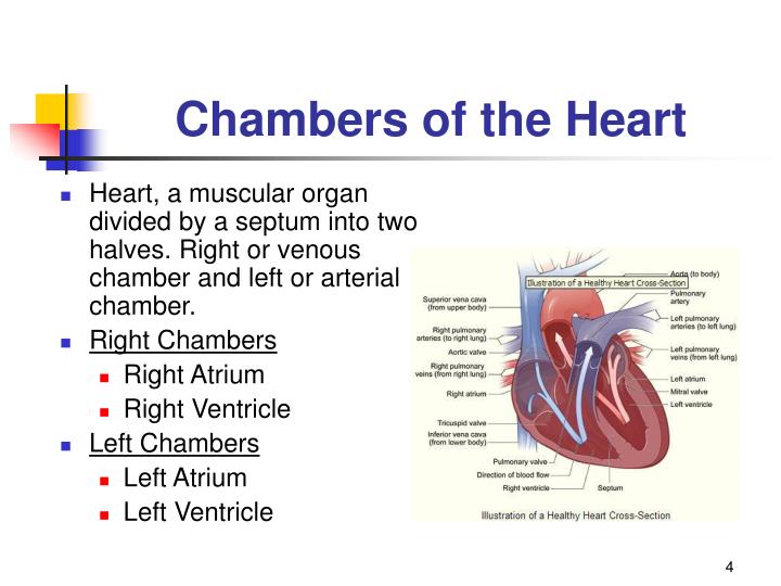
The upper chambers, the right and left atria, receive incoming blood. The lower chambers, the more muscular right and left ventricles, pump blood out of the heart. The heart valves, which keep blood flowing in the right direction, are gates at the chamber openings.
Which Chamber of the heart is most muscular?
The ventricle is the largest and most muscular chamber of the heart. When filled with blood, it constricts, forcing the blood through the bulbus arteriosus. Click to see full answer.
What is the smallest Chamber of the heart?
The right atrium is the smallest chamber of the heart. It is under the least pressure. The following are ranges of pressures in the heart chambers during the heart cycle of systole and diastole, lowest during diastole (atrial or ventricular) and highest during systole (atrial or ventricular) What are the 4 chambers of the heart?
Which chamber of the heart is most anterior?
right ventricleThe right ventricle in the normal heart is the most anteriorly situated cardiac chamber since it is located immediately behind the sternum. It also marks the inferior border of the cardiac silhouette.
Is the right ventricle posterior?
The right ventricle is the most anteriorly positioned chamber of the heart, sitting directly posterior to the sternum. The distinct anatomical features of the right ventricle create an approximately 10-fold difference in vascular resistance between the right and left ventricular systems.
What is the posterior of the heart?
The base of the heart is probably better termed its posterior surface. It is not the most inferior surface of the organ but rather the most superior. It assumed the term because it is thought to resemble the base of the pyramid or cone which extends obliquely to the left to the apex of the heart.
Which atrium is posterior?
right atriumThe posterior part of the right atrium is termed the sinus venarum; also, it includes most of the lateral wall of the chamber. It has a relatively smooth surface compared to the anterior part. The posterior and anterior walls merge at the crista terminalis.
Is the left ventricle posterior?
The left ventricle is situated posterior to the right ventricle, and like its counterpart comprises an inlet portion, apical trabeculae, and an outlet portion [3].
Is the 4th ventricle anterior or posterior?
The fourth ventricle is a diamond-shaped cavity located posterior to the pons and upper medulla oblongata and anterior-inferior to the cerebellum. The superior cerebellar peduncles and the anterior and posterior medullary vela form the roof of the fourth ventricle.
How can you tell the anterior and posterior of the heart?
How can you tell which side of the heart is the anterior surface and which side is the posterior surface? The anterior is the side that the apex is pointing to. The posterior surface lies opposite to the apex.
What is left atrium posterior?
The posterior wall of the left atrium, including its inferior part, is related to the esophagus and its nerves (vagal nerves), the thoracic aorta, and the coronary sinus (Figure 1C).
What is posterior to the chest?
0:073:57Resp - Examination of the posterior chest - YouTubeYouTubeStart of suggested clipEnd of suggested clipOk can I get you to put your arms. Around.MoreOk can I get you to put your arms. Around.
Is aortic valve posterior?
The aortic valve is located posterior to the pulmonary valve and the commissure where the anterior two cusps join together points toward the pulmonary valve. It is these two sinuses that contain the origin of the coronary arteries.
Which atrium is anterior?
The anterior mediastinal aspect of the right lung is anteriorly related to the right atrium. The structures separating the two are the pleura and pericardium. The sinuatrial node, which is responsible for regulating the automaticity of the myocardium, is located in the posterior wall of the right atrium.
Where is the right ventricle located?
The right ventricle is one of the heart's four chambers. It is located in the lower right portion of the heart below the right atrium and opposite the left ventricle.
Where is the posterior ventricle?
The posterior (occipital) horn of the lateral ventricle extends posteromedially into the occipital lobe, and like other parts of the lateral ventricle it has a roof, lateral wall and a medial wall.
Where is the right and left ventricle located?
the heartThe top chambers of the heart are called the left atrium and right atrium. The bottom chambers of the heart are the left ventricle and right ventricle, which have thicker walls. The right atrium receives blood from the veins that has already circulated through the body and pumps it over to the right ventricle.
What is posterior to the fourth ventricle?
Superiorly, it connects to the third ventricle through a thin canal called the cerebral aqueduct of Sylvius. It is surrounded anteriorly by the pons and medulla, posteriorly by the cerebellum, and inferiorly by the spinal canal and spinal cord.
How many chambers are there in the heart?
The heart is made up of four chambers. The superior chambers consist of the right atrium and left atrium ( plural, atria: L., corridor ). which lie primarily on the posterior side of the heart.
Which ventricle pumps blood through the pulmonary system?
The left and right ventricle. The right ventricle pumps blood through the pulmonary circulatory system and the thicker-walled left ventricle pumps blood through the long systemic circulatory system. Internally, the two ventricles are separated by a thick myocardial wall called the interventricular septum.
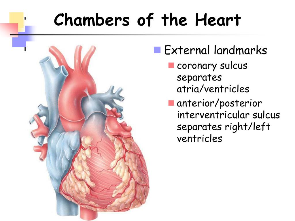
What is the groove of the anterior ventricular sulcus?
The anterior interventricular sulcus. On the posterior surface of the heart, the ventricles are separated by the posterior interventricular sulcus (or groove), which contains the posterior interventricular artery, middle cardiac vein, and adipose tissue. The posterior interventricular sulcus.
What is the appendage of the heart called?
The right and left atrium of the heart. Extending anteriorly from each thin-walled atrium is a small, ear shaped appendage called an auricle ( l., auricula, little ear) that expands the volume of the chamber. Blood drains into the atria from the pulmonary and systemic circulatory systems. The auricles of the heart.
What is the name of the wall that separates the ventricles?
Internally, the two ventricles are separated by a thick myocardial wall called the interventricular septum.
What system drains blood into the atria?
Blood drains into the atria from the pulmonary and systemic circulatory systems. The auricles of the heart. Making up the lower chambers are the right ventricle and left ventricle ( L., ventriculus, a little belly ), which are much larger than the atria.
Where is the heart located?
It is found in the middle mediastinum, wrapped in a two-layered serous sac called the pericardium.
Where does blood flow through the heart?
Blood flows from the atria into the ventricles through the atrioventricular orifices (right and left)–openings in the atrioventricular septa. These openings are periodically shut and open by the heart valves, depending on the phase of the heart cycle.
What are the semilunar valves in a cadaver?
Heart valves in a cadaver. Semilunar valves prevent backflow from the great vessels to the ventricles. The pulmonary semilunar valve is between the right ventricle and the opening of the pulmonary trunk. It has three semilunar cusps/leaflets: anterior/non-adjacent, left/left adjacent, and right/right adjacent.
What are the two leaflets that separate the atria from the ventricles?
Heart valves. Heart valves separate atria from ventricles, and ventricles from great vessels. The valves incorporate two or three leaflets (cusps) around the atrioventricular orifices and the roots of great vessels.
What is the margin of the right atrium?
The right margin is the small section of the right atrium that extends between the superior and inferior vena cava . The left margin is formed by the left ventricle and left auricle. The superior margin in the anterior view is formed by both atria and their auricles. The Inferior margin is marked by the right ventricle.
How big is the ascending aorta?
Because they are large in size; the diameter of the ascending aorta is 2.1 centimeters, which is like the size of an American nickel (five-cent coin), and they all carry blood to and from the heart. Oh, not to mention that the aorta gives off branches which supply the entire body with oxygenated blood.
How many surfaces does the heart have?
Heart anatomy. The heart has five surfaces: base (posterior), diaphragmatic (inferior), sternocostal (anterior), and left and right pulmonary surfaces. It also has several margins: right, left, superior, and inferior: The right margin is the small section of the right atrium that extends between the superior and inferior vena cava .
How many chambers does the heart have?
Log In. The heart consists of four chambers: the two atria and the two ventricles. Blood returning to the heart enters the atria, and is then pumped into the ventricles. From the left ventricle, blood passes into the aorta and enters the systemic circulation. From the right, it enters the pulmonary circulation via the pulmonary arteries.
Which ventricle forms the apex of the heart?
In the anatomical position, the left ventricle forms the apex of the heart, as well as the left and diaphragmatic borders. Much like the right ventricle, it can be divided into an inflow portion and an outflow portion.
What is the septal wall in the right atrium?
The septal wall in the right atrium is marked by a small oval-shaped depression called the fossa ovalis. This is the remnant of the foramen ovale in the fetal heart, which allows right to left shunting of blood to bypass the lungs. It closes once the newborn takes its first breath.
What is a bridge in the heart?
Bridges – attached to the ventricle at both ends, but free in the middle. The most important example of this type is the moderator band, which spans between the interventricular septum and the anterior wall of the right ventricle. It has an important conductive function, containing the right bundle branches.
Which ventricles of the heart receive blood from the atria and pump it into the outflow vessels?
The left and right ventricles of the heart receive blood from the atria and pump it into the outflow vessels; the aorta and the pulmonary artery respectively.
What is the right atrium?
Extending from the antero-medial portion of the chamber is the right auricle (right atrial appendage) – a muscular pouch that acts to increase the capacity of the atrium.
What is the left ventricle?
In the anatomical position, the left ventricle forms the apex of the heart, as well as the left and diaphragmatic borders. Much like the right ventricle, it can be divided into an inflow portion and an outflow portion. Inflow Portion.
