Which gland is present in small intestine Class 10?
The digestive gland present within the walls of the ileum is known as intestinal gland. They secrete digestive juices which help in the digestion of food by breaking down the complex substances into a simpler form and they are absorbed by the villi.
What are the glands of small intestine?
Intestinal glands - Crypts of Lieberkuhn They are of two types namely, crypts of Lieberkuhn and Brunner's glands. They are tubular structure that occur throughout the small intestine between the villi. They secrete digestive enzyme and mucus. The crypts have at the base paneth cells and argentaffin cells.
Which gland is present in large intestine?
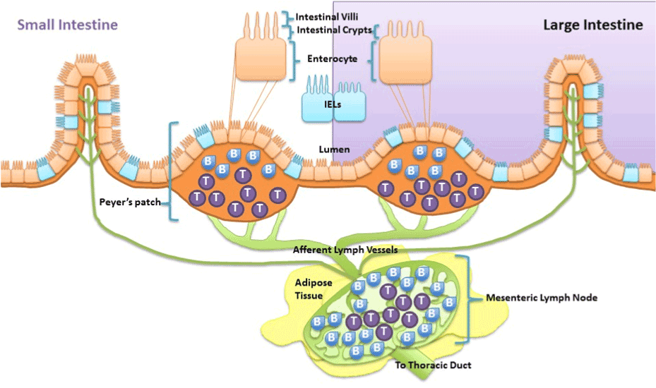
In histology, an intestinal gland (also crypt of Lieberkühn and intestinal crypt) is a gland found in between villi in the intestinal epithelium lining of the small intestine and large intestine (or colon).
Which digestive gland helps the small intestine?
The pancreas delivers the digestive juice to the small intestine through small tubes called ducts. Liver. Your liver makes a digestive juice called bile that helps digest fats and some vitamins. Bile ducts carry bile from your liver to your gallbladder for storage, or to the small intestine for use.
Is lymph present in small intestine?
In the intestine, lymphatic capillaries, or lacteals, are located exclusively in intestinal villi, whereas collecting lymphatic vessels are present in the mesentery. 7 The term gut lymphatics used throughout this review refers to both lacteals in the intestinal villi and lymphatic vessels in the submucosa.
Are Brunner's glands in the small intestine?
Polyps of the Small Intestine Brunner's glands are predominantly located in the duodenal submucosa, although they may focally transgress the muscularis mucosae and extend into the lamina propria.
Is small intestine the largest gland?
Liver: The largest gland in the human body is the liver. It is a reddish-brown organ that lies on the right side of the abdomen. It weighs about 1.5 - 1.6 kg.
What are the 3 digestive glands?
The alimentary canal is associated with the digestive glands in the human digestive system. Three major glands play an important role in the digestion....These are as follows:Salivary gland.Liver.Pancreas.
What is Brunner's gland?
Brunner's gland, which was accurately described by Brunner in 1688, is a gland in the submucosa of the duodenum (1), which has a main physiological function of secreting an alkaline-based mucus to protect the duodenal lining from the acid secreted in the stomach.
What is Lieberkühn gland?
LEE-ber-keen) Tube-like gland found in the lining of the colon and rectum. Glands of Lieberkuhn renew the lining of the intestine and make mucus. Also called colon crypt.
What are the 4 digestive glands?
Glands contributing digestive juices include the salivary glands, the gastric glands in the stomach lining, the pancreas, and the liver and its adjuncts—the gallbladder and bile ducts.
Which endocrine gland is between the stomach and small intestine?
The pancreasThe pancreas is unique in being both an endocrine gland and an exocrine gland. The endocrine function is the secretion of substances such as insulin, which metabolizes sugar. The exocrine function involves the secretion of digestive enzymes into the duodenum.
What are the 4 digestive glands?
Glands contributing digestive juices include the salivary glands, the gastric glands in the stomach lining, the pancreas, and the liver and its adjuncts—the gallbladder and bile ducts.
What are the 3 glands in the digestive system?
The alimentary canal is associated with the digestive glands in the human digestive system. Three major glands play an important role in the digestion....These are as follows:Salivary gland.Liver.Pancreas.
What are the 3 types of glands in the stomach?
—The gastric glands are of three kinds: (a) pyloric, (b) cardiac, and (c) fundus or oxyntic glands. They are tubular in character, and are formed of a delicate basement membrane, consisting of flattened transparent endothelial cells lined by epithelium.
What are 2 major glands of the intestinal secretion?
Both the cardiac and pyloric glands secrete mucus, which coats the stomach and protects it from self-digestion by helping to dilute acids and enzymes.
Which part of the small intestine is responsible for digesting food?
Each segment of the small intestine has a different function, including: The duodenum receives partially digested food (called chyme) through the pylorus (from the stomach), receives digestive enzymes from the pancreas and liver to continue to break down ingested food. In addition, iron is absorbed in the duodenum.
What is the proximal end of the small intestine?
Anatomy. The small intestine is made up of thee sections, including the duodenum, the jejunum and the ileum. On its proximal (near) end, the small intestine—beginning with the duodenum—connects to the stomach. On its distal (far) end, the ileum—the last segment of the small intestine—connects to the large intestine (colon).
What is the longest part of the digestive system?
The small intestine (commonly referred to as the small bowel) is a tubular structure/organ that is part of the digestive system. In fact, it is the longest portion of the digestive system, approximately 20 to 25 feet in length. 1 The reason it is referred to as the “small” intestine, is because its lumen (opening) is smaller in diameter ...
Why is the small intestine called the small intestine?
It is referred to as the “small” intestine because its lumen (opening) is smaller in diameter (at approximately 2.5 centimeters or 0.98 inches) than the large intestine ( colon ).
What is the ampulla of Vater?
The ampulla of Vater is an important landmark that serves as the site where the bile duct and the pancreatic duct empty their digestive juices (containing enzymes that help to break down ingested food) into the duodenum.
What is the function of the small intestine?
The primary function of the small intestine is to break down and absorb ingested nutrients while mixing and moving the intestinal contents (consisting of gastric juices and partly digested food) along the digestive tract into the colon. magicmine/iStock/Getty Images.
How to treat intestinal atresia?
The treatment of intestinal atresia involves a surgical procedure to correct the problem. The type of operation depends on where the obstruction is located.
What are the layers of the digestive tract?
Be able to describe the layers in the wall of the digestive tract (mucosa, submucosa, muscularis externa and adventitia/serosa), and explain how they differ in the small and large intestines.
What is the mucosa of the colon?
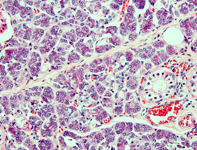
The mucosaof the colon is lined by a simple columnar epithelium with a thin brush border and numerous goblet cells. Note that there arenoplicae or villi. The crypts of Lieberkühn are straight and unbranched and lined largely with goblet cells. In many regions the mucus is partially preserved and stains with hematoxylin. At the base of the crypts, undifferentiated cells and endocrine cells are present; however, Paneth cells are notusually present. The appearance of the lamina propriais essentially the same as in the small intestine: Leukocytes are abundant and the isolated lymphoid nodules present in this tissue extend into the submucosal layer (survey the left lower area of slide 176). The muscularis mucosaeis a bit more prominentcompared to the small intestine, and consists of distinct inner circular and outer longitudinal layers. The submucosaof this specimen is particularly well fixed such that you may better appreciate the mixture of irregular connective and adipose tissue, numerous blood vessels, and several excellent examples of ganglion cells and nerves of the submucosal plexus. The muscularis externaof the large intestine is different from that of the small intestine in that the outer longitudinal layer of smooth muscle varies in thickness and forms three thick longitudinal bands, the taeniae coli(taenia= worm). This section happened to be cut such that a piece of one of these longitudinal bands may be seen.
What is the 211 small intestine?
211 Small intestine - Base of villus from rat jejunum (Simple Columnar Epithelium) View Virtual EM Slide. You can see that this type of epithelium, which is lining the lumen of the jejunum of the small intestine, is a simple epithelium. It is only one cell layer thick and columnar, as the cells are rather tall.
What are the three sublayers of the mucosa?
Note that the mucosa consists of three sub-layers: epithelium. lamina propria ( or lamina propria mucosa –"propria" means "belonging to". muscularis mucosae ( or lamina muscularis mucosae –"mucosae" here is not plural, but genitive, so this literally means "muscular layer of the mucosa")
Which layer of the mucosa is clearly demarcated from the submucosa?
The mucosa, which is clearly demarcated from the submucosa by the prominent muscularis mucosae layer, frequently shows heavy lymphocytic infiltrationin the lamina propria.
Where are the glands in the GI tract?
Lets begin with the pharynx. The pharynx has no muscularis mucosa or submucosa and its glands can be found imbedded in layers of muscle beneath the epithelium. The esophagus is unique because it is one of two places in the gut where you will ever see submucosal glands. Stratified non-keratinizing squamous epithelium and glands in the submucosa (called esophageal glands proper) is characteristic of esophagus. In the stomach you can see various sized glands, all of which are located in the lamina propria, at the base of the gastric pits. These glands contain parietal, chief and enteroendocrine cells. The duodenum is the second place in the GI tract with submucosal glands (Brunner’s glands). Unlike the esophagus, however, the duodenum has villi and intestinal glands in the lamina propria, like the rest of the small intestine (the submucosal glands of the duodenum are of secondary importance to the glands found in the lamina propria). The presence or absence of submucosal glands is a key difference between duodenum and the rest of the small intestine. In the remainder of the small intestine, glands (crypts) are located at the base of the intestinal villi in the lamina propria. These glands contain Paneth cells (which secrete lysozyme) and enteroendocrine cells. The colon, on the other hand, has no villi and has straight glands which are made up of abundant mucus secreting goblet cells.
How many types of enteroendocrine cells are there?
Note that there are about 20 different types of enteroendocrine cell, and you are NOT expected to be able to identify a specific type of enteroendocrine cell (e.g. the "S" cells described above), but you should know the general histological characteristics and functions of enteroendocrine cells as a whole.
What are the functions of the salivary gland?
Functions of Salivary Gland. Salivary glands secrete saliva, which contains salivary amylase or ptyalin, lysozyme, water, electrolyte and mucus. Parotid glands secrete saliva in more quantity. Salivary amylase is the digestive enzyme that breaks down carbohydrates into its simpler form.
What are the functions of the intestinal glands?
a. It secretes intestinal juice or succus entericus, which contains various enzymes such as peptidase, sucrase, maltase, lactase and intestinal lipase that helps in complete digestion of food. b.
What are the three types of tubular glands?
2. Gastric Glands- There are many tubular glands present in the mucosa of the stomach.#N#It is of three types- cardiac glands , pyloric glands and fundic glands.#N#Fundic glands have three different types of cells, namely-#N#a. Chief Cells or peptic cells- It secretes digestive enzymes in their inactive forms called proenzymes like pepsinogen and prorennin. Gastric amylase and lipase are also secreted.#N#Pepsinogen is activated to pepsin which helps in the digestion of proteins. Rennin helps in milk coagulation.#N#Gastric amylase helps in the digestion of carbohydrate, while gastric lipase contributes in the digestion of fats.#N#b. Oxyntic cells or parietal cells – These cells secrete HCl and intrinsic factor of Castle.#N#It helps in the absorption of B 12 in the ileum. HCl makes the medium of the stomach acidic. These cells are numerous in the sidewalls of the gastric gland.#N#c. Goblet Cells – These secrete mucus and are present throughout the epithelium.#N#d. Endocrine cells- These are present at the base of the gastric glands.#N#i. Gastrin Cells – These secrete and store the gastrin hormone, which stimulates the gastric glands to secrete gastric juice.#N#ii. Argentaffin Cells – These produce the hormone serotonin, somatostatin and histamine. The Serotonin hormone is a vasoconstrictor. Somatostatin suppresses gastric secretion. Histamine helps in the dilation of walls of blood vessels.#N#e. Stem cells – These increase its number when it has to repair the damaged gastric epithelium like during ulcer or gastritis.
What is the role of renin in milk coagulation?
b. Oxyntic cells or parietal cells – These cells secrete HCl and intrinsic factor of Castle. It helps in the absorption of B 12 in the ileum.
What do digestive glands secrete?
They secrete enzymes to the nearby target organs to digest food. Let us educate ourselves more about various Digestive Glands present in the human body and their functions in detail.
What enzymes are in HCl?
1. It secretes gastric juice, which contains proteolytic enzymes, HCl and mucus.#N#2. Proteolytic enzymes like pepsin digests protein into its simpler forms.#N#3. HCl makes the environment of the stomach acidic which helps in killing the germs of the stomach coming along with food.#N#4. HCl also activates the inactive enzymes.
Which cells secrete digestive enzymes?
a. Chief Cells or peptic cells- It secretes digestive enzymes in their inactive forms called proenzymes like pepsinogen and prorennin. Gastric amylase and lipase are also secreted. Pepsinogen is activated to pepsin which helps in the digestion of proteins. Rennin helps in milk coagulation. Gastric amylase helps in the digestion ...
What are the three types of salivary glands?
Three pairs of salivary glands are present. They are the parotid, submandibular and sublingual glands. Parotid glands: The largest of the salivary glands are parotid glands. They are located one on each side of the face, just below and in front of the ears.
How much gastric juice is secreted daily?
About 2-3 litres of gastric juice is secreted daily by these gastric glands in adults. At least three different types of gastric glands are present in the gastric mucosa. These are: parietal cells (oxyntic cells), chief cells, and mucous cells. The parietal cells supply the hydrochloric acid of the gastric juice.
Where are the submandibular glands located?
Submandibular glands: These are located embedded in the mucous membrane on the floor of the buccal cavity, under the tongue. Duct of these glands open into the sublingual part of the mouth under the tongue. It secretes mixture of the serous fluid and mucus.
What is the pH of saliva?
The salivary glands secrete saliva which is a viscous fluid. The saliva of man is a viscous, colorless, cloudy, and opalescent liquid. The optimum pH value is 6.8 with a range of 5.6 to 7.6 and specific gravity ranges from 1.002 to 1.008. It is uniformly secreted in small quantities to keep the buccal cavity moist.
What are the functions of the digestive glands?
Structure and Functions of Human Digestive glands: The glands that secrete digestive juices for the digestion of food are termed digestive glands. Besides the number of gastric glands present in the lining of the stomach, there are many other related digestive glands that pour their secretions into the alimentary canal.
What are the three buffering systems?
It consists of three buffering systems, bicarbonate, phosphate, and mucin of which the bicarbonate is most important. When the salivary flow increases particularly at the time of eating, the concentration of bicarbonate and the buffering system rises. 2. Gastric glands: The wall of the stomach has various gastric glands.
What are the functions of the pancreas?
Thus, the pancreas serves two main functions: i) secretion of pancreatic juice which contains digestive enzymes and.

Anatomy
Small Intestine Function
- Overall, the function of the small intestine is to: 1. Churn and mix ingested food, making it into chyme 2. Move the food along its entire length and into the colon 3. Mix ingested food with mucus, which makes it easier to move 4. Receive digesting enzymes from the pancreas and liver via the pancreatic and common bile ducts 5. Break down food with digestive enzymes, making i…
Associated Conditions
- Common conditionsassociated with the small intestine include: 1. Celiac disease 2. Crohn’s disease 3. Inflammatory bowel disease 4. Irritable bowel syndrome (IBS) 5. Small bowel bacterial overgrowth (SIBO) 6. Peptic ulcers, which involve the stomach and duodenum 7. Intestinal infections 8. Intestinal bleeding 9. Intestinal cancer, such as duodenal cancer 10. Intestinal obstr…
Treatment
- The various treatment modalities for disorders of the small intestine include: 1. Surgical treatment, for conditions such as bowel obstructions or cancer 2. Intestine transplant, an infrequently performed procedure for acute (severe, short-term) cases of intestinal failure resulting from loss of blood flow to the intestines caused by a blockage or clot in a major artery …
Tests
- Many common tests are used to diagnose conditions of the small intestine. These include:6 1. Bacterial culture: This may be done on stool to look for infectious organisms. 2. Abdominal X-ray: This looks at the diameter of the small intestine to see if it is dilated. Also, fluid levels in the small intestine can be viewed to make sure there is no obstruction. 3. Esophagogastroduodenoscopy (…
Summary
- The small intestine extends from the stomach to the colon. It is made up of three sections called the duodenum, the jejunum, and the ileum. The small intestine has many functions including mixing ingested food, breaking it down, moving it into the colon, and absorbing nutrients. Certain conditions are associated with the small intestine and many can impact how well nutrients are a…