Full Answer
What is a non invasive technique for brain imaging?
Noninvasive Technique. Noninvasive techniques include functional magnetic resonance imaging (fMRI), magnetoencephalography (MEG), and electroencephalography (EEG). From:Statistical Signal Processing for Neuroscience and Neurotechnology, 2010.
What is a non-invasive procedure?
These are defined as any medical procedure which does not break the skin. Obviously, this is a massively large category. Imaging studies, including x-rays, ultrasound, MRI, and CT scans are all examples of non-invasive procedures. ECGs are also non-invasive.
What are non-invasive skin cancer imaging techniques?
Non-invasive imaging techniques offer an in-depth skin examination, and the ongoing advancements in these technological advancements offer doctors and patients a new tool in diagnosing and treating skin cancers. 4 In addition, optical imaging techniques can be helpful in skin cancer removal procedures, such as Mohs surgery.
What is computer imaging (tomography)?
(Also known as Tomography. Computer imaging (tomography) refers to several non-invasive diagnostic-imaging tests that use computer-aided techniques to gather images of the heart. A computer creates three-dimensional (3-D) images that can show blockages caused by calcium deposits you may have in your coronary arteries.
How does this non-invasive imaging technique for skin cancer work?
These forms of light create images of bodily structures without causing damage to the cells, which can occur with ionizing radiation used in other imaging techniques. Many optical imaging techniques use lasers that produce short light pulses, which reflect back the different structures to create a picture. 1
Why is confocal imaging used for skin cancer?
Non-invasive imaging for skin cancer, like confocal microscopy, can be used to speed up the process, because the frozen sectioning and processing time needed for a traditional microscope isn’t necessary with the confocal microscope. 4. Optical imaging technology provides a new tool to diagnose skin cancers early, when they are most treatable.
Why is confocal microscopy so popular?
Despite its high cost, the confocal microscope is gaining popularity due to its ability to non-invasively diagnose skin diseases. Confocal microscopy is especially useful in differentiating between atypical moles (nevi) and melanoma and diagnosing basal cell carcinoma, squamous cell carcinoma, and actinic keratosis. 4
What is the best way to diagnose skin cancer?
One of the latest technologies in medicine is optical imaging which is a non-invasive imaging technique for a skin cancer diagnosis. Optical imaging is the use of technology to look at structures of the body using visible light and photons. Unlike x-rays, optical imaging uses non-ionizing radiation, which greatly reduces the patient’s radiation exposure. Optical imaging techniques can differentiate between healthy soft tissues and those which might be cancerous, and the technology is non-invasive, meaning no surgical cuts or biopsies are needed to make a diagnosis. These new techniques can be particularly useful in skin cancer detection, as well as aiding in the diagnosis of other skin conditions. 1,2
Can skin cancer be detected by biopsy?
Currently, most skin cancers are first examined visually by a doctor during a skin examination and suspicious areas are then biopsied. Skin biopsy is the gold standard for diagnosis of skin cancers, allowing doctors to remove the suspicious lesion and examine the tissue under a microscope with special stains that can distinguish between cancerous and healthy tissue. However, biopsies are invasive, possibly painful, and can cause scarring. Non-invasive imaging techniques offer an in-depth skin examination, and the ongoing advancements in these technological advancements offer doctors and patients a new tool in diagnosing and treating skin cancers. 4
What is computer imaging?
Computer imaging (tomography) refers to several non-invasive diagnostic-imaging tests that use computer-aided techniques to gather images of the heart. A computer creates three-dimensional (3-D) images that can show blockages caused by calcium deposits you may have in your coronary arteries. Reason for Test.
What is an echocardiogram?
Echocardiogram (echo) A hand-held device placed on the chest that uses high-frequency sound waves (ultrasound) to produce images of your heart's size, structure and motion. Provides valuable information about the health of your heart. Helps gather information about abnormal rhythms ( arrhythmias) in the heart.
What is the purpose of a heart test?
Reason for Test. Helps diagnose coronary artery disease (CAD). Helps diagnose the possible cause of symptoms such as chest pain ( angina ).
What is noninvasive ultrasound?
Noninvasive techniques are available for evaluating myocardial contractility. Echocardiography allows examination of cardiac function by reflected ultrasound. Echocardiography can detect alterations in contractility resulting from a variety of pathologic conditions that affect the myocardium, pericardium, and cardiac valves. Echocardiography has been successfully used in detecting drug-induced myocardial alterations in dogs.
What is a brain imaging procedure?
A noninvasive procedure using radio waves and a strong magnetic field for two- and three-dimensional imaging of living tissue, including the brain.
What is the most accurate noninvasive blood pressure measurement?
Three noninvasive methods are used in clinical practice. The traditional methods using auscultation, palpation, or the flush technique are insensitive, especially when the stroke volume is low. The Doppler ultrasound method is reliable for the measurement of systolic values, but it cannot accurately detect the diastolic pressure ( Emery and Greenough, 1992 ). Automatic oscillometry is the most commonly used noninvasive technique that provides an accurate, reproducible, and convenient estimate of blood pressure (including the mean blood pressure) in most newborns, provided that the appropriate size cuff is used with a cuff width–to–arm circumference ratio of 0.44 to 0.55 ( Low et al, 1995; Sonesson and Broberger, 1987 ). Oscillometry is the only noninvasive method that actually measures mean blood pressure instead of calculating it. In very low-birth-weight (VLBW) newborns, however, as well as in cases of extreme hypotension, some inaccuracy is possible ( Rasoulpour and Marinelli, 1992 ).
How to measure left ventricular function?
Left ventricular function may be assessed by the measurement of systolic time intervals. The following intervals can be measured: total electromechanical systole (QS 2 ), left ventricular ejection time (LVET), and left ventricular pre-ejection period (PEP = QS 2 – LVET). Changes in systolic time intervals diverge from normal at the same time and in the same direction as hemodynamic parameters monitored by invasive techniques. However, correlations between these two methodologies are not perfect because they reflect different functions, i.e., time periods rather than pressure or volume changes. An increase in the PEP:LVET ratio is an indication of the onset of left ventricular dysfunction. Concurrent use of the systolic time intervals with echocardiology can be advantageous because the two methods are complementary by providing information on the isometric and ejection phases of left ventricular contraction.
Can a transesophageal echocardiography probe be used in children?
Transesophageal echocardiography probes with continuous- wave Doppler capability are now commercially available in sizes suitable for use in infants and small children. Muhiudeen and colleagues 94 studied 35 adult patients undergoing major cardiac or vascular surgery and found only a modest correlation between cardiac outputs from transesophageal echocardiography (TEE) pulsed Doppler and thermodilution. Tibby and colleagues 137 studied 100 children 4 days of age to 18 years who were ventilated in the ICU and evaluated transesophageal Doppler ultrasonography as a means of deriving cardiac output. In comparison with femoral artery thermodilution, they found a good correlation. Cardiac output was determined using an equation that factored in the patient's height squared. Notably, this technique looks at descending aortic flow and does not consider great vessel flow. Transtracheal Doppler cardiac output measurements do not correlate well with thermodilution cardiac output in adults. 124 In children, despite new technology, positioning can be difficult and interpretation of data requires experience. 103
Is oscillometry a noninvasive method?
Oscillometry is the only noninvasive method that actually measures mean blood pressure instead of calculating it. In very low-birth-weight (VLBW) newborns, however, as well as in cases of extreme hypotension, some inaccuracy is possible ( Rasoulpour and Marinelli, 1992 ). View chapter Purchase book. Read full chapter.
What is the difference between invasive and noninvasive procedures?
The Difference between Invasive and Noninvasive Procedures. Doctors have a wide range of procedures, tests, and tools available for diagnosing and treating disease. Some of these procedures are more easily done and not as hard on the patient – while others are difficult and present greater risk to the patient.
What are some examples of minimally invasive procedures?
Other common examples of minimally invasive procedures include endoscopy, colonoscopy, PET scans, angioplasty, ...
What are the two types of procedures?
In a very broad and general sense, procedures can be put in to two categories – invasive procedures and non-invasive procedures .
Is MRI invasive or noninvasive?
Imaging studies, including x-rays, ultrasound, MRI, and CT scans are all examples of non-invasive procedures. ECGs are also non-invasive. Non-invasive procedures are not always just good for making a diagnosis (as in the examples above) – sometimes they are used as treatment. The best example of this is radiotherapy used to treat cancer.
Is a scar removed from a tumor invasive?
This is a huge category and includes just about all major surgery and many diagnostic tests. If it leaves a scar, it’s most likely an invasive procedure. Invasive procedures may be required to remove a tumor, repair a broken bone, or stop internal bleeding.
Do you have to cut a patient for radiation?
There is no need to cut the patient as the radiation beam can be applied from the outside. Aside from this example, there are dozens of types and sub-types of non-invasive procedures. Doctors have another category of medical procedures – known as “minimally invasive”.
Is minimally invasive surgery necessary?
Doctors have another category of medical procedures – known as “minimally invasive”. Sometimes it is necessary to make an incision in a patient, but that incision is not large and doesn’t require a long healing time. These types of procedures are becoming more common.
What is non invasive surgery?
A medical procedure is defined as non-invasive when no break in the skin is created and there is no contact with the mucosa, or skin break, or internal body cavity beyond a natural or artificial body orifice.
What is shock wave lithotripsy?
Extracorporeal shock wave lithotripsy is a non-invasive treatment of stones in the kidney, gallbladder or liver, using an acoustic pulse. For centuries, physicians have employed many simple non-invasive methods based on physical parameters in order to assess body function in health and disease ( physical examination and inspection ), ...
What are noninvasive methods of diagnosing diseases?
Various noninvasive techniques to diagnose diseases using sweat and gas biomarkers using nanosensors have been reviewed. Sweat, exhaled breath, and abdomen gas analysis are very important diagnostic methods because they are noninvasive and facile.
What is a skin microscopy?
Overview of Skin Macroscopy. Digital dermoscopy is a noninvasive technique designed to assist dermatologists in diagnosing pigmented skin lesions. Dermoscopy images are acquired through a digital dermatoscope, which also acts as a filter and magnifier.
What is a macroscopy image?
Macroscopy images are commonly known as macroscopic images, and may be obtained using digital cameras. The characteristics of these digital cameras may vary from device to device, going from embedded digital cameras in smartphones to more sophisticated acquisition systems.
Why is macroscopy so challenging?
The analysis of macroscopic images represents a very challenging task because images are usually acquired from different distances and with a variety of illumination conditions. Furthermore, these images may have poor resolution and the presence of artifacts such as hair, reflections, and shadows. On the other hand, macroscopy may allow the development of systems to classify skin lesions using simple photographs. Systems for establishing a priority order for specialized consultation in remote areas and smartphone applications to detect malignant cases of melanocytic skin lesions are examples of emerging applications that shall advance macroscopy.
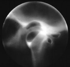
What is virtual endoscopy?
Virtual endoscopy (VE) is a non-invasive technique that amplifies the perception of cross-sectional images, acquired by axial computed tomography (CT), in the 3-D space, providing precise spatial relationships of pathological regions and their surrounding structures. The use of appropriate software and relative algorithms is responsible for the production of virtual reality images that allow endoluminal navigation through a hollow system and simulation of conventional endoscopy. Endoluminal, as well as extraluminal, adjacent structures in all directions can be presented with VE. Body regions inaccessible to or incompatible with conventional endoscopy can also be depicted by VE. In general, VE is capable of providing information on many hollow anatomical structures and has already been used for the exploration of trachea, colon, aorta, brain ventricles, nasal cavity and paranasal sinuses. 54,56–62
How accurate is oculoplethysmography?
All of these procedures are relatively new in the diagnostic field. Oculoplethysmography had a combined accuracy of 74% in detecting normal or abnormal vessels. This is probably due to an inherent problem with the procedure and the interpretation of the results. Also, difficulty was encountered with a good number of patients during placement of the eye-cups. On various occasions this technique failed to detect a totally occluded internal carotid which was probably due to good collateral circulation.
What are the steps of a mcroscopy system?
Macroscopy-based systems can be divided into four steps: (a) preprocessing , where the input image is processed to facilitate the skin lesion segmentation ; (b) segmentation , where the goal is to delimit the skin lesion region in the input image; (c) feature extraction , where the segmented skin lesion is represented by its features; and (d) classification, where the skin lesion is classified as benign or malignant based on the lesion features extracted. Fig. 1 provides an overview of such systems.
How does a gated SPECT imaging test work?
Gated SPECT imaging assists in the recognition of artifacts caused by soft tissue (obesity, breast) and significantly improved the specificity of the test. Gated SPECT imaging should be routinely performed in all laboratories. More recently, methods for attenuation correction have become available and not only enhance specificity through the identification of artifacts but also may detect more extensive areas of ischemia. Attenuation correction is now recommended by nuclear cardiology leaders, but has not been universally accepted due to the high cost of equipment and the lack of reimbursement for this supplemental method.
What is nuclear cardiology?
Nuclear cardiology has experienced explosive growth during the past decade, founded on the solid evidence that this discipline provides the clinician with an accurate assessment of patients with suspected ischemic heart disease.
