The adequate somatosensory stimulus (i.e., the stimulus to which a somatosensory neuron is most sensitive) is either a mechanical force, a temperature change, tissue damage, or a chemical action. The discriminative touch and proprioceptive systems are most sensitive to mechanical force.
What is the somatosensory system?
The somatosensory system is the network of neural structures in the brain and body that produce the perception of touch, as well as temperature, body position ( proprioception ), and pain. [1] It is a subset of the sensory nervous system, which also represents visual, auditory, olfactory, and gustatory stimuli.
What is adequate stimulus in somatosensory system?
The locations of cutaneous (somatosensory) receptors in hairy and non-hairy (glabrous) skin. The Adequate Stimulus. The adequate somatosensory stimulus (i.e., the stimulus to which a somatosensory neuron is most sensitive) is either a mechanical force, a temperature change, tissue damage, or a chemical action.
What are the Somatosensory receptors for pain?
These free nerve endings are considered to be the somatosensory receptors for pain resulting from muscle, tendon, joint, or ligament damage and are not considered to be part of the proprioceptive system. In this chapter, you have learned about somatosensory stimuli and the receptors of three components of the somatosensory systems.
How does a somatosensory neuron respond to touch?
Consequently, a "warm" somatosensory neuron will not respond to cooling of the skin or to a touch stimulus that does not "warm" the skin. The somatosensory receptor and its central connections determine the modality specificity of the neurons forming a somatosensory pathway. Tactile Stimuli.

Which are examples of somatosensory senses?
Somatosensation is the group of sensory modalities that are associated with touch, proprioception, and interoception. These modalities include pressure, vibration, light touch, tickle, itch, temperature, pain, proprioception, and kinesthesia.
What are the 3 somatosensory pathways?
A somatosensory pathway will typically have three neurons: primary, secondary, and tertiary. The cell bodies of the three neurons in a typical somatosensory pathway are located in the dorsal root ganglion, the spinal cord, and the thalamus.
What are the four main somatosensory skin sensations?
The somatosensory system includes the following cutaneous or skin senses: pressure (touch), pain (nociception), vibration and temperature, position sense (proprioception), and body movement (kinesthesis).
What is somatosensory quizlet?
Somatosensory System: This system encodes and makes sense of temperature, touch and body position in space. Glabrous skin: Skin that does not have hair, eg.
What are the two main somatosensory pathways?
The somatosensory system consists of the two main paired pathways that take somatosensory information up to the brain: the medial lemniscal or posterior pathway, and the spinothalamic or anterolateral pathway.
What is meant by somatosensory?
The somatosensory system is the part of the sensory system concerned with the conscious perception of touch, pressure, pain, temperature, position, movement, and vibration, which arise from the muscles, joints, skin, and fascia.
What makes up the somatosensory system?
The somatosensory system consists of primary, secondary, and tertiary neurons. Sensory receptors housed in the dorsal root ganglia project to secondary neurons of the spinal cord that decussate and project to the thalamus or cerebellum.
What are somatosensory receptors?
Somatosensory Receptor(s): a cell or group of cells specialized to detect changes in the environment and trigger impulses in the sensory nervous system. ( OxfordMed) Specialized to respond to a particular physical property, such as "touch," "light," or "temperature." (
What are the five sensory receptors?
The following is a detailed discussion of major sensory receptor types.Receptors of vision. The retinal is the principal molecule of vision in the retina. ... Receptors of hearing. ... Receptors of balance. ... Receptors of taste. ... Receptors of smell. ... Receptors on the skin.
Which body part is most sensitive to somatosensory stimuli quizlet?
Stimulus varies (pressure, temperature, vibration) and the organ affected by somatosensory information is skin.
What is the purpose of the somatosensory system?
The somatosensory system is also known as the somatic senses, touch or tactile perception. Anatomically speaking, the somatosensory system is a network of neurons that help humans recognize objects, discriminate textures, generate sensory-motor feedback and exchange social cues.
How does sensory information reach the somatosensory cortex quizlet?
The posterior column pathway brings sensory information to the post central gyrus of the parietal lobe, which houses the primary somatosensory cortex.
What are the types of sensory pathways?
PathwaysDorsal Columns.Spinothalamic Tracts.Spinocervicothalamic Tracts.Dorsal Spinocerebellar Tract.Cuneo-cerebellar Tract.Ventral Spinocerebellar Tract.
What are the sensory pathways?
Sensory pathways consist of the chain of neurons, from receptor organ to cerebral cortex, that are responsible for the perception of sensations. Somatosensory stimuli activate a chain of neurons starting with the peripheral first-order (1°) afferent and ending in the cerebral cortex (e.g., Figure 4.1).
Which pathways are made up of three neurons?
The dorsal column system (sometimes referred to as the dorsal column–medial lemniscus) and the spinothalamic tract are two major pathways that bring sensory information to the brain (Figure 14.5. 1). The sensory pathways in each of these systems are composed of three successive neurons.
Which are characteristic of somatosensory pathways?
What are characteristic of somatosensory pathways? They are all ascending pathways Each pathway transmits information to different regions of the brain The sensation of an itch and discriminative touch are transmitted in the same pathway.
Where are somatosensory neurons located?
Peripheral Somatosensory Neurons. The cell bodies of the first-order (1°) somatosensory afferent neurons 2 are located in posterior root or cranial root ganglia (i.e., are part of the peripheral nervous system, Figure 2.1). The 1° afferents are pseudounipolar cells. The cell body gives rise to a single process that divides to form a peripheral axon and a central axon. The peripheral axon travels to and ends in the skin, muscle, tendon or joint and the central axon travels to and ends in the central nervous system.
What do somatosensory systems monitor?
The somatosensory systems also monitor the temperature of the body, external objects and environment, and provide information about painful, itchy and tickling stimuli. The sensory information processed by the somatosensory systems travels along different anatomical pathways depending on the information carried.
How is the sensitivity of a somatosensory receptor determined?
As was noted earlier, the sensitivity (modality specificity) of the somatosensory receptor is determined by its location and by the structure of the non-neural tissue surrounding the 1° afferent terminal. The following describes the most commonly observed cutaneous receptors.
What is the modality specificity of the somatosensory system?
Modality Specificity in the Somatosensory System. The somatosensory systems process information about, and represent, several modalities of somatic sensation ( i.e., pain, temperature, touch, proprioception). Each of these modalities can be divided into sub-modalities, as shown in Table 1 (e.g., pain into sharp, pricking, cutting pain; dull, burning pain; and deep aching pain). Discriminative touch is also subdivided into touch, pressure, flutter and vibration. Each of these sensations (i.e., sub-modalities) is represented by neurons that exhibit modality specificity. That is, when a somatosensory neuron is stimulated naturally (e.g., by skin warming) or artificially (e.g., by electrical stimulation of the neuron), the sensation perceived is specific to the information normally processed by the neuron (i.e., warm skin). Consequently, a "warm" somatosensory neuron will not respond to cooling of the skin or to a touch stimulus that does not "warm" the skin. The somatosensory receptor and its central connections determine the modality specificity of the neurons forming a somatosensory pathway.
How are the sensitivity of the receptors to specific stimuli determined?
The sensitivity of the receptors to specific stimuli (e.g., touch verses muscle stretch) is determined by the location of the receptor and by the non-neural tissue surrounding the 1° afferent terminal (Figure 2.6). The locations of cutaneous (somatosensory) receptors in hairy and non-hairy (glabrous) skin.
Which corpuscle is most sensitive to stretch?
The Ruffini corpuscles are oriented with their long axes parallel to the surface of the skin and are most sensitive to skin stretch. Stretching the skin (Figure 2.17) stretches the collagen fibers within the Ruffini corpuscle, which compresses the axon terminals. As the collagen fibers remain stretched and the axon terminals remain compressed during the skin stretch, the Ruffini corpuscle's 1° afferent axon produces a sustained slowly adapting discharge to maintained stimuli.
Which stimulus is most sensitive to mechanical force?
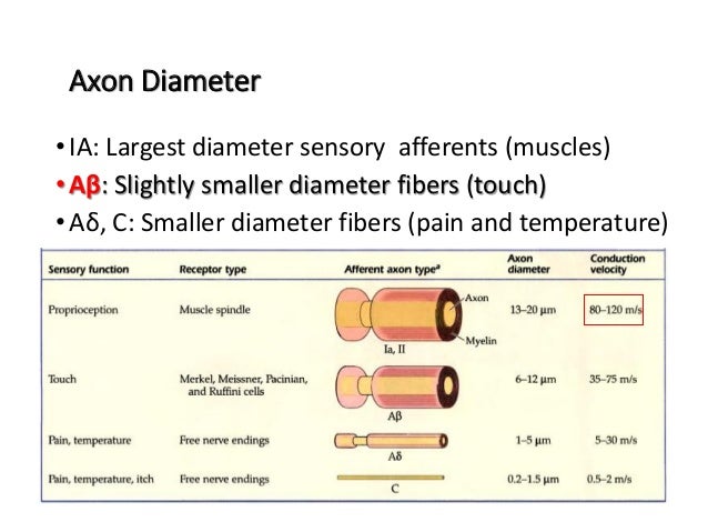
The adequate somatosensory stimulus (i.e., the stimulus to which a somatosensory neuron is most sensitive) is either a mechanical force, a temperature change, tissue damage, or a chemical action. The discriminative touch and proprioceptive systems are most sensitive to mechanical force.
Which spinal cord pathway is responsible for somatosensory stimulation?
The spinal–supraspinal pathways responsible for somatosensory stimulation mainly comprise the posterior spinal cord column pathway and the spinothalamic pathway. Most peripheral thick myelinated afferent fibers activated by the discriminative touch and sense of vibration enter the ipsilateral dorsal column–medial lemniscus tract (posterior column pathway) and emerge into the contralateral spinothalamic pathway. In contrast, the thinly myelinated or unmyelinated afferent fibers activated by pain and temperature are carried up by the contralateral spinothalamic tract to supraspinal levels (spinothalamic pathway). These impulses are further relayed to the thalamus and ultimately sent to the primary somatosensory cortex. In addition, these impulses are also relayed to other brain areas, including the brain stem, periaqueductal gray (PAG), and paraventricular nucleus (PVN) of the hypothalamus, via collateral connections (Hendelman, Humphreys, & Skinner, 2010) ( Fig. 14.1 ).
What is the effect of somatosensory stimulation on CTS?
Somatosensory stimulation activates CTS in 10% to 20% of cases and evokes giant somatosensory evoked spikes (GSES) that, like spontaneous CTS, appear in children with or without seizures and disappear with age.
Which stimulation increases SWS in cats?
during electrical stimulation of the olfactory bulb, which increases SWS in cats.
What changes occur in the auditory system during sleep?
The auditory system single neuronal firing exhibits a variety of changes along the different nuclei and primary cortical loci linked to the sleep–wakefulness cycle in many ways: (a) increasing or decreasing firing on passing to sleep, (b) firing during sleep as during wakefulness, (c) changing the discharge pattern, (d) exhibiting theta rhythm phase-locking, with no auditory neurone stopping its firing on passing to sleep. Furthermore, Edeline et al. (2001) reported changes in the receptive field of cortical auditory neurones.
How are sound and sleep related?
Sleep and sound are closely related. Environmental noises as well as regular, monotonous auditory stimuli, e.g., mother lullaby, are influences impeding or facilitating sleep.
What causes sleep imbalances?
Thus, the induced changes in the waking and sleep networks lead to imbalances not simply explained by passive sleep production but also by introducing sensory sleep–active influences. A diversity of approaches supports this notion:
How long does it take for a bilateral lesions of the vestibular nucleus to clear?
bilateral lesions of some vestibular nuclei that abolish rapid eye movements during PS up to 36 days;
Where are somatosensory receptors located?
Somatosensory Receptors. While receptors for the other senses are localized in compact sense organs (the ears for hearing, the eyes for sight , the nose for smell), receptors for touch and its kindred senses are distributed all over the skin and inside the body. Many of these receptors are essentially nerve endings encapsulated in the cells ...
What is the function of the somatosensory cortex?
Actually, the brain’s work of integration goes a lot further—vision, hearing, smell, and taste meld with somatosensory information to create a multidimensional yet unified perception of what is going on in and around you. Pain pathways facilitate rapid response. While some signals go all the way to the somatosensory cortex, others connect at the thalamus—a kind of switchboard area of the brain—to regions that ready us for action, such as the emotion-regulating limbic system and structures that control heart rate and respiration.
Why is the somatosensory cortex important?
The brain can interpret where sensations are coming from because the somatosensory cortex is organized to reflect the way the body is laid out —a kind of “body map.” The map isn’t scaled to body size, but reflects sensitivity: the hands and face are relatively small physically, but because they are highly sensitive to touch, the parts of the somatosensory cortex that represent them are disproportionately large compared to those devoted to other body parts.
How does touch affect the brain?
There are separate but parallel receptors and nerve pathways for the sensations of temperature, body position and movement, and pain.
What are the mechanical receptors?
Many of these receptors are essentially nerve endings encapsulated in the cells of surrounding skin, muscle, or other tissue that have been modified into structures to convey physical forces (pressure, stretching, motion) to them. Stimulating these mechanical receptors allows the flow of charged particles into the nerve, transforming the physical force
Which part of the brain is responsible for voluntary movements?
The Body in Motion. It is no accident that the somatosensory cortex is located directly adjacent to the motor cortex, which initiates voluntary movements. Both external and internal sensations provide essential information to guide when and how we move.
What is the function of a nociceptor?
Nociceptors are specialized, too: different ones respond to different kinds of tissue injury or distress, to register sharp, dull, or aching pain.
What are the somatosensory receptors?
Somatosensory receptors are distributed throughout the entire body, rather than being concentrated at specialized locations. Different parts of the body will also have different concentrations of each type of receptor. The various types of receptors are able to sense various kinds of stimuli such as pressure against the skin, limb position, distention of the bladder, and body temperature. If a stimulus becomes so strong that it may be harmful, the somatosensory system is also responsible for feeling pain (nociception).
What causes somatosensory impairment?
Conditions such as cerebral palsy, nerve injuryor laceration, stroke, and nervous system disorders for instance spinal cordand braininjury, can result in somatosensory impairments. Symptoms of somatosensory impairments may include, but are not limited to: Disrupted movement coordination.
What is Somatosensation?
Somatosensation is a mixed sensory category, and is mediated, in part, by the somatosensory and posterior parietal cortices. They underlie the ability to identify tactile characteristics of our surroundings, create meaning about sensations, and formulate body actions related to the sensations. These various sensations contribute to the somatic aspects of the body scheme as a basis for interacting with our environments.
Why is it important to be aware of somatosensation deficits?
It is important to be aware of possible somatosensation deficits with your patients, as difficulties in tactile and proprioceptive discrimination can limit a person's spontaneous hand use and the ability to manipulate and grip objects, affecting quality of life and perhaps more importantly, safety .
What is somatosensation in psychology?
Somatosensation is a mixed sensory category, and is mediated, in part, by the somatosensory and posterior parietal cortices. They underlie the ability to identify tactile characteristics of our surroundings, create meaning about sensations, and formulate body actions related to the sensations[1].
What are the five senses?
The body functions and interacts with its surrounding environment through the simultaneous inputs of our five senses; gustation (taste), ocular (vision), olfaction (smell), vestibular (balance) and auditory (hearing), respectively. However, it is often forgotten that we also have a "sixth sense", understood to be our sense of somatosensation. Somatosensation is an overarching sense which includes the sub-modalities of:
What is the function of mechanoreceptors?
1. Mechanoreceptors: Detects mechanical changes or deformations in tissues.
What is somatosensory cortex dysfunction?
Somatosensory Cortex Dysfunction. Damage to the somatosensory cortex can result in mostly mild deficits, and symptoms of damage are dependant upon which area was damaged. Damage to this could result from lesions to one or more areas, sometimes as a result of a stroke.
Why is the primary somatosensory cortex important?
Likewise, the primary somatosensory cortex can help us judge the shapes of objects with our eyes closed and be able to identify objects through touch.
What are the three types of somatosensory neurons?
Somatosensory pathways are typically comprised of three neurons: primary, secondary, and tertiary. The primary neurons are the sensory receptors within the periphery of the somatosensory cortex which are able to detect various stimuli such as touch or temperature. The secondary neurons are located within the spinal cord and brainstem ...
Which cortex is responsible for tactile object recognition?
The secondary somatosensory cortex is believed to be involved in tactile object recognition and memory. It is suggested that whilst the primary area receives peripheral sensory information, it requires the secondary area to store, process, and retain this information.
Which part of the brain receives projections from the thalamus?
The primary somatosensory cortex receives projections from nuclei of the thalamus of the brain. These nuclei receive fibers from the contralateral half of the body, meaning the opposite side of the body from which the area is located in the brain. Overall, the primary somatosensory cortex is responsible for the processing ...
Where is the secondary somatosensory cortex located?
The secondary somatosensory cortex is located adjacent to the primary somatosensory cortex in the upper part of the lateral sulcus, a fissure in the cortex that separates the frontal and parietal lobes from the temporal lobes.
Which part of the brain is responsible for receiving and processing sensory information from across the body?
The somatosensory cortex is a region of the brain which is responsible for receiving and processing sensory information from across the body, such as touch, temperature, and pain. This cortex is located within the which is located in the postcentral gyrus of the parietal lobe, and lies behind the primary motor cortex of the frontal lobe.
What is a somatosensory receptor?
Somatosensory Receptor (s): a cell or group of cells specialized to detect changes in the environment and trigger impulses in the sensory nervous system. (OxfordMed) Specialized to respond to a particular physical property, such as "touch," "light," or "temperature." (Kandel, 447)
Which sensory receptors respond to fluttering sensations?
Pacinian Corpuscles: respond to fluttering sensations. (Kolb, 371) Important sensory receptors involved with the sense of touch, located beneath the skin. When stimulated by pressure, it converts the stimulation into a neural message that is relayed to the brain. (Hockenbury, 102)
What is the haptic receptor?
Haptic Receptor (s): consists of a "dendrite" attached to a hair or to connective tissue or a dendrite encased in a capsule of tissue. Mechanical stimulation of the hair, tissue, or capsule activates special "channels" on the dendrite, which in turn initiate an "action potential." (Kolb, 370)
What is the thermoreceptor?
Thermoreceptor (s): sensitive to warmth, cold, excessive heat, or excessive cold. (Patestas, 139) Cellular receptors which mediate the sense of temperature. Thermoreceptors in vertebrates are mostly located under the skin. In mammals there are separate types of thermoreceptors for cold and for warmth and “nociceptors” which detect cold or heat extreme enough to cause pain. (MeSH)
What is the receptor that responds to the stimuli responsible for the sensation of pain?
Nociceptor (s): a receptor that responds to the stimuli responsible for the sensation of pain. (OxfordMed) Detects piercing pain, heat pain, chemical pain, "joint" pain, deep tissue pain, tickle, and itch. (Blakeslee, 8) Found in the skin, “muscles,” and internal "organs." (Hockenbury, 102) Rapidly adapting receptors that are sensitive to noxious or painful stimuli. Located at the terminations of lightly "myelinated" "free nerve endings" or unmyelinated fibers. (Patestas, 139)
What is the function of an exteroceptor?
Exteroceptor (s): specialized to detect sensory information from the external environment (such as "visual," "olfactory," "gustatory," "auditory," and tactile stimuli). Located close to the body surface. Sensitive to touch (light stimulation of the skin surface), "pressure" (in the deep layers of the skin, or deeper parts of the body), temperature, "pain," and "vibration." (Patestas, 139)
Which sensory information leads to conscious awareness of the stimulus?
B) Sensory information that leads to conscious awareness of the stimulus is called transduction.
What is sensory receptor?
E) Some sensory receptors are modifications of the peripheral endings of efferent neurons. Click card to see definition 👆. Tap card to see definition 👆. C) The term "sensory unit" refers to a group of receptors that receive a particular stimulus and the afferent neuron associated with those receptors.
What is a sensory unit?
C) The term "sensory unit" refers to a group of receptors that receive a particular stimulus and the afferent neuron associated with those receptors.
What does "adequate stimulus" mean?
D) The term "adequate stimulus" means that a stimulus is strong enough to be detected.
Which part of the brain can activate heat sensing?
D) Chemoreceptors can activate heat sensing parts of the brain.
Where are both sensations grouped together?
D) both sensations are grouped together in the somatosensory cortex.