How do the diaphragm and phrenic nerve contract?
Diaphragm (innervated by phrenic nerve) and external intercostal muscles (innervated by segmental intercostal nerves) contract, creating a negative pressure around the lung. Air rushes into the lungs in order to equalise the pressure.
How are the external intercostal muscles innervated?
The external intercostal muscles receive innervation from intercostal nerves of corresponding intercostal spaces, which are the ventral rami of thoracic spinal nerves. The external intercostal muscles have an extensive vascular supply. They are supplied by the muscular branches of the anterior and posterior intercostal arteries .
What is the movement of the diaphragm during breathing?
Mechanics of breathing When we inhale the intercostal muscles (between the ribs) and diaphragm contract to expand the chest cavity. The diaphragm flattens and moves downwards and the intercostal muscles move the rib cage upwards and out.
What is the movement of the rib cage and diaphragm?
The movement of the rib cage is controlled by the intercostal muscles and the diaphragm is a skeletal muscle. During inspiration, the diaphragm and the external intercostal muscles contract causing an increase in the thoracic cavity volume. The contraction of the diaphragm accounts for approximately 75% of the air movement during normal breathing.
Which nerve controls the diaphragm and intercostal muscles?
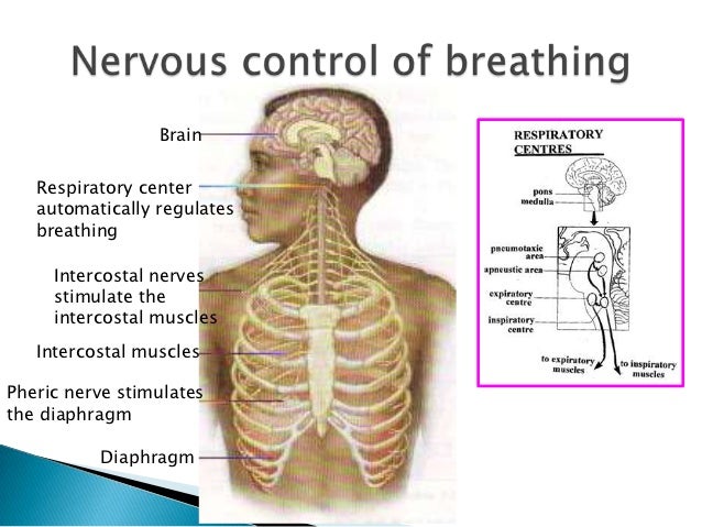
The phrenic nerves provide motor innervation to the diaphragm and work in conjunction with secondary respiratory muscles (trapezius, pectoralis major, pectoralis minor, sternocleidomastoid, and intercostals) to allow respiration.
What nerve stimulates the intercostal muscles to contract?
Intercostal muscles are stimulated to contract by the intercostals nerves. The external intercostals muscles and the diaphragm contract simultaneously during inspiration, resulting in the enlargement of the thoracic cavity in all directions.
What nerves stimulate the diaphragm and cause it to contract?
Your phrenic nerve plays a critical role in your respiratory system to aid breathing. It's the only nerve in your nervous system that provides motor (movement) function to your diaphragm. It sends signals that cause your diaphragm to expand and contract. These movements allow your lungs to inhale and exhale air.
When the diaphragm and external intercostal muscles contract the?
During inspiration the diaphragm and the external intercostal muscles contract, causing an increase in the thoracic cavity volume. The contraction of the diaphragm accounts for approximately 75% of the air movement during normal breathing.
What causes diaphragm to contract?
Upon inhalation, the diaphragm contracts and flattens and the chest cavity enlarges. This contraction creates a vacuum, which pulls air into the lungs. Upon exhalation, the diaphragm relaxes and returns to its domelike shape, and air is forced out of the lungs.
What nerve Innervates the external intercostal muscles?
intercostal nervesExternal intercostal muscles They are innervated by the anterior rami of spinal nerves T1-T11, i.e. the intercostal nerves of the corresponding intercostal space.
What is the nerve that controls your diaphragm?
The phrenic nerve controls function of the diaphragm muscle - the primary muscle involved in breathing. It tells the diaphragm when to contract, allowing the chest cavity to expand and triggering the inhalation of air into the lungs.
How does the phrenic nerve stimulate the diaphragm?
The phrenic nerves send a signal to the diaphragms stimulating them to breathe. People who have problems with the brain or spinal cord at times do not send the signals well to breathe. Diaphragm pacing can use the phrenic nerves to send the signals to a person's diaphragm muscles to contract and take a breath in.
What is the nerve supply of the diaphragm?
The phrenic nerve provides the primary motor supply to the diaphragm, the major respiratory muscle. It passes motor information to the diaphragm and receives sensory information from it. There are two phrenic nerves, a left and a right one. Image 1: The Phrenic nerve, showing course to diaphragm.
When the diaphragm and external intercostal muscles contract what happens quizlet?
contraction of both the diaphragm (the diaphragm flattens) and the external intercostals (pulls the ribs up and out) will increase the volume of the thoracic cavity. This will cause air to move into the lungs (inspiration). You just studied 50 terms!
When the diaphragm and external intercostals muscles contract Which of the following actions does not occur?
carry air toward each lung. When the diaphragm and external intercostal muscles contract, which of the following actions does NOT occur? A. the intrapulmonic pressure decreases.
What happens when the external intercostal muscles contract?
During inhalation, the diaphragm is relaxed, allowing the lungs to expand. The innermost intercostal muscles relax, while the external intercostal muscles contract, causing the chest cavity to expand. This expansion allows the lungs to fill with air, due to the negative pressure created by the extra space.
What is the role of the vagus nerve?
The vagus nerve is responsible for the regulation of internal organ functions, such as digestion, heart rate, and respiratory rate, as well as vasomotor activity, and certain reflex actions, such as coughing, sneezing, swallowing, and vomiting (17).
Are intercostal nerves sympathetic?
Upon arising, each intercostal nerve is connected to its corresponding sympathetic ganglion (of the sympathetic trunk) by pre- and postganglionic branches (rami communicantes).
Where is the intercostal brachial nerve?
The intercostobrachial nerve (ICBN) is a cutaneous nerve that provides sensation to the lateral chest, medial aspect of the upper arm, and the axilla.
What does the subcostal nerve do?
Subcostal nerve (T12). The subcostal nerve provides sensory innervation to the region under the umbilicus and also provides motor innervation to the pyramidalis and quadratus lumborum muscles.
Which cells produce mucus that traps dust particles and other debris?
d. Goblet cells produce mucus that traps dust particles and other debris
What cell creates a sweeping motion that propels mucus toward the throat?
a. Goblet cells create a sweeping motion that propels mucus toward the throat.
Which nerves innervate the external intercostal muscles?
The external intercostal muscles receive innervation from intercostal nerves of corresponding intercostal spaces, which are the anterior rami of thoracic spinal nerves .
Where are the external intercostal muscles located?
The external intercostal muscles are located superficially to the internal intercostal muscles, separated from them by a thin fascia. Superficial to the external intercostal muscles lie the pectoralis major and minor, serratus anterior and the upper part of the rectus abdominis muscle.
What is the most superficial set of muscles that occupy the 11 intercostal spaces?
External intercostal muscles (Musculi intercostales externi) The external intercostal muscles are the most superficial set of muscles that occupy the 11 intercostal spaces. Their name is derived from their spatial relationship with other intercostal muscles, since they are found superficially to the internal and innermost intercostals.
How many pairs of muscles are there in the intercostal muscle?
The external intercostal muscles consist of 11 pairs of muscles. Every external intercostal originates from the sharp inferior costal border of one rib. The fibers of each muscle course inferomedially and insert along the outer lip of the superior border of the immediate rib below.
What muscles are involved in forced inspiration?
Being one of the accessory respiratory muscles, the external intercostals elevate ribs during forced inspiration. This increases the transverse and anteroposterior diameter of the lungs, which in turn decreases the intrapleural pressure. This process expands the lungs and facilitates the entry of air into them.
Which artery supplies the muscles of the upper 6 intercostal spaces?
They are supplied by the muscular branches of the anterior and posterior intercostal arteries . The anterior intercostal arteries supplying the muscles of the upper 6 intercostal spaces arise from the internal thoracic artery directly, whereas the lower 5 arise from its branch; the musculophrenic artery.
Which muscles are associated with the thoracic wall?
The intercostal muscles, together with serratus posterior, levatores costarum, subcostal, and transversus tho racis muscles comprise the muscles of the thoracic wall . The external intercostal muscles elevate the ribs during forced inhalation and are functionally classified as the accessory respiratory musculature.
Which nerves stimulate the inspiratory muscles?
During inspiration (breathing in), nerve impulses are sent via the phrenic and intercostal nerves which stimulate the inspiratory muscles, the external intercostal and diaphragm, causing them to contract, this stimulation lasts for approximately two seconds, after which, the inspiratory muscles relax and expiration occurs.
Which muscles are stimulated to lift the ribs and sternum further?
In addition to the external intercostal muscles and diaphragm, the sternocleidomastoid, scalene and pectoralis minor are stimulated to lift the ribs and sternum further, increase the volume of the thoracic cavity, allowing an increase in the depth of breathing. Breathing frequency is also increased during exercise due to ...
What is the function of the medulla oblongata during exercise?
During exercise, the proprioceptors detect a rise in movement and therefore oxygen demand, and send a nerve impulse to the medulla oblongata, which stimulates the sympathetic nervous system to increase breathing rate and depth. During exercise, the depth of breathing is increased through the stimulation of three additional muscles.
What muscles move the ribs?
Mechanics of breathing. When we inhale the intercostal muscles (between the ribs) and diaphragm contract to expand the chest cavity. The diaphragm flattens and moves downwards and the intercostal muscles move the rib cage upwards and out. This increase in size decreases the internal air pressure and so air from the outside ...
How does breathing rate work?
Breathing rate is all controlled by chemoreceptors within the main arteries which monitor the levels of Oxygen and Carbon Dioxide within the blood. If oxygen saturation falls, ventilation accelerates to increase the volume of Oxygen inspired. If levels of Carbon Dioxide increase a substance known as carbonic acid is released into ...
What do chemoreceptors do during exercise?
During exercise, the chemoreceptors detect a rise in carbon dioxide, a by-product of increased respiration, and a reduction in oxygen. The chemoreceptors, send a nerve impulse to the medulla oblongata, which subsequently stimulates the sympathetic nervous system (the pedals) to increase breathing rate and depth.
Where is the rate of inhalation and exhalation controlled?
The rate at which we inhale and exhale is controlled by the respiratory centre, within the Medulla Oblongata in the brain. Inspiration occurs due to increased firing of inspiratory nerves and so the increased recruitment of motor units within the intercostals and diaphragm. Exhalation occurs due to a sudden stop in impulses along ...
Which pathway is responsible for the transmission of signals back to the medulla?
The efferent neural pathway then follows, with relevant signals transmitted back from the cerebral cortex and medulla via the vagus and superior laryngeal nerves to the glottis, external intercostals, diaphragm, and other major inspiratory and expiratory muscles. The mechanism of a cough is as follows:
What is the auricular branch of the vagus nerve that causes coughing?
Stimulation of the auricular branch of the vagus nerve supplying the ear may also elicit a cough. This ear-cough reflex is also known as Arnold's reflex. Weakness of the respiratory muscles, tracheostomy, or vocal cord pathology (including paralysis or anesthesia) may prevent effective clearing of the airways.
What is the cough reflex?
Jump to navigation Jump to search. The cough reflex has both sensory ( afferent) mainly via the vagus nerve and motor ( efferent) components. Pulmonary irritant receptors (cough receptors) in the epithelium of the respiratory tract are sensitive to both mechanical and chemical stimuli.
What is the mechanism of coughing?
The mechanism of a cough is as follows: Diaphragm (innervated by phrenic nerve) and external intercostal muscles (innervated by segmental intercostal nerves) contract , creating a negative pressure around the lung. Air rushes into the lungs in order to equalise the pressure.
Where do the receptors travel?
When triggered, impulses travel via the internal laryngeal nerve, a branch of the superior laryngeal nerve which stems from the vagus nerve (CN X), to the medulla of the brain. This is the afferent neural pathway.
Where are the receptors located in the respiratory system?
The cough receptors or rapidly adapting irritant receptors are located mainly on the posterior wall of the trachea, pharynx, and at the carina of trachea, the point where the trachea branches into the main bronchi. The receptors are less abundant in the distal airways, and absent beyond the respiratory bronchioles.
Which organs are sensitive to light?
The bronchi and trachea are so sensitive to light touch that slight amounts of foreign matter or other causes of irritation initiate the cough reflex. The larynx and carina are especially sensitive. Terminal bronchioles and even the alveoli are sensitive to chemical stimuli such as sulfur dioxide gas or chlorine gas.
Which organs pass through the diaphragm?
The diaphragm separates the thoracic and abdominal cavities. Several large organs (i.e., the esophagus, aorta, and vena cava) pass through the diaphragm. The skeletal muscle fibers of this dome-shaped muscle pass radially from the lower ribs, coastal cartilages, and vertebrae and insert on a thick central tendon.
Where are the external intercostals?
The external intercostals are the most posterior of the two, extending from the vertebrae to the costal cartilages, whereas the internal intercostals extend from the angles to the ribs to the sternum. Sign in to download full-size image. Figure 8. Intercostal muscles.
What are the respiratory muscles?
The respiratory section includes the lungs and associated muscle groups. Two major muscle groups promote inhalation (breathing in). One of these major muscle groups is the diaphragm, which lies at the base of the rib cage. Another major muscle group is the external intercostals. This group of muscles lifts and expands the rib cage, allowing for exhalation (breathing out). The internal intercostals pull down on the rib cage and push air out of the lungs. The internal intercostals are the most important respiratory muscles for normal speech and singing, for they are the muscles that propel air out through the mouth and nose. In general, the greater the pressure of the escaping air, the louder one’s voice.
What happens during the inhalation-exhalation cycle of speech?
After the lungs have filled with air, the rib cage recoils elastically from a state of complete or near-complete expansion. During this period, the external intercostals (which promote inhalation) remain active until receptors within them indicate that the lungs have been fully filled. After the elastic recoil process has continued for a while and the lung volume has decreased to a critical value (but one well above complete emptying of the lung), the external intercostals shut off, based on feedback from stretch receptors within them. This event signals the internal intercostals to contract, which allows for active exhalation and production of the next utterance. Because the internal intercostals do not contract until the lungs have begun to contract, they take advantage of the inertia of lung contraction.
How does air move in the lungs?
We have briefly discussed in a previous section that air movement into the lungs is caused by changes in the lung volume. Following Boyle’s law, the gas pressure must change inversely with the change in volume (assuming that the temperature is constant and there is no change in the amount of gas within the system). Here we will briefly discuss some of the mechanics of breathing that bring about the changes in volume. Recall that we have stated that during inspiration, the rib cage moves outward and upward and the diaphragm moves downward. The movement of the rib cage is controlled by the intercostal muscles and the diaphragm is a skeletal muscle. During inspiration, the diaphragm and the external intercostal muscles contract causing an increase in the thoracic cavity volume. The contraction of the diaphragm accounts for approximately 75% of the air movement during normal breathing. During strenuous activity, the increase in the speed of rib movement and the increase in rib displacement are controlled by the pectoralis minor, the scalene, and the sternocleidomastoid muscles, which completely accounts for the increased volume of the thoracic cavity. During expiration, the contraction of the internal intercostal muscles brings the rib cage back to its normal position and the abdominal muscles contract to assist the internal intercostal muscles and to force the diaphragm upward.
What are the muscles of the thoracic wall?
The most important of these muscles are the external intercostals, internal intercostals, serratus posterior, traversus thoracis, and diaphragm. The intercostal muscles fill spaces between the ribs (Figure 8 ).
Which muscles are involved in the inhalation of air?
This action expands the thoracic cavity and draws air into the lungs. Portions of both intercostal muscles and the superior serratus posterior muscles will act to pull the ribs upward and outward, also expanding the thoracic cavity during inhalation.
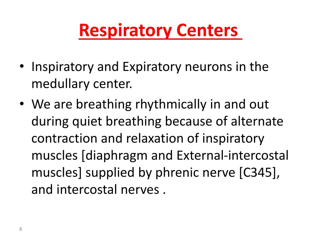
Mechanics of Breathing
Breathing Rate
- The rate at which we inhale and exhale is controlled by the respiratory centre, within the Medulla Oblongata in the brain. Inspiration occurs due to increased firing of inspiratory nerves and so the increased recruitment of motor units within the intercostals and diaphragm. Exhalation occurs due to a sudden stop in impulses along the inspiratory nerves. Our lungs are prevented from exc…
Regulation of Breathing
- Respiration is controlled by the autonomic nervous system, which enables us to alter our breathing without thinking about it. The autonomic nervous system consists of two branches, the sympathetic nervous system (the pedals) and the parasympathetic nervous system (the breaks). At rest, we inspire approximately 500 ml of air per breath and on average we breathe 12-15 time…
Regulation of Breathing at Rest
- Medulla oblongata controls breathing
- Phrenic and intercostal nerves stimulate the external intercostal muscles and diagram
- Stimulation causes these muscles to contract
- Contraction of these muscles results in inspiration