which of the following structures is contained within the mediastinum
by Tevin Sporer
Published 3 years ago
Updated 2 years ago
The mediastinum is a division of the thoracic cavity
thoracic cavity
thoracic cavity, also called chest cavity, the second largest hollow space of the body. It is enclosed by the ribs, the vertebral column, and the sternum, or breastbone, and is separated from the abdominal cavity (the body's largest hollow space) by a muscular and membranous partition, the diaphragm.
; it contains the heart, thymus gland, portions of the esophagus and trachea, and other structures. For clinical purposes it is traditionally divided into the anterior, middle, posterior, and superior regions.
What structures are contained within the mediastinum?
The mediastinum houses many vital structures including the heart, great vessels, trachea, and essential nerves. It also functions as a protected pathway for structures traversing from the neck, superiorly, and into the abdomen, inferiorly.
What structures are contained within the mediastinum quizlet?
The esophagus, heart, thymus gland, and trachea are found in the mediastinum. The lungs are not found in the mediastinum.
What are the four important structures located in the mediastinum?
The main mediastinal contents are the heart, esophagus, trachea, thoracic nerves and systemic blood vessels.
Which of the following is not contained within the mediastinum?
Answer and Explanation: The organ which is not present in the mediastinum is the A. lungs.
What cavity lies within the mediastinum?
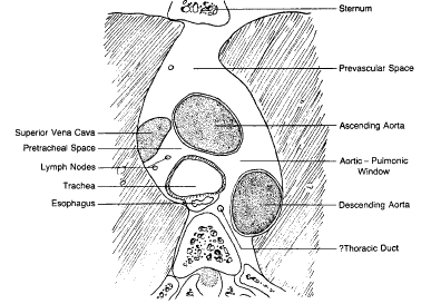
the thoracic cavityThe mediastinum is the central (midline) portion of the thoracic cavity. It contains all of the extrapulmonary structures of the cranial and caudal thoracic cavity, including esophagus, trachea, thymus, lymph nodes, nerves, and major blood vessels.
Where does the mediastinum lie within the chest quizlet?
it is the thoracic region between the two lungs, between the sternum and vertebrae.
Is the pericardium in the mediastinum?
The middle mediastinum contains the heart covered by the pericardium. The serous pericardium contains the pericardium cavity, which facilitates the free movement of the heart. Therefore, the mediastinum and pericardial cavity are two compartments that protecting organs in the human body.
Where is the mediastinum located and what is its function?
The mediastinum is an important region of the body located between the lungs. Structures that lie in this region include the heart, the esophagus, the trachea, and large blood vessels including the aorta. The mediastinum is also home to lymph nodes.
Is the lungs in the mediastinum?
The area between the lungs. The organs in this area include the heart and its large blood vessels, the trachea, the esophagus, the thymus, and lymph nodes but not the lungs.
Are the ribs in the mediastinum?
The posterior mediastinum includes the descending aorta, adjacent osseous structures (the spine and ribs), nerves, roots, spinal cord, esophagus, azygous and hemiazygous veins, and some lymph nodes.
Does the mediastinum contain the lungs?
mediastinum, the anatomic region located between the lungs that contains all the principal tissues and organs of the chest except the lungs.
Does the mediastinum contains the pericardial cavity?
The Pericardial and Pleural Cavities along with the Mediastinum make up the Thoracic Cavity. The boundaries of the Thoracic Cavity are the Ribs (and Sternum), Vertebral Column, and the Diaphragm. The Diaphragm seperates the Thoracic Cavity from the Abdominal Cavity.
What does the mediastinum separate?
The mediastinum completely separates the right and left pleural cavities. As in humans and other mammals the esophagus lies dorsal (posterior) to the trachea and its bifurcation and to the pericardium and ventral (anterior) to the thoracic (descending) aorta (Figure 4.8D.).
What specific portion of the mediastinum contains the heart?
Middle: The middle mediastinum is the largest portion, and contains the heart, blood vessels including those that travel from the lungs to the heart, and lymph nodes.
35 hours ago
· X-rays travelling through lung tissue cast less of a shadow than those passing through the soft tissues of the mediastinum or abdomen. Consolidation. In the case of pneumonia, air within the alveoli is replaced by inflammatory exudate and pus. Affected alveoli become as dense as soft tissue structures such as the heart. In the lobes which abut ...
31 hours ago
The following is a list of some of the structures of the respiratory tree: secondary bronchi tertiary bronchi bronchioles primary bronchi The order in which air passes through these structures beginning at the trachea is A) 4, 1, 2, 3. B) 3, 2, 1, 4. C) 2, 3, 1, 4. D) 4, 1, 3, 2. E) 1, 2, 3, 4.
26 hours ago
Circulation of blood within an organ or tissue in adequate amounts to meet the cells' oxygen, nutritional, and waste-removal needs is termed _____. Select one: A. coagulation B. perfusion C. hemorrhage D. hypoperfusion. B. The severity of bleeding should be based on all of the following findings, EXCEPT: Select one: A. clinical signs and symptoms. B. poor general appearance. C. …
16 hours ago
Also examine the mediastinum. Its borders should be clear, although some haziness may be present at the angle between the heart and diaphragm. 1,8 If the mediastinum appears enlarged, consider disorders that could cause this, such as an aortic aneurysm. 5. D: Diaphragm. The diaphragm is dome-shaped and has the same density as water. The right ...
21 hours ago
The outside of the foreskin is a continuation of the skin on the shaft of the penis, but the inner foreskin is a mucous membrane like the inside of the eyelid or the mouth. The mucocutaneous zone occurs where the outer and inner foreskin meet. Like the eyelid, the foreskin is free to move after it separates from the glans, which usually occurs before or during puberty.
12 hours ago
A chest radiograph, called a chest X-ray (CXR), or chest film, is a projection radiograph of the chest used to diagnose conditions affecting the chest, its contents, and nearby structures. Chest radiographs are the most common film taken in medicine. Like all methods of radiography, chest radiography employs ionizing radiation in the form of X-rays to generate images of the chest.
17 hours ago
· However, when describing structures within the skull, a dorsal structure is closer to the top of the skull, and a ventral structure is closer to the base of the skull. Other Directional Terms: Ipsilateral means on the same side— e.g. left arm is ipsilateral (on the same side) to the left leg. Intermediate – in between—your heart is intermediate to your lungs. Visceral – may be …
28 hours ago
· Normally, it enhances avidly following the administration of iodinated contrast. Ectopic thyroid tissue may be detected in the tongue near the foramen cecum (90 %) and along the midline between the thyroid isthmus and posterior tongue, lateral neck, mediastinum, and oral cavity. The most frequent location is the base of the tongue (Figs. 16 ...
21 hours ago
· Structure and function of a ganglion Ganglia are oval in structure and contain neuronal cell bodies (somata), satellite cells (a type of glial cell), and a protective connective tissue layer.Autonomic and sensory ganglia are histologically similar, with the former containing multipolar neurons, and the latter usually containing unipolar or pseudo unipolar neurons.