Which strokes cause homonymous hemianopia?
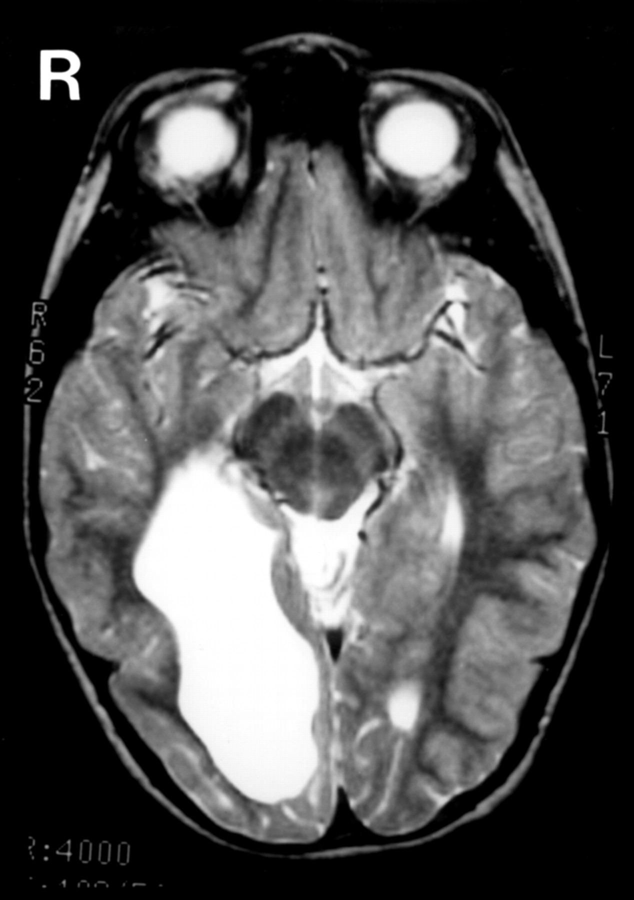
3 Homonymous hemianopia is a loss of the right or left halves of the visual field of both eyes (Figure 1a, 1b) and usually occurs as a result of a middle cerebral or posterior cerebral artery stroke affecting either the optic radiation or visual cortex of the occipital lobe (Figure 2).
Where does homonymous hemianopia occur?
In homonymous hemianopsia, an injury to the left part of the brain results in the loss of the right half of the visual world of each eye. An injury to the right part of the brain produces loss of the left side of the visual world of each eye.Apr 26, 2021
What causes left homonymous hemianopsia?
Homonymous hemianopsia (or homonymous hemianopia, HH) is a field loss deficit in the same halves of the visual field of each eye. This condition most commonly results from stroke for adults, or tumors/lesions for patients under the age of 18.Jul 26, 2021
Where is the lesion in left homonymous hemianopia?
Left Homonymous Hemianopia: This results from lesions to the optic tract in route towards the lateral geniculate body of the thalamus (location 3) as well as lesions right after the radiating fibers leave the lateral geniculate body (location 5). These lesions are often caused by strokes or neoplasms.
Why is macula spared in homonymous hemianopia?
Causes. The favored explanation for why the center visual field is preserved after large hemispheric lesions is that the macular regions of the cortex have a double vascular supply from the middle cerebral artery (MCA) and the posterior cerebral artery (PCA).
What is a homonymous hemianopia?
Introduction. Homonymous hemianopia (HH) involves vision loss on the same side of the visual field in both eyes. This type of visual field loss is indicative of a lesion involving the visual pathway posterior to the chiasm.Sep 22, 2014
What is crossed homonymous hemianopia?
Crossed-quadrant homonymous hemianopsia is the homonymous loss of two opposite quadrants of the visual field which is a rare occurrence. Cases of crossed-quadrant homonymous hemianopsia are rare. Visual loss may be sudden or gradual.Dec 5, 2021
What is incongruous homonymous hemianopia?
Summary. Optic disorder in which the visual field defects in both eyes are completely symmetric in extent and intensity are defined as Congruous homonymous hemianopia.
What causes homonymous hemianopia?
Stroke is the most common cause of homonymous hemianopia (HH) in adults, followed by trauma and tumors. Associated signs and symptoms, as well as visual field characteristics such as location and congruity, can help determine the location of the causative brain lesion. HH can have a significant effect on quality of life, ...
Which lesions are more likely to cause inferior visual field defects with sloping borders superiorly?
In contrast, parietal lesions are more likely to cause inferior visual field defects with sloping borders superiorly. Lesions isolated to the upper, cuneus gyrus or the lower, lingual gyrus result in an inferior or superior quadrantanopia, respectively (Figure 1D).
What is HH in medical terms?
Homonymous hemianopia (HH) involves vision loss on the same side of the visual field in both eyes. This type of visual field loss is indicative of a lesion involving the visual pathway posterior to the chiasm.
What is the difference between a congruous and incongruous visual field defect?
A congruous visual field defect is identical between the two eyes, whereas an incongruous defect differs in appearance between the eyes. For lesions behind the LGN, visual field defects are generally more congruous if the lesion is located more posteriorly along the visual pathway. However, exceptions do occur.
What is the most common cause of HH?
The potential causes of HH are dependent on the age of the patient. The most common cause of HH in adults is stroke. Approximately 8%–10% of stroke patients have permanent HH, and 52%–70% of hemianopias are caused by stroke. 1,2As the population ages and stroke patients live longer, the incidence of stroke and resultant HH is likely to increase.3.
What is the treatment for visual deficits?
Depending on the patient needs, treatment of visual deficits can include prismatic correction to expand the remaining visual field, compensatory training to improve visual search abilities, and vision restoration therapy to improve the vision itself. One type of therapy does not preclude other intervention methods.
What is HH in a blind hemifield?
Damage to the occipital lobe usually does not produce other neurologic manifestations. Some patients may experience photopsias or other hallucinations in the blind hemifield.
What is a homonymous hemianopia?
Homonymous hemianopsia (or homonymous hemianopia, HH) is a field loss deficit in the same halves of the visual field of each eye. This condition most commonly results from stroke for adults, or tumors/lesions for patients under the age of 18. [1] Often, the cause of HH is located at the occipital lobe, followed by an injury to the optic radiations or optic tract. [1] HH can also be characterized as contralateral hemianopsia (unilateral involvement at the optic tract, lateral geniculate nucleus, optic radiations, or occipital cortex opposite to the side of field loss) in contrast to bitemporal hemianopsia (involvement at the optic chiasm).
What are the most common homonymous hemianopsia deficits?
The most common homonymous hemianopsia deficits are from occipital lobe lesions. These deficits typically present without other associated neurologic symptoms. However, the patterns may vary. Often these lesions include macular sparing as a result of the dual blood supply and bilateral macular representation at the occipital cortex. At the occipital pole, most posteriorly, homonymous scotomas are produced. Most anteriorly, temporal crescent loss and other similar peripheral vision patterns occur. If the lesion extends anteriorly enough to involve the left corpus callosum, patients may present with alexia without agraphia (if the lesion spares the angular gyrus). Bilateral occipital lobe lesions will produce bilateral homonymous hemianopsia of various types. Most notably is Anton’s syndrome involving a complete bilateral homonymous hemianopsia (cortical blindness) with which the patient also experiences anosognosia, being unaware of their blindness. With bilateral occipital lesions, it is possible that visual acuity may be impacted. [1] [19] [20]
What are the risk factors for homonymous hemianopsia?
Vascular insult being most frequent, patients should be aware of their possible stroke risk. Atherosclerosis, hypertension, diabetes, smoking history, obesity, heavy alcohol intake, and previous vascular injury are all factors. [28]
What is a partial quadrantanopia?
Homonymous quadrantanopia, often partial but may be complete, is a visual field deficit with a superior or inferior component in addition to the nasal or temporal involvement. This presents itself as a loss of ¼ of the visual field. Most often, damage to the superior or inferior banks of the primary visual cortex (e.g., infarct) will present as a bilateral quadrantanopia. Quadrantanopia may also result from unilateral superior or inferior optic radiation involvement. For instance, right-sided Meyer loop (inferior radiation) damage would cause a loss of vision in the superior-left quadrant bilaterally. [13]
What is visual field confrontation test?
Visual field confrontation test is the most commonly used screening tool to assess for visual field defects. Typically, the patient will cover one eye and be asked to look at a stationary object (the wall or instructor’s finger) while noting the number of fingers visible in their periphery. These tests, however, are instructor dependent and have low sensitivity. Another option is the automated or manual (Goldmann, tangent screen) perimetry test. This test provides information not only on the field loss but also on the size and form of the deficit. [21]
Is hemianopsia complete or partial?
Corresponding projections to the retinal ganglion cells of the visual fields can be vertically divided into temporal and nasal halves. With respect to homonymous hemianopsia, the field deficit can be described as complete or partial/incomplete. The involvement of the entire half is complete hemianopsia, whereas any other loss is partial.
Can retrochiasmal visual pathway cause hemianopsia?
Any injury to the retro chiasmal visual pathway may result in homonymous hemianopsia. As a result, in addition to the various derivations of cancer, other diseases exist, which may cause such a visual field deficit. [25]
