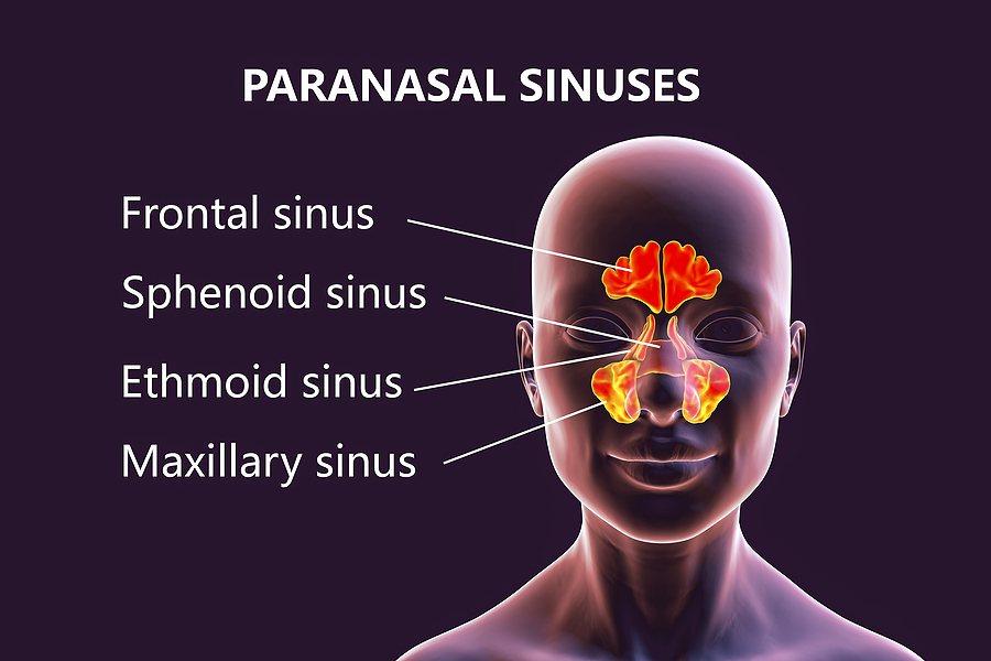
Paranasal sinuses. Paranasal sinus disease is common and on occasion can become life-threatening if not treated in a timely fashion. At birth the maxillary sinuses and ethmoid air cells are present but hypoplastic. The sphenoid sinus develops around 4 years of age secondary to pneumatization of the sphenoid bone.
What is the development of paranasal sinuses?
Anatomy of the Paranasal Sinuses. Development. The maxillary and ethmoid sinuses are present at birth, starting to form around the 3rd or 4th month of gestational development [10]. They further develop over the first few years of life [11].
Do all mammals have paranasal sinuses?
All mammals have paranasal sinuses, although not all mammals have four. Paranasal sinuses in dogs number just three – the frontal and sphenoidal sinus, and the maxillary recess. Rabbits do not have frontal or ethmoidal sinuses but a dorsal conchal sinus, maxillary sinus, and sphenoidal sinus. What Are the Functions of Paranasal Sinuses?
Is paranasal sinus disease life threatening?
Paranasal sinus disease is common and on occasion can become life-threatening if not treated in a timely fashion. At birth the maxillary sinuses and ethmoid air cells are present but hypoplastic.
When do sinuses develop in the human body?
The maxillary and ethmoid sinuses are present at birth, starting to form around the 3rd or 4th month of gestational development [10]. They further develop over the first few years of life [11]. Rudimentary sphenoid sinuses are there at birth, forming (pneumatizing) completely by the age of 5 years [6].

Which sinus is formed after birth?
Ethmoid sinus The ethmoid sinuses arise in the ethmoid bone, forming several distinct air cells between the eyes. They are a collection of fluid-filled cells at birth that grow and pneumatize until the age of 12.
Are all sinuses present at birth?
Consistent with other studies, all the paranasal sinuses were present at birth, but most were undeveloped. At birth, the sinuses—from largest to smallest—are ethmoid, maxillary, sphenoid, and frontal.
Which sinuses are Pneumatized at birth?
Results: The ethmoidal sinuses was the first pneumatized in 100% (46/46) of newborn children. And 45.7% (21/46) of maxillary sinuses showed pneumatization during the first month of life and 97.8% (45/46) were pneumatized at 7 - 12 months. The pneumatized sphenoid sinuses was first identified as early as 4 months.
Which sinus group is well developed and aerated at birth?
At birth, only the maxillary sinus and the ethmoid sinus are developed but not yet pneumatized; only by the age of seven they are fully aerated. The sphenoid sinus appears at the age of three, and the frontal sinuses first appear at the age of six, and fully develop during adulthood.
Is the maxillary sinus rudimentary at birth?
At birth, it is a rudimentary aerated or fluid-filled slit orientated longest in the anteroposterior dimension with a volume of 60–80 mm3, situated inferomedial to the orbit. Partial or complete opacification of the maxillary sinus in the first few years of life is normal.
Are sphenoid sinuses full size at birth?
The sphenoidal sinuses are minute at birth; their main development takes place after puberty.
At what age does the sphenoid sinus develop?
This sinus does not develop until around 7 years of age. Sphenoid sinus. Located deep in the face, behind the nose.
What sinuses are present in rudimentary form at birth and are fully formed in adolescence?
sphenoid sinuses are present in a rudimentary form at birth; they enlarge appreciably after the 8th year and become fully formed during adolescence.
What are the 4 paranasal sinuses?
Paranasal sinuses are named after the bones that contain them: frontal (the lower forehead), maxillary (cheekbones), ethmoid (beside the upper nose), and sphenoid (behind the nose).
At what age does the frontal sphenoid and ethmoid sinuses develop?
The maxillary and ethmoid sinuses are present at birth. Pneumatization of the sphenoid sinuses begins at about 2 to 3 years of age and is usually complete by about age 5. Frontal sinus pneumatization varies considerably, beginning at about 3 to 7 years of age and finishing by age 12 years.
At what age are all of the sinuses completely developed?
Results: The maxillary sinus, present at birth, increases in size until the end of the 18th year. The growth pattern includes changes in vertical, horizontal and antero-posterior directions. No bilateral dimorphism was observed, but gender-related differences were found in children over the age of 8 years.
What paranasal sinuses is usually sufficiently well developed and aerated at birth to be shown radiographically?
The maxillary sinuses are usually sufficiently well developed and aerated at birth to be shown radiographically. The other groups of sinuses develop more slowly; by age 6 or 7 years, the frontal and sphenoidal sinuses are distinguishable from the ethmoidal air cells, which they resemble in size and position.
How many sinuses are we born with?
There are four sets of paired sinuses. The maxillary sinuses are located beneath the cheeks and under the eyes. The frontal sinuses are above the eyes behind the forehead.
Can you be born without frontal sinuses?
The paranasal sinuses are air filled spaces located within the bones of face and skull. The frontal sinus is absent at birth and develops after the age of 2 years. Frontal sinus is absent bilaterally in 3–4 to 10 % of population [1].
Can babies have sinus?
It's possible, but rare, for babies to get sinus infections because their sinuses aren't fully formed. Sinusitis can be caused by either a virus or bacteria. Some children get recurring sinus infections.
When do frontal sinuses appear?
The frontal sinuses are the last to appear, around 7-8 years of age , with the pneumatization being complete only after the individual reaches late adolescence [8, 10]. Due to this, the shape and size of the sinuses usually vary from person to person in adulthood [8].
Which cranial bones are paired sinuses?
Three of the cranial bones, namely, the frontal, ethmoid, and sphenoid bones, and the paired facial bone maxilla, each contain a paired sinus: Frontal Sinus (within the frontal bones): Starting from the lower middle part of the forehead, these two sinuses reach over the eyes and eyebrows [3]. Maxillary Sinus (within the maxillary bones): The ...
What is the purpose of the nasal mucus lining?
The mucus lining of the sinuses helps in purifying the air, as all dust and other harmful particles stick to the mucus, to be brushed out of the body through the nasal cavity [6].
What are the cavities in the skull called?
Air-filled cavities located within specific facial and skull bones are known as paranasal sinuses [1]. Humans have four paired paranasal sinuses , frontal, maxillary, sphenoid, and ethmoid, all extending from the respiratory area of the nasal cavity [2], and named after the bones they are found in. Paranasal Sinuses.
What are the symptoms of a sinus infection?
Characteristic symptoms include a sinus headache, facial pain or tenderness, and feeling of a stuffy nose [16]. Most commonly affecting the maxillary sinuses [17], inflammation or infection in this area may cause pain in the maxillary teeth as both are innervated by the maxillary nerve [9].
What are the structures that cover the inner surfaces of the sinuses?
Like the nasal cavity walls, all the sinuses have mucus [14], and cilia (tiny hair-like structures) covering their inner surfaces. The mucus produced within them is continually swept into the nasal cavity by the cilia [8].
What causes mucus to accumulate in the sinuses?
Any blockage in a sinus passage may cause mucus to accumulate, also leading to bacterial or viral infections [18].
How many paranasal sinuses are there?
Paranasal Sinuses. There are four paired paranasal sinuses, one on either side of the midline. They develop from ridges in the lateral nasal wall by the eighth week of embryogenesis and continue pneumatization until early adulthood. Each sinus is named after the bone in which it is located.
What is the frontal sinus?
Each frontal sinus lies within the frontal bone. The frontal sinus is rudimentary at birth and begins to grow by age 6, continuing until the late teens. The frontal sinuses on two sides are separated by the interfrontal sinus septum. The anterior wall is the thick anterior table, and the posterior wall is the thinner posterior table that separates the sinus from the cranial cavity. The floor is the anterior roof of the orbit. The frontal sinus drains through the frontal sinus ostium and the frontal recess into the middle meatus (see Fig. 23-2 ).
What is the OMU in sinuses?
The OMU is the common drainage pathway of the anterior sinuses, the key area in the pathophysiology of chronic sinusitis and the center of interest since the advent of FESS. It is a narrow anatomic region bounded by the middle turbinate medially, the lamina papyracea laterally, and the basal lamella posteriorly. This region and all its components are best visualized on coronal CT sections (see Fig. 23-1 ). The OMU comprises the uncinate process, hiatus semilunaris, ethmoid infundibulum, and the ethmoid bulla (bulla ethmoidales).
What are the three turbinates in the nasal cavity?
The lateral wall of the nasal cavity shows three (sometimes four) constant projections—the superior, middle, and inferior turbinates—that divide the lateral nasal wall into three meati lateral to the turbinates—the superior, middle, and inferior meati, respectively. A fourth turbinate (supreme turbinate) may be present, and the supreme meatus is the space above the superior turbinate and lateral to the supreme turbinate. The turbinates and meati are best seen on coronal CT sections ( Fig. 23-1 ). The middle turbinate and meatus form the most important anatomic area in the lateral wall of the nose. The attachments of the middle turbinate have anatomic and surgical relevance and provide fairly good stability to the turbinate. Anteriorly the turbinate runs in the sagittal plane and attaches superiorly to the cribriform plate (see Fig. 23-1 ). As it continues behind, it turns laterally into the coronal plane and attaches to the lamina papyracea to form the basal lamella that divides the ethmoid cells into the anterior and posterior ethmoid cells. The middle meatus contains the drainage pathways of the anterior ethmoid air cells, the maxillary and frontal sinuses (osteomeatal unit [OMU]). This region is most commonly involved in the pathophysiology of chronic rhinosinusitis and will be described in greater detail in the following sections.
What are the two main groups of sinuses?
The paranasal sinuses are broadly divided into two major groups: the anterior sinuses and the posterior sinuses . The anterior sinuses comprise the frontal sinus, anterior ethmoid air cells, and maxillary sinus. These drain into a common area centered in the middle meatus, the osteomeatal unit (OMU). The posterior sinuses are the posterior ethmoid air cells and the sphenoid sinuses. These drain into the sphenoethmoid recess.
Where are the ethmoid sinuses located?
Each ethmoid sinus is located within the ethmoid labyrinth and has a honeycomb appearance. It is present at birth and continues to grow until about 12 years of age. The ethmoid labyrinth is situated inferolateral to the olfactory fossa, which is bounded by the horizontal and vertical lamellae of the cribriform plate. The roof of the sinus is formed by the fovea ethmoidales of the frontal bone, which joins the vertical lamella of the cribriform plate (see Fig. 23-1 ). The medial wall is formed by the lateral surface of the middle and superior turbinate, the lateral wall by the lamina papyracea (see Fig. 23-1 ), and the posterior wall by the anterior wall of the sphenoid sinus ( Fig. 23-2 ). The ethmoid sinus is divided into multiple discrete cells by bony lamellae that extend laterally to the lamina papyracea and superiorly to the fovea ethmoidales. Of these, the basal lamella divides the ethmoid air cells into anterior and posterior groups of ethmoid cells. The anterior ethmoid air cells drain into the ethmoid infundibulum, and the posterior ethmoid air cells drain into the superior meatus (see Figs. 23-1 and 23-2 ). The pneumatization of the ethmoid air cells shows the highest variation among all the sinuses. There are two primary types of ethmoid air cells: intramural and extramural. Intramural cells remain within the ethmoid bone and include the ethmoid bulla, suprabullar cell, and frontal bullar cell. The extramural cells develop external to the ethmoid labyrinth and include the agger nasi cell, frontal cell, supraorbital ethmoid cell, Haller cell, and Onodi cell. These are considered separately in the section on anatomic variants.
Where is the maxillary sinus located?
Each maxillary sinus is located within the maxillary bone and is the largest and most constant paranasal sinus. It is present at birth and shows a bimodal growth pattern between ages 1 and 3 and 7 and 18 years. The roof of the sinus is formed by the floor of the orbit and has a canal for the second branch of fifth cranial nerve (see Fig. 23-1 ). The medial wall is formed by portions of the ethmoid, palatine, and lacrimal bones. A fairly large part of the medial wall, however, does not contain any bone and is formed by connective tissue and sinus mucosa. This membranous area is called the fontanelle and can break down secondary to sinus infection, forming an accessory ostium of the sinus. The accessory ostium is nonfunctional and drains the sinus only when the main ostium is blocked. The primary maxillary sinus ostium is located on the highest part of the medial wall of the maxillary sinus (see Fig. 23-1 ). The floor of the sinus is formed by the alveolar process of the maxilla and palatine bones. The posterolateral wall separates it from the pterygomaxillary fissure medially and infratemporal fossa laterally (see Fig. 23-23A, B, and D ). The maxillary sinus drains through the primary ostium into the ethmoid infundibulum (see Fig. 23-1 ).
What are the symptoms of cavernous sinus disease?
Symptoms of cavernous sinus disorders include double vision, drooping eyelid, and facial pain or numbness. Signs of cavernous sinus disease may include limitation of ocular movement in the distribution of the third, fourth, or sixth cranial nerves; facial pain or numbness (typically in the V1 distribution); partial or complete ptosis; dilated pupil (third nerve involvement); or Horner's syndrome (ipsilateral miosis or ptosis) (Figure 1). If the lesion extends from the cavernous sinus to involve the intracranial or intraorbital optic nerve, ipsilateral visual loss may occur. Patients with combined ocular sympathetic (Horner's syndrome) and parasympathetic (third nerve palsy) denervation may have a midsized, poorly reactive pupil. Table 1 lists the causes and characteristic features of cavernous sinus lesions.
What causes cavernous sinus thrombosis?
Staphylococcus aureus is the most common infectious organism, with Streptococcus the second leading cause. Gram-negative rods and anaerobes may also lead to cavernous sinus thrombosis.
Can a sinus infection cause a dentoalveolar abscess?
Acute dentoalveolar pathology of the maxillary posterior teeth can often be accompanied by signs and symptoms consistent with sinus disease. In addition, acute dentoalveolar inflammation or infection (dental abscess) can cause secondary maxillary sinus inflammation or infection.
Is cavernous sinus meningioma a therapeutic problem?
Cavernous sinus meningioma is a challenging therapeutic problem. There is growing evidence supporting the use of radiation as the primary treatment in many situations because for the most part these lesions are not amenable to gross total resection without significant morbidity. Rapidly progressive visual loss or ophthalmoplegia may be indications for more aggressive treatment, including a combination of surgery for tissue confirmation and debulking and/or radiation. Close observation and serial neuroimaging are required.
