What nerves are involved in the pupillary light reflex?
Mar 21, 2020 · The pupillary light reflex is the normal constriction of the pupils when bright light shines on the retina. It is controlled by a subcortical reflex arc. The sensory afferent link of the reflex arc is the optic nerve (CN II), and the motor efferent link is the oculomotor nerve (CN III). What nerve dilates the pupil? sympathetic nerves
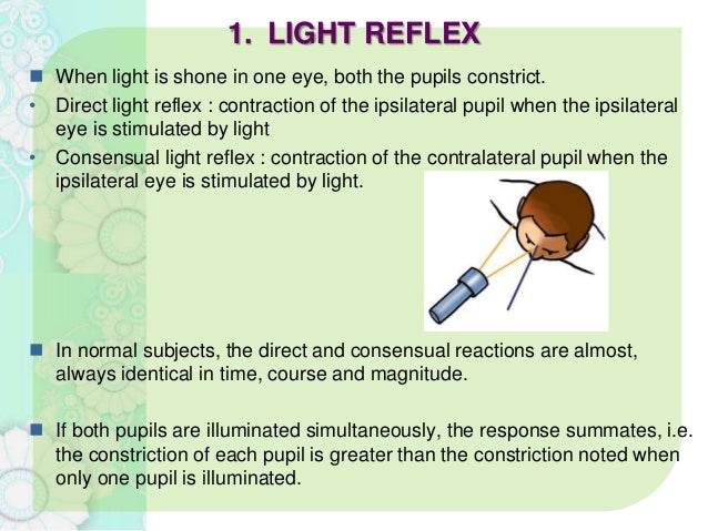
What are direct and consensual pupillary light reflexes?
Jan 01, 2022 · The pupillary light reflex requires the CN II, III and central brain stem connections. Light shined in one eye stimulates retinal photoreceptors which are carried by axons to terminate at pretectum (pretectal nucleus). Which Cranial Nerve Controls Eye Movement The optic nerve is the third cranial nerve (CN III).
What causes abnormal pupillary light reflex?
Which Two Nerves Are Involved In The Pupillary Reaction? The pupillary light reflex is a very important part of vision that many people don't know about. When you look at something with your eyes, it stimulates retinal photoreceptors and ganglion cells in one eye which sends messages along their axons to travel through the optic nerve until they reach an area called …
What is the pathway of the pupillary dark reflex?
Jan 01, 2022 · Which Two Nerves Are Involved In The Pupillary Reaction? The pupillary light reflex is an important part of your eyesight. It requires CN II, III and central brain stem connections for stimulation retinal photoreceptors with respective fibers going through the optic nerve into our brains' pretectum (pretective nucleus).
What nerves are involved in pupil dilation?
These axons then enter the orbit upon the short and long ciliary nerves (branches of V1, the ophthalmic division of CN V - the trigeminal nerve) to synapse on the dilator pupillae muscle, causing pupillary dilation.
What nerve is tested in the pupillary reflex?
optic nervePupillary light reflex is used to assess the brain stem function. Abnormal pupillary light reflex can be found in optic nerve injury, oculomotor nerve damage, brain stem lesions, such as tumors, and medications like barbiturates.
What cranial nerve is responsible for pupillary dilation?
Oculomotor NerveMotor nerve- Oculomotor Nerve-Controls most eye muscles. Works closely with Cranial Nerves 4 & 6. Controls eye movement, pupil dilation, and pupillary constriction. It also controls the muscles that elevate the upper eyelids.
What is cranial nerve II?
The optic nerve is the second cranial nerve (CN II) responsible for transmitting visual information. The optic nerve contains only afferent (sensory) fibers, and like all cranial nerves is paired.Nov 14, 2021
What do the vagus and sympathetic nerves do?
The sympathetic side increases alertness, energy, blood pressure, heart rate, and breathing rate. The parasympathetic side, which the vagus nerve is heavily involved in, decreases alertness, blood pressure, and heart rate, and helps with calmness, relaxation, and digestion.Jun 28, 2017
How sympathetic nervous system dilates pupils?
Pupil dilation is mediated by a sympathetic output acting in opposition to parasympathetically mediated pupil constriction. While light stimulates the parasympathetic output, giving rise to the light reflex, it can both inhibit and stimulate the sympathetic output.Dec 18, 2018
Which Two Nerves Are Involved In The Pupillary Reaction?
The pupillary light reflex requires the CN II, III and central brain stem connections. Light shined in one eye stimulates retinal photoreceptors which are carried by axons to terminate at pretectum (pretectal nucleus).
Which Cranial Nerve Controls Eye Movement
The optic nerve is the third cranial nerve (CN III). It allows movement of eye muscles, constriction of pupil and focusing on objects.
