
The deep layers of the anterolateral abdominal wall are supplied by the following:
- Superior epigastric artery, a terminal branch of the internal thoracic artery. ...
- Inferior epigastric artery and deep circumflex iliac artery, both branches from the external iliac artery, supply the inferior part of the wall. ...
- The tenth and eleventh intercostal arteries and subcostal artery supply the lateral part of the abdominal wall.
What muscles make up the rectus sheath?
The rectus sheath, also called the rectus fascia, is formed by the aponeuroses of the transverse abdominal and the internal and external oblique muscles. It contains the rectus abdominis and pyramidalis muscles. It can be divided into anterior and posterior laminae.
What is rectus sheath haematoma?
Rectus sheath haematoma- This is a condition where a haematoma forms within the rectus sheath, around the rectus abdominis muscle, it is usually due to damage to the superior and inferior epigastric arteries or their branches.
What is the difference between rectus sheath and posterior sheath?
Rectus sheath. The anterior sheath is made of the fibers of the EAO that lie over the area where the rib cage ends. The posterior sheath consists of the fibers of the IAO and the transversus abdominis. The posterior sheath is absent lower down the abdomen, but the anterior sheath is present here as a combination of fibers from all three muscles...
What is the arcuate line of the rectus abdominis?
The line between the upper three quarters and the lower quarter of the rectus abdominis muscle is called the arcuate line . Inferior to the arcuate line, the lower quarter of the rectus abdominis muscle is covered by the rectus sheath on its anterior surface only, while the posterior surface is in direct contact with the transversalis fascia.
Which of the following arteries travel in the rectus sheath?
The rectus abdominis muscle has two main vascular pedicles (Figure 37-3). The deep superior epigastric artery, a terminal branch of the internal mammary artery, enters the rectus sheath from the posterior aspect behind the seventh costal cartilage.
Which two arteries meet in rectus sheath?
The superior and inferior epigastric arteries anastomose with each other at the level of umbilicus after entering into the rectus sheath. The space between the right and left rectus abdominis muscles is filled with the thickening of the anterior wall of the rectus sheath.
What structures are found in the rectus sheath?
The rectus sheath, also called the rectus fascia, is formed by the aponeuroses of the transverse abdominal and the internal and external oblique muscles. It contains the rectus abdominis and pyramidalis muscles.
What makes up the anterior rectus sheath?
Gross anatomy The rectus sheath is composed of the aponeuroses of transversus abdominis, external oblique and internal oblique muscles, which form anterior and posterior layers of the sheath that fuse laterally at the linea semilunaris and in the midline at the linea alba.
What is the arcuate line in the rectus abdominis muscle sheath?
What is the arcuate line? The arcuate line, also known as the semicircular line of Douglas, is a curved line found posterior to the rectus abdominis muscle bilaterally, between the umbilicus and the pubic symphysis. This anatomical finding may not always be present, and its exact position may vary.
What is Hesselbachs triangle?
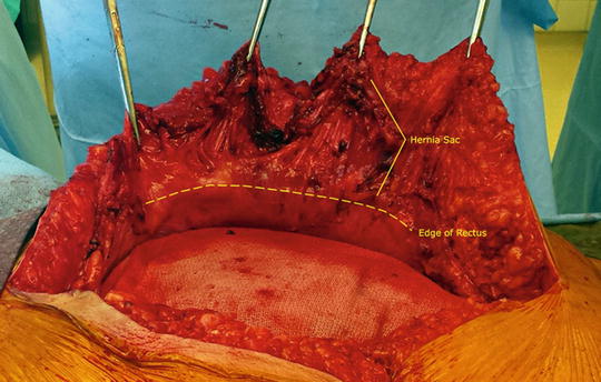
The Hesselbach triangle, also called the inguinal triangle, is a region of the lower, anterior abdominal wall, or groin, that was first described by Frank Hesselbach, a German surgeon and anatomist, in 1806. It describes a potential area of weakness in the abdominal wall, through which a hernia can protrude.
What arteries supply abdominal wall?
The major arteries of the anterolateral abdominal wall are the superior epigastric, inferior epigastric, musculophrenic, subcostal, and posterior intercostal arteries, deep circumflex iliac artery, superficial circumflex iliac artery, and superficial epigastric artery.
What is rectus sheath block?
A bilateral rectus sheath block provides analgesia to the anteromedial abdominal wall and periumbilical area (spinal dermatomes T9, T10, and T11). The technique blocks the anterior cutaneous branches of the intercostal nerves, and therefore, it is well suited for postoperative analgesia for midline abdominal incisions.
What makes up the posterior sheath?
posterior sheath is made up of a combination of the aponeuroses of the internal abdominal oblique IAO and the transversus abdominis muscles TA. Notice that the aponeurosis of the internal abdominal oblique splits around the two sides of the rectus abdominis muscle.
What is the function of Circle of Willis?
Structure and Function The circle of Willis acts to provide collateral blood flow between the anterior and posterior circulations of the brain, protecting against ischemia in the event of vessel disease or damage in one or more areas.
What is in the circle of Willis?
Overview. The Circle of Willis is the joining area of several arteries at the bottom (inferior) side of the brain. At the Circle of Willis, the internal carotid arteries branch into smaller arteries that supply oxygenated blood to over 80% of the cerebrum.
Which part of the artery is responsible for vasoconstriction and vasodilation?
tunica mediaAnswer and Explanation: The tunica media is the layer of an artery which is responsible for vasoconstriction and vasodilation. Within this layer the vessel wall is smooth muscle. When the smooth muscle contracts, that is called vasoconstriction and the diameter of the artery decreases.
What is the largest artery found in the body?
the aortaHow large is the aorta? The aorta is the largest blood vessel in your body. It's more than 1 foot long and an inch in diameter at its widest point.
What is the rectus sheath?
Rectus sheath. The rectus sheath is made up of two parts, known as the posterior sheath and the anterior sheath. These sheaths are made of fibers of the transversus abdominis, internal abdominal oblique (IAO), and external abdominal oblique (EAO), which are muscles of the abdomen.
Where do the muscles come together?
These muscles come together at the linea alba, which is a tendon-like tissue that runs down the middle of the abdomen. The compositions of the posterior sheath and the anterior sheath differ from one abdominal wall to another.
Is the posterior sheath present in the abdomen?
The posterior sheath is absent lower down the abdomen, but the anterior sheath is present here as a combination of fibers from all three muscles of the abdomen.
Where is the rectus sheath?
The rectus sheath extends from the inferior costal margin and the costal cartilages of ribs 5 to 7 to the pubic crest. The composition of the anterior and posterior rectus sheath will differ according to its position superior or inferior to the arcuate line. The arcuate line is an area of demarcation visible from the peritoneal surface of the abdominal wall, residing one-third the distance between the umbilicus and the pubis. The arcuate line can be a sharp demarcation, or it can be a gradual transition zone where the fibers of the posterior sheath gradually disappear. [1]
What muscles are in the rectus sheath?
The rectus abdominis and pyramidalis muscles are contained within the rectus sheath as paired, midline abdominal wall muscles. The pyramidalis muscle can be absent in 20% of people. Contraction of the rectus abdominis and pyramidalis muscles causes flexion of the lumbar spine. Abdominal wall muscles also play a significant role in intra-abdominal pressure and provide support to the axial skeleton.
What is a rectus sheath block?
A rectus sheath block (RSB) involves placing a local anesthetic in the plane between the posterior sheath and the rectus abdominis muscle. This type of block works well for midline abdominal pain near the umbilicus. A transversus abdominis plane block (TAP) is also an option. In this method, a local anesthetic is placed in the neurovascular plane, more posteriorly on the nerves, in the posterior lumbar triangle. However, the TAP block affects a broader area than the rectus sheath block, thus making it a viable option for a greater range of surgical procedures. [10]
What muscle is used for flap repair?
The rectus abdominis muscle also has utility in many muscle flap repairs, so knowledge of the vascular supply of the muscle and its innervation are essential considerations in plastic surgery. [5]
What are the superior and inferior arteries?
The inferior epigastric artery has its origin as a branch of the external iliac artery. The superior and inferior epigastric arteries form anastomoses with each other to varying degrees, thus allowing for collateral flow along the midline abdominal wall. These arteries, along with the superior and inferior epigastric veins, run within the posterior rectus sheath. Additionally, small tributaries of the lower six internal intercostal arteries contribute to the blood supply of the rectus abdominis muscle and rectus sheath. [4]
Where is the transversus abdominis muscle located?
One recent study noted the transversus abdominis muscle within the rectus sheath, especially superiorly near the costal margin. The presence of the transversus abdominis muscle in the posterior rectus sheath decreases as the sheath descends caudally. [2]
Which nerve innervates the rectus abdominis muscle?
The ventral rami of the spinal nerves supplying each myotome innervate the rectus abdominis muscles and sheath. The thoracoabdominal nerves arising from spinal segments T7 to T11 and the subcostal nerve (T12) innervate the rectus abdominis muscle. The subcostal nerve innervates the pyramidalis muscle. [5]
What is the rectus sheath?
The rectus sheath, also called the rectus fascia, is formed by the aponeuroses of the transverse abdominal and the internal and external oblique muscles. It contains the rectus abdominis and pyramidalis muscles.
Which muscle passes in front of the rectus?
Below this level, the aponeuroses of all three muscles (including the transversus) pass in front of the rectus.
Which ligament is seen from the front?
The interfoveolar ligament, seen from in front.
Which fat is separated from the peritoneum?
extraperitoneal fat. parietal peritoneum. The rectus, in the situation where its sheath is deficient below, is separated from the peritoneum only by the transversalis fascia, in contrast to the upper layers, where part of the internal oblique also runs beneath the rectus.
Which artery runs within the spermatic cord in males and supplies its layers, as well as the cremaster?
Cremasteric artery: runs within the spermatic cord in males and supplies its layers, as well as the cremaster muscle. The cremasteric artery in females is rudimentary and accompanies the round ligament of uterus.
What vessels supply the skin and muscles of the anterior abdominal wall?
The inferior epigastric vessels give off several branches that supply the skin and muscles of the anterior abdominal wall, the deep structures of the abdominal wall and the spermatic cord. It terminates at the level of the umbilicus by anastomosing with the superior epigastric artery. This article will discuss the anatomy and function ...
Which branch of the obturator artery is larger than the obturator artery?
A common variation, that occurs in about 20% of people, involves the pubic branch that is larger than the obturator artery and takes over the majority of the blood flow to the obturator artery. The pubic branch of the inferior epigastric artery is then considered as the site of origin of the obturator artery, and is referred to as ...
Which artery gives off muscular branches?
Along its course, the inferior epigastric artery gives off several branches, including muscular branches, anastomotic branches, cutaneous branches, the cremasteric artery and a pubic branch.
Which artery supplies the parietal peritoneum?
Along its course, the inferior epigastric artery gives off several branches, including muscular branches, anastomotic branches, cutaneous branches, the cremasteric artery and a pubic branch. Muscular branches: supply the parietal peritoneum, rectus abdominis, the medial portions of the transversus abdominis, internal abdominal oblique ...
Where does the transversalis fascia pierce?
Below the arcuate line, the inferior epigastric vessels pierce the transversalis fascia on each side of the rectus sheath and enter the rectus abdominis muscle.
Which artery forms the lateral border of the Hesselbach triangle?
The inferior epigastric artery, together with its accompanying veins, forms the lateral border of the Hesselbach’s triangle. This is an important anatomical landmark which outlines the site of the occurrence of direct inguinal hernias.
What is the most valuable laboratory study for rectus sheath hematoma?
The most valuable laboratory studies for the evaluation of rectus sheath hematoma are hemoglobin/hematocrit and coagulation studies. As previously mentioned, over half of patients would be expected to demonstrate a decline in hemoglobin of greater than or equal to 0.4 g/dL. This is neither sensitive nor specific and thus cannot reliably indicate the presence or severity of a hematoma. However, following the trend of serial hemoglobin values can help establish a trajectory for the patient’s course.
How common is rectus sheath hematoma?
Overall, rectus sheath hematoma accounts for only about 1% to 2% of all causes of acute abdominal pain. [2] In a 2016 review by Sheth et al. evaluating a series of patients with rectus sheath hematoma, women were more likely than men to develop a rectus sheath hematoma, with a ratio of 1.7 to 1. This is consistent with epidemiologic data demonstrated in other studies. The mean age of patients in Sheth’s review was 67 years. The overall mortality rate associated with rectus sheath hematoma is less than 2% in the most recent publications. [3]
What are the risk factors for rectus sheath hematoma?
A few well-documented risk factors are associated with rectus sheath hematoma development. The greatest risk is in those who are therapeutically anticoagulated. In Sheth’s review, almost 70% of patients were on some form of anticoagulation. Naturally, as the prevalence of chemical anticoagulation increases, one may reason that the incidence of rectus sheath hematoma also rises. However, there is a paucity of data in the modern literature to reflect this. In the same series, nearly 60% of patients with rectus sheath hematoma also had chronic kidney disease stage III or greater. Other risk factors, in order of descending prevalence, include abdominal wall injections, steroid or immunosuppressant therapy, cough, femoral puncture, and antiplatelet therapy. [3]
What causes a hematoma in the rectus sheath?
Rectus sheath hematoma occurs as a result of injury to an epigastric artery or its perforating branches within the rectus muscle. Recall that the blood supply to the rectus abdominis muscles originates from the superior and inferior epigastric arteries. The superior epigastric artery arises from the internal thoracic artery and travels caudally within the rectus sheath to anastomose with the inferior epigastric artery. The inferior epigastric artery, deriving from the external iliac, travels cephalad along the posterior surface of the rectus muscle, where it lacks the protection of a posterior rectus sheath until it reaches the arcuate line. The arcuate line is located a third the distance between the umbilicus and the pubic symphysis. [1]
How much of blood volume is lost in a hemorrhagic shock?
According to the widely accepted classification of hemorrhagic shock, patients generally do not manifest any hemodynamic changes associated with hemorrhage until at least 15% to 30% of blood volume is lost. While this certainly is a possibility in the setting of rectus sheath hematoma, it is the case in only about 1% to 13% of patients. [4] Thus the absence of these changes, such as tachycardia, hypotension, or orthostasis, should not lessen the clinician’s suspicion for the diagnosis.
Can INR be used for rectus sheath hematoma?
All patients with a suspected rectus sheath hematoma should have baseline coagulation studies at the time of their initial evaluation. Again, a large majority of patients with rectus sheath hematoma are taking some form of therapeutic anticoagulation. For those taking warfarin, the INR can help steer the decision to administer a reversal agent. With the increasing use of antiplatelet therapies and novel oral anticoagulants (NOAC), the INR may be less informative but should still be obtained as part of a coagulation panel.
Can an epigastric artery rupture?
Epigastric artery rupture can occur when there is direct abdominal trauma or excessively forceful contraction of the abdominal wall. The resulting hematoma may present differently depending on whether it arises in the upper or lower abdomen. When the superior epigastric artery is injured, the resulting hematoma is typically smaller and quickly tamponaded within the rectus sheath. On the other hand, rupture of the inferior epigastric artery is not as easily controlled, owing to the lack of posterior sheath. This allows the development of a larger and generally more clinically significant hematoma. [1]
Overview
Structure
The rectus sheath can be divided into anterior and posterior laminae. The arrangement of the layers has important variations at different locations in the body.
For context, above the sheath are the following two layers:
1. Camper's fascia (anterior part of Superficial fascia)
2. Scarpa's fascia (posterior part of the Superficial fascia)
Clinical significance
The rectus sheath is a useful attachment for surgical meshes during abdominal surgery. This has a higher risk of infection than many other attachment sites.
Additional images
• The Cremaster
• The interfoveolar ligament, seen from in front.
External links
• Anatomy figure: 35:04-02 at Human Anatomy Online, SUNY Downstate Medical Center - "Incisions and the contents of the rectus sheath."
• Anatomy photo:35:10-0103 at the SUNY Downstate Medical Center - "Anterior Abdominal Wall: The Rectus Abdominis Muscle"
• Anatomy image:7180 at the SUNY Downstate Medical Center - anterior layer