What was the first CT scanner?
- Prototype of the EMI CT1010 body scanner installed at Atkinson Morley's Hospital (London, UK)
- First commercial PET scanner installed (Los Angeles, USA)
- first scans from EMI body scanner shown, at first International Conference on CT (Bermuda)
- research body scanner installed at Northwick Park Hospital (London, UK)
What is the history of the CT scan?
CT scanners were first introduced in 1971 with a single detector for brain study under the leadership of Sir Godfrey Hounsfield, an electrical engineer at EMI (Electric and Musical Industries Ltd). Thereafter, it has undergone multiple improvements with an increase in the number of detectors and decrease in the scan time.
Who invented the first image scanner?
When was the first scanner invented? raman October 10, 2011 Comments Off. on When was the first scanner invented? Russell Kirsch and his team at the US National Bureau of Standards in the year 1957 built the first image drum scanner. He scanned the first image of his three month old son. Technology.
What was the first CAT scan?
The first CT system that could make images of any part of the body and did not require the "water tank" was the ACTA (Automatic Computerized Transverse Axial) scanner designed by Robert S. Ledley, DDS, at Georgetown University. This machine had 30 photomultiplier tubes as detectors and completed a scan in only nine translate/rotate cycles, much ...
When was CT scans invented?
The first computed tomography image – a CT scan – of the human brain was made 50 years ago, on Oct. 1, 1971. Hounsfield never made it to Egypt, but his invention did take him to Stockholm and Buckingham Palace.
Why was CT scan invented?
So, what was the catalyst for his work with the CT scan? According to Hounsfield, the idea to invent such technology came to him while on vacation. At the time, all he wanted was to reconstruct a 3D picture of a box. He intended to achieve this by re-imagining the object as a series of slices.
Where was first CT scan done?
In 1967 Sir Godfrey Hounsfield invented the first CT scanner at EMI Central Research Laboratories using x-ray technology. In 1971 the first patient brain CT was performed in Wimbledon, England but it was not publicized until a year later.
What came first CT or MRI?
Moniz, a neurologist, accomplished the first cerebral arteriogram in 1927. Oldendorf himself developed the basis for computerized tomography (CT) in 1961 and the technique was applied to clinical diagnosis by an electrical engineer, Hounsfield, in 1973. Finally, magnetic resonance imaging (MRI) was introduced.
What was used before CT scans were invented?
Before computed tomography, tomographic images could be made by radiography using focal plane tomography, representing a single slice of the body on radiographic film. This method was proposed by the Italian radiologist Alessandro Vallebona in the early 1900s.
What is a CT scan and why is it used?
A computerized tomography (CT) scan combines a series of X-ray images taken from different angles around your body and uses computer processing to create cross-sectional images (slices) of the bones, blood vessels and soft tissues inside your body. CT scan images provide more-detailed information than plain X-rays do.
Why are CT scans important?
Why are CT scans important? CT scans are a valuable diagnostic tool. They are able to detect some conditions that conventional X-rays cannot because CT scans can show a 3D view of the section of the body being studied. CT scans are also useful for monitoring a patient's progress during or after treatment.
Did South Africa invent the CT scan?
In November 1975 South African physicist and inventor Robert Ledley was granted patent #3,922,552 for a Diagnostic X-ray System also known as a CAT-Scan.
Who is the inventor of CT scan?
Godfrey Hounsfield Godfrey Hounsfield, a biomedical engineer contributed enormously towards the diagnosis of neurological and other disorders by virtue of his invention of the computed axial tomography scan for which he was awarded the Nobel Prize in 1979.
When did Robert Ledley invent the Acta machine?
Then, in 1973, Dr. Ledley introduced one of the most powerful diagnostic aides since the discovery of X-rays in 1895. He called his invention the automatic computerized transverse axial scanner (ACTA). It was, in effect, the first machine capable of producing cross-sectional images of any part of the body.
When was the first CT scan used?
Early CT scanner in use, 1980. The first detailed picture of a living brain was taken by a CT scanner in 1971. CT stands for computerized tomography. CT scanners, a type of X‑ray machine, became important for diagnosis within hospitals during the late 20th century.
When was the first whole body CT scanner invented?
In 1975 the first whole-body CT scanner was built. CT scanners are now used to take pictures of virtually any part of the body. CT scanning owed a lot to pop band The Beatles in the 1960s. Hounsfield worked for EMI, the company that owned The Beatles’ music.
What is a CT scanner?
Unlike X‑ray machines, CT scanners send multiple X‑ray beams through the body at different angles. This is called tomography. Detectors inside the machine record how the beams pass through sections of the body. A computer uses complex mathematics to process these measurements and construct an internal image of the body, displayed on a monitor.
What was the first CT scanner?
The first CT system that could make images of any part of the body and did not require the "water tank" was the ACTA (Automatic Computerized Transverse Axial) scanner designed by Robert S. Ledley, DDS, at Georgetown University. This machine had 30 photomultiplier tubes as detectors and completed a scan in only nine translate/rotate cycles, much faster than the EMI-Scanner. It used a DEC PDP11/34 minicomputer both to operate the servo-mechanisms and to acquire and process the images. The Pfizer drug company acquired the prototype from the university, along with rights to manufacture it. Pfizer then began making copies of the prototype, calling it the "200FS" (FS meaning Fast Scan), which were selling as fast as they could make them. This unit produced images in a 256×256 matrix, with much better definition than the EMI-Scanner's 80×80. It took it about 20 seconds to acquire one slice, which made body scans possible, as the patient had to hold his/her breath until the slice was acquired. That is the main reason why the EMI scanner could not do body scans. The 5 minutes to acquire one slice was much too long. Typically, the operator, after completing the whole series of slices, would then process the images, photograph them onto films, and archive the raw images onto magnetic tape. This had to be done because the computer did not have the storage capacity for more than one study at a time. This meant that in a large, busy hospital, the CT operator was a very busy person. This machine required a lot of maintenance to keep it running. The PDP11/34 computer did everything from controlling the gantry and the scanning process to processing the raw data into finished images. Yet it had only 64 KB of memory and a 5 MB hard disk, which held both the operating program and the acquired raw data. The hard disk consisted of two 12" platters, one internal and fixed, the other platter was contained in a round cartridge and was removable.
When was X-ray computed tomography invented?
The history of X-ray computed tomography goes back to at least 1917 with the mathematical theory of the Radon transform In October 1963, William H. Oldendorf received a U.S. patent for a "radiant energy apparatus for investigating selected areas of interior objects obscured by dense material". The first clinical CT scan was performed in 1971 using ...
How was tomography made before computed tomography?
Before computed tomography, tomographic images could be made by radiography by focal plane tomography , representing a single slice of the body on radiographic film. This method was proposed by the Italian radiologist Alessandro Vallebona in the early 1900s. The idea is based on simple principles of projective geometry: moving synchronously and in opposite directions the X-ray tube and the film, which are connected together by a rod whose pivot point is the focus; the image created by the points on the focal plane appears sharper, while the images of the other points annihilate as noise. This is only marginally effective, as blurring occurs in only the "x" plane. This method of acquiring tomographic images using only mechanical techniques advanced through the mid-twentieth century, steadily producing sharper images, and with a greater ability to vary the thickness of the cross-section being examined. This was achieved through the introduction of more complex, multidirectional devices that can move in more than one plane and perform more effective blurring. However, despite the increasing sophistication of focal plane tomography, it remained ineffective at producing images of soft tissues. With the increasing power and availability of computers in the 1960s, research began into practical computational techniques for creating tomographic images, leading to the development of computed tomography (CT).
How long does it take to scan X-rays?
The first production X-ray CT machine (in fact called the "EMI-Scanner") was limited to making tomographic sections of the brain, but acquired the image data in about 4 minutes (scanning two adjacent slices), and the computation time (using a Data General Nova minicomputer) was about 7 minutes per picture.
How fast can a Siemens scanner take an image?
In 2008 Siemens introduced a new generation of scanner that was able to take an image in less than 1 second, fast enough to produce clear images of beating hearts and coronary arteries.
What is Siemens scanning?
In 2021, the FDA approved Siemens' photo counting scanner. The scanner counts individual x-ray photons that pass through a patient and discriminates their energy, increasing the detail supplied to the reader. The technique also reduces the amount of x-rays needed for a scan.
Where was the first EMI scanner installed?
In the U.S., the first installation was at the Mayo Clinic. As a tribute to the impact of this system on medical imaging the Mayo Clinic has an EMI scanner on display in the Radiology Department. Allan McLeod Cormack of Tufts University in Massachusetts independently invented a similar process, and both Hounsfield and Cormack shared the 1979 Nobel Prize in Medicine.
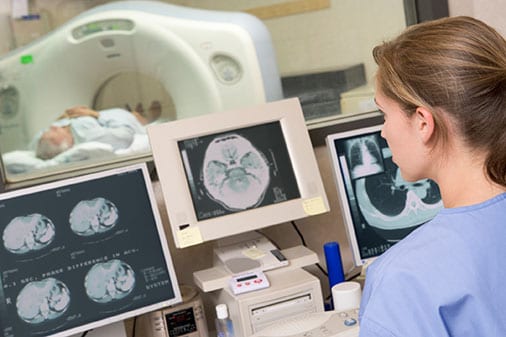
What is the name of the person who performs a CT scan?
The personnel that perform CT scans are called radiographers or radiology technologists . CT scanners use a rotating x-ray tube and a row of detectors placed in the gantry to measure X-ray attenuations by different tissues inside the body.
What is a CT scan?
A CT scan or computed tomography scan (formerly known as computed axial tomography or CAT scan) is a medical imaging technique used in radiology to get detailed images of the body noninvasively for diagnostic purposes.
What is photon counting in CT?
Photon counting computed tomography is a CT technique currently under development. Typical CT scanners use energy integrating detectors; photons are measured as a voltage on a capacitor which is proportional to the x-rays detected. However, this technique is susceptible to noise and other factors which can affect the linearity of the voltage to x-ray intensity relationship. Photon counting detectors (PCDs) are still affected by noise but it does not change the measured counts of photons. PCDs have several potential advantages, including improving signal (and contrast) to noise ratios, reducing doses, improving spatial resolution, and through use of several energies, distinguishing multiple contrast agents. PCDs have only recently become feasible in CT scanners due to improvements in detector technologies that can cope with the volume and rate of data required. As of February 2016, photon counting CT is in use at three sites. Some early research has found the dose reduction potential of photon counting CT for breast imaging to be very promising. In view of recent findings of high cumulative doses to patients from recurrent CT scans, there has been a push for sub-mSv CT scans, a goal that has been lingering.
What is EBT in CT?
Electron beam tomography (EBT) is a specific form of CT in which a large enough X-ray tube is constructed so that only the path of the electrons, travelling between the cathode and anode of the X-ray tube, are spun using deflection coils. This type had a major advantage since sweep speeds can be much faster, allowing for less blurry imaging of moving structures, such as the heart and arteries. Fewer scanners of this design have been produced when compared with spinning tube types, mainly due to the higher cost associated with building a much larger X-ray tube and detector array and limited anatomical coverage.
What is CT scan of the abdomen?
CT is an accurate technique for diagnosis of abdominal diseases like Crohn's disease, GIT bleeding, and diagnosis and staging of cancer, as well as follow-up after cancer treatment to assess response . It is commonly used to investigate acute abdominal pain.
What is CT angiography?
Main article: Computed tomography angiography. Computed tomography angiography (CTA) is a type of contrast CT to visualize the arteries and veins throughout the body. This ranges from arteries serving the brain to those bringing blood to the lungs, kidneys, arms and legs.
Why is CT important?
Since its introduction in the 1970s, CT has become an important tool in medical imaging to supplement X-rays and medical ultrasonography. It has more recently been used for preventive medicine or screening for disease, for example, CT colonography for people with a high risk of colon cancer, or full-motion heart scans for people with a high risk of heart disease. Several institutions offer full-body scans for the general population although this practice goes against the advice and official position of many professional organizations in the field primarily due to the radiation dose applied.
Who developed computer assisted tomography?
In the late 1960s, Godfrey Hounsfield began developing computer-assisted tomography, or CAT scanning, an improved form of diagnostic imaging. At Thorn EMI Ltd., he combined his understanding of electronics and radar to create three-dimensional images that illuminated the internal physiology of the human head.
When was the first CAT scanner used?
The first CAT scanner was installed for use in 1971. It provided physicians valuable diagnostic information without potentially hazardous exploratory surgery, revolutionizing medical care. Computer tomography was first used to take images of the skull to study diseases of the brain.
How many patents did Hounsfield have?
Credited with 72 patents, Hounsfield was awarded many honors for his technology, including the 1979 Nobel Prize.
When was the first CT scanner invented?
The first clinical CT scanners were installed between 1974 and 1976. The original systems were dedicated to head imaging only, but "whole body" systems with larger patient openings became available in 1976. CT became widely available by about 1980.
When was CT invented?
CT was invented in 1972 by British engineer Godfrey Hounsfield of EMI Laboratories, England and by South Africa-born physicist Allan Cormack of Tufts University, Massachusetts. Hounsfield and Cormack were later awarded the Nobel Peace Prize for their contributions to medicine and science.
What is CAT scan?
Computed Tomography (CT) imaging is also known as "CAT scanning" (Computed Axial Tomography). Tomography is from the Greek word "tomos" meaning "slice" or "section" and "graphia" meaning "describing".
Why is CT scan faster?
Faster scanning helps to eliminate artifacts from patient motion such as breathing or peristalsis. CT exams are now quicker and more patient-friendly than ever before. Tremendous research and development has been made to provide excellent image quality for diagnostic confidence at the lowest possible x-ray dose.
How long does it take to scan a chest?
An entire chest (forty 8 mm slices) can be scanned in five to ten seconds using the most advanced multi-slice CT system.
When was the first CT scan made?
The first computed tomography image – a CT scan – of the human brain was made 50 years ago, on Oct. 1, 1971. Hounsfield never made it to Egypt, but his invention did take him to Stockholm and Buckingham Palace.
Who invented the whole body scanner?
In 1973, American Robert Ledley developed a whole-body scanner that could image other organs, blood vessels and, of course, bones. Modern scanners are faster, provide better resolution, and most important, do it with less radiation exposure. There are even mobile scanners.
What did Hounsfield do?
Finally, in possibly his most ingenious invention, Hounsfield created an algorithm to reconstruct an image of the brain based on all these layers. By working backward and using one of the era’s fastest new computers, he could calculate the value for each little box of each brain layer. Eureka!
What does the brain look like on an X-ray?
Plain X-rays show marvelous details of bones, but the brain is an amorphous blob of tissue – on an X-ray it all looks like fog . This got Hounsfield thinking about his old idea of finding hidden structures without opening the box.
How many CT scans are performed in 2020?
By 2020, technicians were performing more than 80 million scans annually in the U.S.. Some physicians argue that number is excessive and maybe a third are unnecessary. While that may be true, the CT scan has benefited the health of many patients around the world, helping identify tumors and determine if surgery is needed. They’re particularly useful for a quick search for internal injuries after accidents in the ER.
When did Hounsfield start scanning?
He found a somewhat reluctant neurologist who agreed to help. The team installed a full-sized scanner at the Atkinson Morley Hospital in London, and on Oct. 1, 1971, they scanned their first patient: a middle-aged woman who showed signs of a brain tumor.
How far do X-rays go through the brain?
X-rays beam through each ‘slice’ of brain, oriented at each degree from 1 to 180 in a semicircle. Edmund S. Higgins, CC BY-ND
How is CT used in cancer screening?
Studies have shown that CT can be effective in both colorectal cancer screening (including screening for large polyps) and lung cancer screening.
Where can people get more information about CT?
Additional information about CT imaging is available from the U.S. Food and Drug Administration (FDA), the federal agency that regulates food, drugs, medical devices, cosmetics, biologics, and radiation-emitting products.
What is computed tomography?
Computed tomography ( CT) is an imaging procedure that uses special x-ray equipment to create detailed pictures, or scans, of areas inside the body. It is sometimes called computerized tomography or computerized axial tomography (CAT).
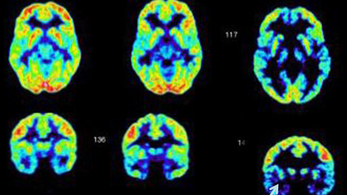
What is combined PET/CT?
Combined PET/CT uses two imaging methods, CT and positron emission tomography ( PET), in one procedure. CT is done first to create anatomic pictures of the organs and structures in the body, and then PET is done to create pictures that provide functional data about the metabolic pathways (chemical reactions that take place in a cell to create and use energy) that are active in tissues or cells. Cancer cells often use different metabolic pathways than normal cells.
What are the risks of CT scans for children?
Radiation exposure from CT scans affects adults and children differently. Children are considerably more sensitive to radiation than adults because of their growing bodies and the rapid pace at which the cells in their bodies divide. In addition, children have a longer life expectancy than adults, providing a larger window of opportunity for radiation-related cancers to develop ( 5 ).
What is being done to reduce the level of radiation exposure from CT?
In response to concerns about the increased risk of cancer associated with CT and other imaging procedures that use ionizing radiation, several organizations and government agencies have developed guidelines and recommendations regarding the appropriate use of these procedures.
What is NCI doing to improve CT imaging?
Researchers funded by NCI are studying ways to improve the use of CT in cancer screening, diagnosis, and treatment. NCI also conducts and sponsors clinical trials that are testing ways to improve CT or new uses of CT imaging technology. Some of these clinical trials are run by the ECOG-ACRIN Cancer Research Group
Overview
Commercial scanners
CT technology has vastly improved. Improvements in speed, slice count, and image quality have been the major focus primarily for cardiac imaging. Scanners now produce images much faster and with higher resolution enabling doctors to diagnose patients more accurately and perform medical procedures with greater precision.
The first commercially viable CT scanner was invented by Sir Godfrey Hounsfield in Hayes, Unite…
Mathematical theory
The mathematical theory behind computed tomographic reconstruction dates back to 1917 with the invention of the Radon transform by Austrian mathematician Johann Radon, who showed mathematically that a function could be reconstructed from an infinite set of its projections. In 1937, Polish mathematician Stefan Kaczmarz developed a method to find an approximate solution to a large system of linear algebraic equations. This, along with Allan McLeod Cormack's theoreti…
Largely replaced techniques
CT replaced the more invasive pneumoencephalography for imaging of the brain, as well as most applications of focal plane tomography.
Before computed tomography, tomographic images could be made by radiography using focal plane tomography, representing a single slice of the body on radiographic film. This method was proposed by the Italian radiologist Alessandro Vallebona in the early 1900s. The idea is based o…
Overview
A computed tomography scan (usually abbreviated to CT scan; formerly called computed axial tomography scan or CAT scan) is a medical imaging technique used to obtain detailed internal images of the body. The personnel that perform CT scans are called radiographers or radiology technologists.
CT scanners use a rotating X-ray tube and a row of detectors placed in a gantry t…
Types
Spinning tube, commonly called spiral CT, or helical CT, is an imaging technique in which an entire X-ray tube is spun around the central axis of the area being scanned. These are the dominant type of scanners on the market because they have been manufactured longer and offer a lower cost of production and purchase. The main limitation of this type of CT is the bulk and inertia of the equipment (X-ray tube assembly and detector array on the opposite side of the circl…
Medical use
Since its introduction in the 1970s, CT has become an important tool in medical imaging to supplement conventional X-ray imaging and medical ultrasonography. It has more recently been used for preventive medicine or screening for disease, for example, CT colonography for people with a high risk of colon cancer, or full-motion heart scans for people with a high risk of heart disease. Several institutions offer full-body scans for the general population although this practice goes ag…
Other uses
Industrial CT scanning (industrial computed tomography) is a process which utilizes X-ray equipment to produce 3D representations of components both externally and internally. Industrial CT scanning has been utilized in many areas of industry for internal inspection of components. Some of the key uses for CT scanning have been flaw detection, failure analysis, metrology, assembly analysis, image-based finite element methods and reverse engineering applications. …
Interpretation of results
The result of a CT scan is a volume of voxels, which may be presented to a human observer by various methods, which broadly fit into the following categories:
• Slices (of varying thickness). Thin slice is generally regarded as planes representing a thickness of less than 3 mm. Thick slice is generally regarded a…
Advantages
CT scanning has several advantages over traditional two-dimensional medical radiography. First, CT eliminates the superimposition of images of structures outside the area of interest. Second, CT scans have greater image resolution, enabling examination of finer details. CT can distinguish between tissues that differ in radiographic density by 1% or less. Third, CT scanning enables multiplanar reformatted imaging: scan data can be visualized in the transverse (or axial), coronal, …
Adverse effects
The radiation used in CT scans can damage body cells, including DNA molecules, which can lead to radiation-induced cancer. The radiation doses received from CT scans is variable. Compared to the lowest dose x-ray techniques, CT scans can have 100 to 1,000 times higher dose than conventional X-rays. However, a lumbar spine x-ray has a similar dose as a head CT. Articles in the media often exaggerate the relative dose of CT by comparing the lowest-dose x-ray techniqu…
Mechanism
Computed tomography operates by using an X-ray generator that rotates around the object; X-ray detectors are positioned on the opposite side of the circle from the X-ray source. As the X-rays pass through the patient, they are attenuated differently by various tissues according to the tissue density. A visual representation of the raw data obtained is called a sinogram, yet it is not suffici…