The spinous process of a typical cervical vertebra is short and bifid posteriorly. It is bifid because it develops from two separate secondary centers of ossification.
Why do cervical vertebrae have a bifid spinous process?
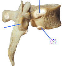
Another feature unique to the cervical vertebrae is the bifid spinous process (See “physiologic variants” section), which may serve to increase surface area for muscle attachment. The spinous process of cervical vertebrae increases as the spinal column descends.
What does bifid mean for cervical vertebrae?
The spinous processes were classified into three categories: “bifid: clearly distinct cleft resulting in two elongated projections,” “partially bifid: two distinct tubercles at the end of the spinous process are present without a cleft,” and “monofid: rounded or flattened.”
What is unique about the cervical vertebrae?
Lastly, cervical vertebrae are known to have the greatest intervertebral disc height, which increases the range of motion. There are three atypical vertebrae found in the cervical region. C1, also known as “atlas,” is unique among all vertebrae in that it lacks both a vertebral body and a spinous process.
Are all cervical spinous processes bifid?
Typically, cervical vertebrae display bifid spinous processes. Nevertheless, this feature may vary both between subjects and even within the vertebrae of the same individual.
What does bifid spinous process mean?
Bifid spinous process – this is where the spinous process splits into two distally. Transverse foramina – holes in the transverse processes. They give passage to the vertebral artery, vein and sympathetic nerves[2]. The main role of the cervical spine is to support and promote the movement of the head and neck.
Does C7 have bifid process?
The spinous process of C7 usually projects directly posteriorly. Unlike typical cervical vertebrae, the spinous process of C7 is not bifid. The funicular portion of the ligamentum nuchae attaches to the single posterior tip of the C7 spinous process.
What 2 features do cervical vertebrae have that no other vertebrae have?
The main anatomical characteristics of a typical cervical vertebra that separate it from other types of vertebrae are the small size, transverse foramina, saddle-shaped body, and bifid spinous process (Fig. 1.7. 18).
Why do only cervical vertebrae have transverse foramen?
Transverse foramina are only present in the cervical vertebrae. These foramina allow the passage of the vertebral artery and vein. The vertebral arteries arise from the first part of the subclavian artery.
Is C2 bifid?
Gross anatomy The axis is formed by a body with the attached dens, two lateral masses, a posterior neural arch (formed by the pedicle and a thick lamina), and a large spinous process, which is commonly bifid.
Where do bifid spinous process occur and what are they?
The spinous process of a typical cervical vertebra is short and bifid posteriorly. It is bifid because it develops from two separate secondary centers of ossification. This morphology is unique to cervical spinous processes.
What is the purpose of the spinous process?
The lumbar spinous processes (LSP) have an important anatomical and biomechanical function protecting the neural structures in the spinal canal, and as an anchor for the interspinous and supraspinous ligaments, and the intersegmental paraspinal muscles.
Where are bifid spinous processes found?
The spinous (spinal) process projects in a posterior direction from the junction of the lamina of a vertebra. Each region has a characteristic shape. When we palpate vertebrae, it is the spinous process that we detect. We find that most cervical vertebrae have a bifid spinous process.
Is c2 bifid?
Gross anatomy The axis is formed by a body with the attached dens, two lateral masses, a posterior neural arch (formed by the pedicle and a thick lamina), and a large spinous process, which is commonly bifid.
Where is the bifid spinous process located?
Cervical RegionThe Cervical Region It is bifid because it develops from two separate secondary centers of ossification. This morphology is unique to cervical spinous processes.
What are the typical cervical vertebrae?
Typical Vertebrae: C3, C4, C5, and C6 Cervical vertebrae C3 through C6 are known as typical vertebrae because they share the same basic characteristics with most of the vertebrae throughout the rest of the spine.
Does C7 have a transverse foramen?
C7 possesses the standard cervical vertebral features but has some distinct features: spinous process ends in a rounded tubercle and is not bifid. C7 transverse foramina are small, and do not transmit the vertebral artery.
What is spinal bifida?
Spina bifida is a birth defect that occurs when the spine and spinal cord don't form properly. It's a type of neural tube defect. The neural tube is the structure in a developing embryo that eventually becomes the baby's brain, spinal cord and the tissues that enclose them. Normally, the neural tube forms early in pregnancy and it closes by ...
Where is the spinal canal in Spina Bifida?
In this severe type of spina bifida: The spinal canal remains open along several vertebrae in the lower or middle back. Both the membranes and the spinal cord or nerves protrude at birth, forming a sac. Tissues and nerves usually are exposed, though sometimes skin covers the sac.
What is Chiari malformation?
Chiari malformation (kee-AH-ree mal-for-MAY-shun) type II is a common brain abnormality in children with the myelomeningocele type of spina bifida. The brainstem, or lowest part of the brain above the spinal cord, is elongated and positioned lower than usual. This can cause problems with breathing and swallowing.
What is the most severe spinal canal?
Also known as open spina bifida, myelomeningocele is the most severe type. The spinal canal is open along several vertebrae in the lower or middle back. The membranes and spinal nerves push through this opening at birth, forming a sac on the baby's back, typically exposing tissues and nerves.
What is the occulta of the spine?
"Occulta" means hidden. It's the mildest and most common type. Spina bifida occulta results in a small separation or gap in one or more of the bones of the spine (vertebrae). Many people who have spina bifida occulta don't even know it, unless the condition is discovered during an imaging test done for unrelated reasons.
What is the name of the spinal cord that protrudes at birth?
Spina bifida (myelomeningocele) Spina bifida (my elomeningocele) Myelomeningocele is a severe type of spina bifida in which the membranes and the spinal nerves protrude at birth, forming a sac on the baby's back. The exposed nervous system may become infected, so prompt surgery is needed after birth.
How severe is Spina Bifida?
Spina bifida can range from mild to severe, depending on the type of defect, size, location and complications. When necessary, early treatment for spina bifida involves surgery — although such treatment doesn't always completely resolve the problem.
Which cervical vertebrae have the most spinous process?
The seventh cer vical vertebra, also called the vertebra prominens, is commonly considered a unique vertebra and has the most prominent spinous process. When feeling the back of the neck, the C7 vertebra’s spinous process (bony hump) sticks out more than the other cervical vertebrae.
What is the difference between cervical vertebrae and lower cervical vertebrae?
There are some differences among the cervical vertebrae. The vertebrae at the top of the neck tend to be smaller and more mobile while the lower cervical vertebrae are larger to handle greater loads from the neck and head above.
What are the different types of vertebrae?
Cervical vertebrae C3 through C6 are known as typical vertebrae because they share the same basic characteristics with most of the vertebrae throughout the rest of the spine. Typical vertebrae have: 1 Vertebral body. This thick bone is cylindrical-shaped and located at the front of the vertebra. The vertebral body carries most of the load for a vertebra. At most levels of the spine, an intervertebral disc sits between 2 vertebral bodies to provide cushioning and help absorb the shock of everyday movements. 2 Vertebral arch. This bony arch wraps around the spinal cord toward the back of the spine and consists of 2 pedicles and 2 laminae. The pedicles connect with the vertebral body in the front, and the laminae transition into the spinous process (a bony hump) in the back of the vertebra. 3 Facet joints. Each vertebra has a pair of facet joints, also known as zygapophysial joints. These joints, located between the pedicle and lamina on each side of the vertebral arch, are lined with smooth cartilage to enable limited movement between 2 vertebrae. Spinal degeneration or injury to the facet joints are among the most common causes of chronic neck pain.
What are the atypical vertebrae?
Atypical Vertebrae: C1 and C2. C1 and C2 are considered atypical vertebrae because they have some distinguishing features compared to the rest of the cervical spine. C1 Vertebra (the atlas). The top vertebra, called the atlas, is the only cervical vertebra without a vertebral body.
What is the intervertebral disc?
At most levels of the spine, an intervertebral disc sits between 2 vertebral bodies to provide cushioning and help absorb the shock of everyday movements. Vertebral arch. This bony arch wraps around the spinal cord toward the back of the spine and consists of 2 pedicles and 2 laminae.
Which vertebrae are considered typical vertebrae?
Cervical vertebrae C3 through C6 are known as typical vertebrae because they share the same basic characteristics with most of the vertebrae throughout the rest of the spine. Typical vertebrae have:
Where are the facet joints located?
These joints, located between the pedicle and lamina on each side of the vertebral arch, are lined with smooth cartilage to enable limited movement between 2 vertebrae.
What are the features of cervical vertebrae?
The most notable distinction is the presence of one foramen, in each transverse process. These transverse foramina encircle the vertebral arteries and veins. This is true of all cervical vertebrae except C7, whose transverse foramina contain only accessory veins. Another feature unique to the cervical vertebrae is the bifid spinous process (See “physiologic variants” section), which may serve to increase surface area for muscle attachment. The spinous process of cervical vertebrae increases as the spinal column descends. Cervical vertebrae tend to have superior articular facets that face posteromedially. Some studies have shown that more inferior cervical vertebrae have superior facets that face in a posterolateral direction – more akin to those of the thoracic region. Lastly, cervical vertebrae are known to have the greatest intervertebral disc height, which increases the range of motion.
What is the role of the vertebrae?
The spine has several major roles in the body that include: protection of the spinal cord and branching spinal nerves, support for thorax and abdomen, ...
How many vertebrae are in the cervical region?
The cervical region contains seven vertebrae, denoted C1-C7, which are the smallest of the vertebral column. The intervertebral discs, along with the laminae and the articular processes of adjacent vertebrae, create a space through which spinal nerves exit. The cervical vertebrae, as a group, produce a lordotic curve.
What are the roles of the spine?
The spine has several major roles in the body that include: protection of the spinal cord and branching spinal n …. Vertebrae, along with intervertebral discs, compose the vertebral column, or spine. It extends from the skull to the coccyx and includes the cervical, thoracic, lumbar and sacral regions. The spine has several major roles in the body ...
What is the vertebral body?
The vertebral body consists of trabecular bone, which contains the red marrow, surrounded by a thin external layer of compact bone. The arch, along with the posterior aspect of the body, forms the vertebral (spinal) canal, which contains the spinal cord. The arch consists of bilateral pedicles, cylindrical processes of bone ...
Which vertebrae have the greatest disc height?
Lastly, cervical vertebrae are known to have the greatest intervertebral disc height, which increases the range of motion. There are three atypical vertebrae found in the cervical region. C1, also known as “atlas,” is unique among all vertebrae in that it lacks both a vertebral body and a spinous process.
Which vertebrae have superior articular facets?
Cervical vertebrae tend to have superior articular facets that face posteromedially. Some studies have shown that more inferior cervical vertebrae have superior facets that face in a posterolateral direction – more akin to those of the thoracic region.

Structure
Typical Cervical Vertebra
- Vertebral Body
1. The bodies of these four vertebrae are small, and transverse diameter is greater than anterio-posterior and height dimensions. 2. The anterior and posterior surfaces are flattened and of equal depth; the former is placed on a lower level than the latter, and its inferior border is prolonged do… - Vertebral Foramen
1. Large, triangular in shape.
Atypical Cervical Vertebrae
- C1
The Atlas, C1, is the topmost vertebra, and along with the Axis; forms the joint connecting the skull and spine. Its chief peculiarity is that it has no body, and this is due to the fact that the body of the atlas has fused with that of the Axis. - C2
The Axis, C2, forms the pivot upon which the Atlas rotates. The most distinctive characteristic of this bone is the strong odontoid process (dens) that rises perpendicularly from the upper surface of the body. The body is deeper in front than behind, and prolonged downward anteriorly so as t…
Function
- The cervical spine functions to provide mobility and stability to the head while connecting it to the relatively immobile thoracic spine. The movement of nodding the head takes place predominantly through flexion and extension at the joint between the atlas and the occipital bone, the atlanto-occipital joint. However, the cervical spine is comparatively mobile, and some component of thi…
Pathology
- Vertebral Causes of Spinal Pain: 1. Developmental: Scoliosis, Hypermobility, Various uncommon disorders. 2. Degenerative: Disc lesions without root compression, Disc lesions with root compression, Disc lessions with compression of spinal cord or cauda equina, Osteoarthrosis of apophyseal joint, Hyperostosis, Instability. 3. Trauma: Fracture, Stress fracture, Subluxation, Liga…
Assessment
- Physical Examination
Sequence proposed by Maitland for the physical examination of the intervertebral segment: 1.Active tests 1.1. Active movements: in standing, except for rotation which is best tested in sitting. 1.2. Auxiliary tests associated with active movements tests. 2.Passive tests 2.1. Movem… - Radiography, X-ray examination
Radiography is very suitable for scans of bones. It is more appropiate than MRI when considering surgical planning in all myelopathy patients.
Treatment
- Manual Treatment
1. Traction: Traction therapies have been widely adopted in clinics and rehabilitation centers worldwide.Traction therapy, also known as spinal decompression therapy, refers to any medical procedure that applies force along the inferior-superior axis of the spine to extend the cervical a… - Posture Education
Proper posture reduces the loads placed on the spinal segments at end-ranges and returns the spine to a biomechanically efficient position. Changes in posture can be the cause of neck pain.