
What causes necrosis in the body?
Caseous necrosis is caused by different infections in your body. When this happens, your body’s immune system responds to the infection, sometimes leading to necrosis, including caseous necrosis. In caseous necrosis, the invaders are usually bacteria or fungi.
What causes caseous necrosis of the lungs?
Tuberculosis, a disease that causes lung infection, is the main cause of caseous necrosis, but it can also be caused by syphilis or fungal infection. The symptoms of caseous necrosis depend on the location of the cellular death.
What causes caseous necrosis in tuberculosis?
Causes. Frequently, caseous necrosis is encountered in the foci of tuberculosis infections. It can also be caused by syphilis and certain fungi. A similar appearance can be associated with histoplasmosis, cryptococcosis, and coccidioidomycosis.
What is a caseous necrotic area?
3) Caseous necrosis: The term caseous means "cheese-like," which refers to the whitish appearance of the necrotic area. This necrosis takes place in tuberculous infection, and the necrotic area is referred to as a granuloma.
Why does tuberculosis cause caseous necrosis?
Despite much debate over semantics, caseous necrosis is related to DTH. Activated cytolytic T lymphocytes kill M. tuberculosis-infected macrophages, leading to destruction of surrounding tissue.
Why is caseous necrosis cheese like?
As the cells die they disintegrate but are not completely digested and the debris of the disintegrated cells clump together creating soft granular mass that has the appearance of cheese.
What is the meaning of caseous?
cheeselikeadjective. cheeselike, especially in appearance, smell, or consistency: The infant's caseous vomit was reported to the pediatrician.
What cells are found in areas of caseous necrosis?
This is typical of a granuloma associated with tuberculosis in which there is a necrotic center (1) and a rim of lymphocytes, macrophages, and occasional multinucleated giant cells around the periphery.
What causes caseous necrosis?
The main cause of caseous necrosis is tuberculosis, a disease that causes lung infection. This disease is commonly caused by the Mycobacterium tuberculosis bacteria that is spread through the air. Therefore, tuberculosis can be passed by an infected person coughing, sneezing, or even simply breathing on another person.
Where does caseous necrosis occur?
Caseous necrosis is a condition of cellular death that usually occurs in the lungs. When the lung cells die, they begin to take on a crumbly, dull white appearance that resembles cheese. Although caseous necrosis most often occurs in the lungs, it can also happen in other locations of the body such as the kidneys. Lesson.
What is the term for a cellular death that occurs in the lungs but can also happen in other body parts?
Caseous necrosis is a type of cellular death that usually occurs in the lungs but can also happen in other body parts such as the kidneys. This condition causes the cells to take on a crumbly, dull white appearance that resembles cheese.
Why is necrosis not contagious?
However, the bacteria are dormant in most of these people, meaning they're not contagious and don't bring about symptoms. Other possible causes of caseous necrosis are syphilis and fungal infection. Syphilis is a sexually transmitted disease that can cause widespread infection throughout the body.
What are the symptoms of cellular death?
People who have caseous necrosis in their lungs may suffer from coughing, fever, chills, loss of appetite, and weight loss. While those who have affected kidneys may experience edema, back pain, and bloody urine.
What causes a cellular death in the lungs?
This condition causes the cells to take on a crumbly, dull-white appearance that resembles cheese. Tuberculosis, a disease that causes lung infection, is the main cause of caseous necrosis, but it can also be caused by syphilis or fungal infection.
Can a caseous necrosis cause low back pain?
These symptoms may include: One of the main symptoms of caseous necrosis in the lungs is constant coughing. Caseous necrosis in the kidneys can cause low back pain.
What causes necrosis in tuberculosis?
Causes. Frequently, caseous necrosis is encountered in the foci of tuberculosis infections. It can also be caused by syphilis and certain fungi. A similar appearance can be associated with histoplasmosis, cryptococcosis, and coccidioidomycosis.
What does the word "caseous" mean?
Etymology. The word caseous means 'pertaining or related to cheese ', and comes from the Latin word caseus 'cheese'. Necrosis refers to the fact that cells do not die in a programmed and orderly way as in apoptosis .
What happens to cells when they die?
As macrophages release chemicals that digest cells, the cells begin to die. As the cells die they disintegrate but are not completely digested and the debris of the disintegrated cells clump together creating soft granular mass that has the appearance of cheese.
Is tan granulomas a caseous necrosis?
However, in the lung, extensive caseous necrosis with confluent cheesy tan granulomas is typical. The tissue destruction is so extensive that there are areas of cavitation (also known as cystic spaces). See Ghon's complex .
Where is caseous necrosis found?
Caseous necrosis is more frequently found in the mesenteric nodes than in intestinal tissue itself.
Which fungi mimics caseous necrosis?
Most fungal infections elicit a granulomatous response. One of the fungi that mimics caseous necrosis is Histoplasma capsulatum. The granulomas are often isolated or small and scattered, thereby resembling miliary granulomas. The organism can be identified histologically. They are intracellular organisms, with 1–5 μm, round to oval yeast-like bodies, with a small, central round nucleus. Periodic acid–Schiff (PAS) and methenamine–silver (Meth-Ag) stains highlight these organisms.
What is the name of the cell that forms when macrophages fuse?
As the granulomas age, central necrosis occurs; this is usually referred to as caseous necrosis because of the gross cheesy texture of these zones.
What is the necrotizing granulomatous inflammation?
Adrenal TB shows necrotizing granulomatous inflammation including epithelioid histiocytes, giant cells and caseous necrosis. Sometimes bacilli can be evident. Caseous necrosis has been found in about 70% of cases, and thus is an important sign for the diagnosis of adrenal TB.1 However, typical granulomatous inflammation with Langhans giant cells has been found in less than half of cases, which may be related to the local suppressive effect of steroids secreted into the adrenal cortex. If Addison's disease occurs, Langhans giant cells are more commonly seen (Fig. 49.2C ). 1,46,53
What are the pathologies of necrobacillosis?
Pathology. Lesions of necrobacillosis typically include ulceration and caseous necrosis of the skin and subcutaneous tissue. Sometimes necrosis and inflammation of the underlying bone occur as well. Caseous lesions consist of necrotic cellular debris surrounded by a zone of suppurative inflammation.
What is the biopsy of erythema induratum of Bazin?
A biopsy of erythema induratum of Bazin demonstrates a lobular panniculitis on low power.
What causes lymph node enlargement in AIDS patients?
In patients with AIDS, M. avium-intracellulare may cause lymph node enlargement due to massive infiltration of the node by macrophages stuffed with organisms; differential diagnosis includes histiocytosis or large cell lymphoma
What causes necrosis in the body?
Necrosis is caused by a lack of blood and oxygen to the tissue. It may be triggered by chemicals, cold, trauma, radiation or chronic conditions that impair blood flow. 1 There are many types of necrosis, as it can affect many areas of the body, including bone, skin, organs and other tissues. It isn't always a clot or cold ...
What is the type of necrosis that occurs when a clot forms in a blood vessel?
Another type of necrosis happens when a clot, such as a deep vein thrombosis (DVT) forms in a blood vessel and blocks blood flow to an area of the body.
Why do my lungs turn black after a frost bite?
One common type of necrosis is caused by damage from frostbite. During frostbite, the tissues are severely damaged by cold, and if the condition is not treated quickly, the frostbitten areas turn black and die. 2 These black areas are necrotic, or affected by necrosis, and cannot be healed and are typically removed during surgery.
Can a dead tissue be removed from the body?
Necrosis in the death of tissues of the body. Necrosis can be treated, with the dead tissue being removed, but the affected tissue can not be returned to good health. 1
Can a car accident cause necrosis?
Any time blood flow is blocked to an area, or an area is so damaged that blood can not flow to and from it , necrosis may be possible.
Where does necrosis occur?
Necrosis can occur in the centre of granulomas, typically in mycobacterial infection. This is described as caseous necrosis because the macroscopic appearance was considered to be cheese-like.
Where is coagulative necrosis seen?
This pattern of coagulative necrosis is seen in many tissues, for example in the myocardium after infarction (necrosis following ischemia).
What is it called when a cell dies in a lipid-rich tissue?
This can occur when there is a secondary bacterial infection of the dead tissue when it is called wet gangrene. Liquefactive necrosis is common after cell death in lipid-rich tissue such as the brain (cerebral infarction). Cerebral infarct with haemorrhage and liquefaction.
What is the pattern of necrosis that occurs due to degradation of fatty tissue by lipases?
Fat necrosis is a pattern of necrosis that occurs due to degradation of fatty tissue by lipases (released from dead cells) to form chalky deposits.
What happens if you have a severe injury to your cell?
If the injury is too severe or prolonged, death by necrosis occurs. Cell death by necrosis is central to many pathologies, including most of the common diseases.
What is the pattern of cell death that occurs under violent circumstances?
Necrosis is the pattern of cell death that occurs under violent circumstances. These include: hypoxia. extremes of temperature. toxins at high doses. complement. physical trauma. infection with lytic viruses. All of these cause severe disturbance of the cellular environment leading to death.
What is the pattern of cell death that occurs in response to injuries such as hypoxia, extremes of temperature,?
Introduction Part 1 of 10. Necrosis is the pattern of cell death that occurs in response to injuries such as hypoxia, extremes of temperature, toxins, physical trauma, and infection with lytic viruses. The injury to a cell is said to be irreversible if it kills the cell. If the damage is a bit less, the injury is said to be reversible.
What is necrosis in a cell?
The cellular mechanism that leads to necrosis is the loss of cell membrane integrity as a result of exposure to a noxious stimulus; this allows extracellular ions to move inside the cell, followed by fluid leading to eventual swelling of the cell and its organelles. Another cellular mechanism is the disruption of the lysosomal membrane, which leads to the release of proteolytic enzymes into the cell, such as proteases, RNAase, DNAases, and phosphatases. These, when activated in the cytosol, leading to damage to DNA, RNA, and proteins.[8] These enzymes cause the digestion of the cellular components causing cell destruction. Both these mechanisms lead to disruption of the plasma membrane leading to the spilling of intracellular contents into the surrounding tissue. [1]
Why is necrosis important?
Identifying the various types of necrosis and the underlying cause of necrosis can help to target treatment for multiple diseases. Most of the time, identifying the cause of necrosis and treating it is more important than removing the dead tissue. In the case of myocardial infarction, we are aware that necrosis occurs due to hypoxia due to the occlusion of coronary vessels. Therefore treatment is targeted at opening the coronary vessels either by thrombolysis or PCI to restore blood supply. [25]
What causes necrotic death?
Necrotic death is almost always associated with an inflammatory response . Necrotic cells release factors like high mobility group box 1 protein (HMGB1), and hepatoma-derived growth factor (HDGF). [18][19] These factors are sensed by a nod-like receptor protein 3 (NLRP3), which is a core protein of the inflammasome.[20] This results in inflammasome activation and causes the release of the pro-inflammatory cytokine IL1β. NLRP3 inflammasome activation is triggered mainly through ATP produced by mitochondria released from damaged cells.[21] Necrosis does not typically correlate with activation of caspases, and it appears that it causes cell demise in response to damage or pathology, but not during normal development. Despite this, it turns out that a programmed form of necrotic death (termed necroptosis) is very common in vivo, mainly in diverse forms of neurodegeneration and death inflicted by ischemia or infection. Unlike unordered necrosis, necroptosis is a more physiological and programmed type of necroptotic death and shares several key processes with apoptosis. It occurs due to the activation of the kinase domain of the receptor-interacting protein 1 (RIP1) and the assembly of the RIP1/RIP3-containing signaling complex. It is triggered by tumor necrosis factor (TNF) family members, needs caspase 8 inhibition, and assembly of necrosome(RIPK1-RIPK3 complex IIb). [9][19]
What type of necrosis occurs in blood vessels due to the deposition of immune complexes in blood vessel walls?
6) Fibrinoid necrosis: This type of necrosis occurs in blood vessels due to the deposition of immune complexes in blood vessel walls leading to leakage of fibrin. This observed staining appears as a bright pink amorphous material. [16]
What is gangrenous necrosis?
4) Gangrenous necrosis: This is not a morphological pattern but rather a clinical term for ischemic necrosis of the limbs. It has two types i) dry (ischemia leading to coagulative necrosis), and ii) wet (ischemia with superimposed bacterial infection leading to liquefactive necrosis). [14]
What are some examples of necrosis?
Various drugs have been linked to kidney injury including phenylbutazone, ibuprofen, and mefenamic acid.[22] Similarly, alcohol consumption has been studied to lead to hepatic inflammation, necrosis, and steatosis. inflammation has been proposed to be a progression event in the development of alcoholic steatohepatitis. [23][24] Ischemia of the heart leading to myocardial injury, ischemia of the brain leading to stroke, and ischemia of the limbs leading to gangrene are all clinical examples of necrosis. Necrosis hence helps to describe the pathological mechanism of most commonly encountered diseases. [25]
What is the term for a necrotic area?
This necrosis takes place in tuberculous infection, and the necrotic area is referred to as a granuloma.
Overview
Causes
Frequently, caseous necrosis is encountered in the foci of tuberculosis infections. It can also be caused by syphilis and certain fungi.
A similar appearance can be associated with histoplasmosis, cryptococcosis, and coccidioidomycosis.
Etymology
The word caseous means 'pertaining or related to cheese', and comes from the Latin word caseus 'cheese'. Necrosis refers to the fact that cells do not die in a programmed and orderly way as in apoptosis.
Pathophysiology
This begins as infection is recognized by the body and macrophages begin walling off the microorganisms or pathogens. As macrophages release chemicals that digest cells, the cells begin to die. As the cells die they disintegrate but are not completely digested and the debris of the disintegrated cells clump together creating soft granular mass that has the appearance of cheese. As cell death begins, the granuloma forms and cell death continues the inflammatory re…
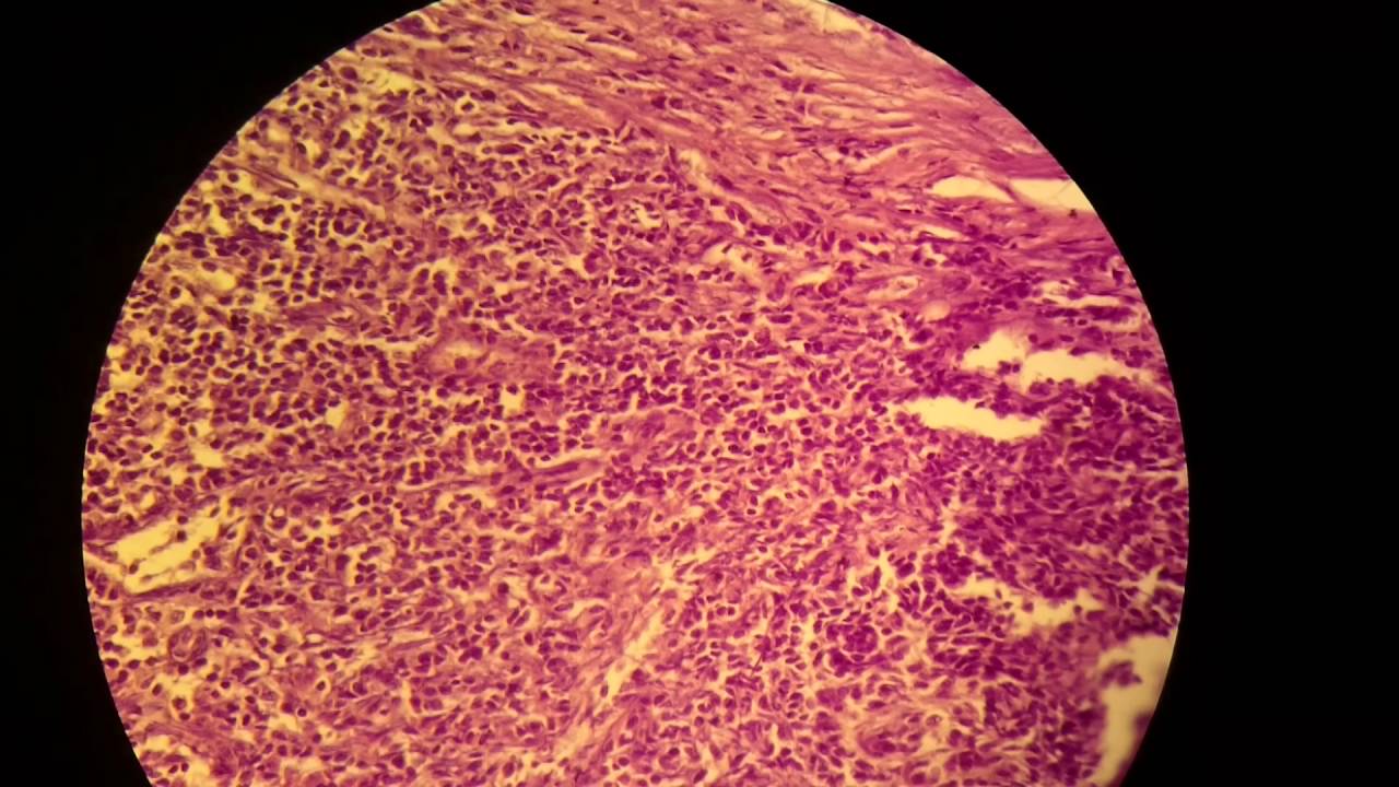
Appearance
In caseous necrosis no histological architecture is preserved. On microscopic examination with H&E staining, it is characterized by acellular pink areas of necrosis surrounded by a granulomatous inflammatory process.
When the hilar lymph node for instance is infected with tuberculosis and leads to caseous necrosis, its gross appearance can be a cheesy tan to white, which is why this type of necrosis i…
External links
• Microscope images of caseous necrosis
• Image of a hilar lymph node demonstrating caseous necrosis
• Image of a caseating granuloma of tuberculosis in the adrenal gland