
What does the semimembranosus muscle do?
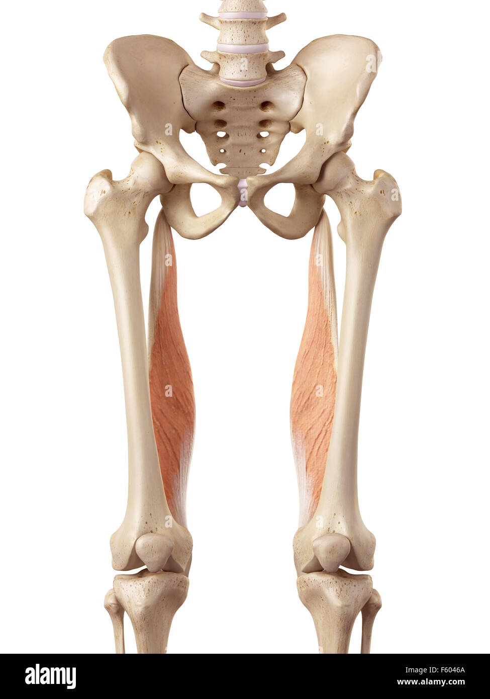
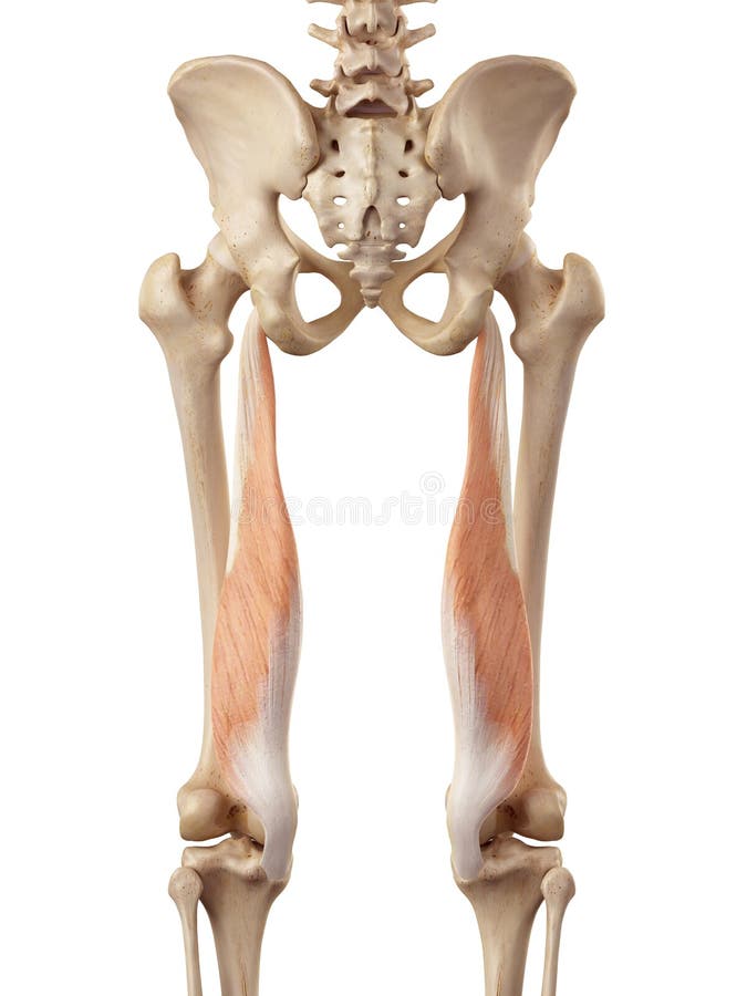
The semimembranosus muscle ( / ˌsɛmiˌmɛmbrəˈnoʊsəs /) is the most medial of the three hamstring muscles in the thigh. It is so named because it has a flat tendon of origin. It lies posteromedially in the thigh, deep to the semitendinosus muscle. It extends the hip joint and flexes the knee joint .
Where does the semimembranosus originate?
Semimembranosus originates from the upper lateral facet on the ischial tuberosity (part of the pelvis) attaching to it via a strong tendon. Fleshy muscles fibers run downwards and slightly medially underneath semitendinosus and biceps femoris.
What is semimembranosus bursitis?
Excess fluid in the knee e.g. from a knee injury, arthritis or gout can seep into the bursa, causing it to swell. In fact arthritis is the most common cause of semimembranosus bursitis behind the knee. Localised Knee Pain: bursitis knee pain typically develops gradually, fluctuates and tends to be a general ache rather than sharp knee pain.
How is the semimembranosus separated from the tibia?
Other fibres pass upwards and laterally to form the oblique popliteal ligament which attaches to the posterolateral femoral condyle. Semimembranosus is separated from the gastrocnemius calf muscle and the tibia via a bursa (small fluid filled sac) to reduce friction.
What does semimembranosus mean?
Medical Definition of semimembranosus : a large muscle of the inner part and back of the thigh that arises by a thick tendon from the back part of the tuberosity of the ischium, is inserted into the medial condyle of the tibia, and acts to flex the leg and rotate it medially and to extend the thigh.
Why is it called the semitendinosus?
Semitendinosus is one of the three muscles that make up the hamstrings muscle group, and it is located at the posterior and medial aspect of the thigh. The semitendinosus is so named due to it having a long tendon of insertion.
What is the common name for semimembranosus muscle?
Collectively semimembranosus, semitendinosus and biceps femoris are referred to as the hamstring muscles. Semimembranosus (along with semitendinosus) occupies the medial aspect of the posterior thigh.
How do you remember the semimembranosus?
0:161:27Hamstrings Muscles Order How to Memorize - YouTubeYouTubeStart of suggested clipEnd of suggested clipTowards me so that's B T M bent stands for biceps femoris T stands for - what it stands forMoreTowards me so that's B T M bent stands for biceps femoris T stands for - what it stands for semitendinosus. And me it's semimembranosus.
What is the difference between semitendinosus and semimembranosus?
The semitendinosus is more superficial than the semimembranosus (with which it shares very close insertion and attachment points). However, because the semimembranosus is wider and flatter than the semitendinosus, it is still possible to palpate the semimembranosus directly.
Why hamstring muscles are called so?
The "ham" of "hamstring" comes from an Old Teutonic word "ham" meaning crooked. This is in reference to the crooked part of the leg, that is the knee. To "hamstring" someone is to cripple them. See Hamstring Injuries and the Hamstring Stretch to prevent hamstring injuries.
What is the role of the semimembranosus?
The semimembranosus muscle flexes and medially rotates the leg at the knee and extends the thigh at the hip joint. A fibrous extension of the muscle called the oblique popliteal ligament extends upward and laterally to provide support to the posterior knee joint.
Is semimembranosus a hamstring muscle?
The semimembranosus muscle is the deepest and inner most of the hamstring muscles, found on the inner (medial) side of the back of the thigh. It runs almost directly below one of the other hamstring muscles, semitendinosus.
What is the action of the semimembranosus?
Actions of Semimembranosus on the leg (tibia & fibula): Flexes leg at knee. b. Medially rotates leg at knee when knee is flexed.
What is the proper name for the hamstring muscle?
The hamstring muscles, or simply the hamstrings, are a group of three long muscles located in the posterior compartment of the thigh, shaping up the surface anatomy of this region. These muscles are the biceps femoris, semimembranosus and semitendinosus muscles.
What movement does the semitendinosus muscle perform?
As a prime mover, semitendinosus extends and internally rotates the thigh, flexes and internally rotates the leg. It also has a postural role, stabilising the pelvic girdle. Hip joint: Thigh extension, thigh internal rotation, stabilizes pelvis. Knee joint: Leg flexion, leg internal rotation.
What actions does the semitendinosus muscle perform?
The semitendinosus muscle, collectively with the other two muscles of the posterior compartment of the thigh, works to extend at the hip and flex at the knee. The semitendinosus muscle, in particular, has the added functionality of assisting the popliteus muscle in rotating the leg internally.
Does Semitendinosus grow back?
It has been demonstrated that the semitendinosus tendon can regenerate after being harvested in its whole length and thickness for anterior cruciate ligament (ACL) reconstruction.
What happens to hamstring after ACL graft?
So, to answer the question: What Happens to My Harvested Hamstring Tendon After ACL Surgery? The answer is in 70% of the patients, the hamstring tendon will regenerate and this occurs within a year after surgery.
Where is the semimembranosis located?
Semimembranosis is one of a group of muscles called the Hamstrings. It is located on the posteromedial side of the thigh deep to Semitendinosus. Its origin is the ischial tuberosity on the inferior pelvis and the insertion is the medial tibial condyle. It's primary action is knee flexion, hip extension and knee internal rotation. In the lower part of the thigh, semitendinosus and semimembranosus together form the upper medial boundary of the popliteal fossa.
Which is harder to palpate, semimembranous or semitendinosus?
Semimembranous lies deep to semitendinosus and is difficult to palpate, but can be felt easier when the knee is flexed.
What is the bursae that separates the muscle from the medial head of the tibia and?
The bursae that separates the muscle from the medial head of the tibia and the medial head of the Gastrocnemius may at times become enlarged with distended fluid. This swelling is termed 'Baker's cyst' (described by Morrant Baker in the 19th century as a cystic mass in the popliteal fossae of children).
Which ligament is formed by the bursae?
The tendon then passes upwards and laterally to form the oblique popliteal ligament. [1] A bursae separate the muscle from the medial head of the tibia and the medial head of the Gastrocnemius[1] Nerve[edit| edit source] Tibial divisionof the Sciatic Nerve(root value L5, S1 and 2).
Which of the following tendons assists with flexion of the knee?
Gracilis, Sartorius, Popliteus, gastrocnemius, and plantaris assist with flexion of the knee.
Where is the semimembranosus muscle located?
Structure. The semimembranosus muscle, so called from its membranous tendon of origin, is situated at the back and medial side of the thigh. It is wider, flatter, and deeper than the semitendinosus (with which it shares very close insertion and attachment points). The muscle overlaps the upper part of the popliteal vessels.
Which part of the sciatic nerve innervates the semimembranosus?
The semimembranosus is innervated by the tibial part of the sciatic nerve. The sciatic nerve consists of the anterior divisions of ventral nerve roots from L4 through S3. These nerve roots are part of the larger nerve network–the sacral plexus.
What muscle is reduced or absent?
The semimembranosus muscle may be reduced or absent, or double, arising mainly from the sacrotuberous ligament and giving a slip to the femur or adductor magnus .
Which muscle is used to medially rotate the knee?
It also helps to medially rotate the knee: the tibia medially rotates on the femur when the knee is flexed. It medially rotates the femur when the hip is extended. The muscle can also aid in counteracting the forward bending at the hip joint.
Which part of the muscle is the tendon of origin?
The tendon of origin expands into an aponeurosis, which covers the upper part of the anterior surface of the muscle; from this aponeurosis, muscular fibers arise, and converge to another aponeurosis which covers the lower part of the posterior surface of the muscle and contracts into the tendon of insertion.
Which ligament is inserted into the posterior lateral condyle of the femur?
The tendon of insertion gives off certain fibrous expansions: one, of considerable size, passes upward and laterally to be inserted into the posterior lateral condyle of the femur, forming part of the oblique popliteal ligament of the knee-joint; a second is continued downward to the fascia which covers the popliteus muscle; while a few fibers join the medial collateral ligament of the joint and the fascia of the leg.
Can semitendinosus be dry needled?
The semitendinosus muscle may be dry needled.
What is the semimembranosus?
Overview. Muscle Group: Semimembranosus is the inner most (medial) hamstring muscle. Action: Knee flexion and rotation, hip extension. Origin: Ischial tuberosity. Insertion: The back of the medial tibial condyle extending to the lateral tibial condyle via the oblique popliteal ligament. Nerve Supply: Tibial division of the sciatic nerve (L5, S1, S2)
Where does semimembranosus originate?
Semimembranosus originates from the upper lateral facet on the ischial tuberosity (part of the pelvis) attaching to it via a strong tendon. Fleshy muscles fibers run downwards and slightly medially underneath semitendinosus and biceps femoris.
What muscles are used to bend the knee?
The primary function of semimembranosus and the other two hamstring muscles is to bend the knee, e.g. when stepping over an object. They can also work to move the hip and when the trunk (back) is bent forwards e.g. when reaching down to pick something off the ground, the hamstrings work to extend the hip joint to help you come up to standing.
Which ligament attaches to the posterolateral femoral condyle?
Other fibres pass upwards and laterally to form the oblique popliteal ligament which attaches to the posterolateral femoral condyle. Semimembranosus is separated from the gastrocnemius calf muscle and the tibia via a bursa (small fluid filled sac) to reduce friction.
Where does the aponeurotic originate?
Location. It originates from the upper lateral facet on the ischial tuberosity (part of the pelvis) attaching to it via a strong tendon. Fleshy muscles fibres run downwards and slightly medially underneath semitendinosus and biceps femoris. Just above the knee joint, the muscle narrows into an aponeurotic ...
Where is the Pes Anserine Bursa?
Pes Anserine bursitis occurs on the inner side of the knee. The pes anserine bursa sits between the medial collateral ligament and the conjoined medial knee tendons of the gracilis, sartorius and semitendinosus muscles.
Why does my bursa sit behind my knee?
Bursitis behind the knee is usually due to knee injuries or arthritis . You can find out all about the common causes, symptoms, diagnosis and treatment options in the Bakers Cyst Knee section.
How many bursas are there in the knee?
There are approximately eleven bursa located around the knee and bursitis can develop at any one of the bursa knee locations.
What is knee bursitis?
Knee bursitis occurs when there is irritation or inflammation in one of the knee bursa . Bursa are small fluid filled sacs that reduce the friction between two surfaces. They are found all over the body, and sit between bones and muscles, a bit like ball bearings.
Signs and symptoms
Symptoms may include swelling behind the knee, stiffness, and pain. If the cyst breaks open, pain may increase, and there may be swelling of the calf. Rupture of a Baker's cyst may also cause bruising below the medial malleolus of the ankle (Crescent sign).
Cause
In adults, Baker's cysts usually arise from almost any form of knee arthritis (e.g., rheumatoid arthritis) or cartilage (particularly a meniscus) tear. Baker's cysts in children do not point to underlying joint disease. Baker's cysts arise between the tendons of the medial head of the gastrocnemius and the semimembranosus muscles.
Diagnosis
Diagnosis is by examination. A Baker's cyst is easier to see from behind with the patient standing with knees fully extended. It is most easily palpated (felt) with the knee partially flexed.
Treatment
Baker's cysts usually require no treatment unless they are symptomatic. It is very rare that the symptoms are actually coming from the cyst. In most cases, there is another disorder in the knee (arthritis, meniscal (cartilage) tear, etc.) that is causing the problem.
Overview

Structure
Function
Clinical significance
See also
External links