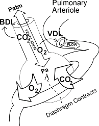
What is the difference between ventilation and perfusion?
Ventilation (V) refers to the flow of air into and out of the alveoli, while perfusion (Q) refers to the flow of blood to alveolar capillaries. Individual alveoli have variable degrees of ventilation and perfusion in different regions of the lungs.
What happens if ventilation and perfusion are not matched?
If ventilation and perfusion are not matched, then gas exchange diminishes, particularly in the case of oxygen. The relationship between ventilation and perfusion is referred to as V/Q, which describes the ratio between ventilation (V) and perfusion (Q) for a particular lung region.
What is ventilation/perfusion ratio (V/Q)?
Ventilation (V) refers to the flow of air into and out of the alveoli, while perfusion (Q) refers to the flow of blood that reaches the alveoli via the capillaries. The ventilation/perfusion ratio (V/Q ratio) is a ratio used to the efficiency and adequacy of the matching of these two variables. In an average 70 kg male:
How is ventilation and perfusion in the lungs measured?
Individual alveoli have variable degrees of ventilation and perfusion in different regions of the lungs. Collective changes in ventilation and perfusion in the lungs are measured clinically using the ratio of ventilation to perfusion (V/Q). Changes in the V/Q ratio can affect gas exchange and can contribute to hypoxemia.

What is the relationship between ventilation and perfusion?
Ventilation (V) refers to the flow of air into and out of the alveoli, while perfusion (Q) refers to the flow of blood to alveolar capillaries. Individual alveoli have variable degrees of ventilation and perfusion in different regions of the lungs.
What happens when there is a mismatch between ventilation and perfusion?
Ventilation-perfusion (V/Q) mismatch occurs when either the ventilation (airflow) or perfusion (blood flow) in the lungs is impaired, preventing the lungs from optimally delivering oxygen to the blood.
What is meant by ventilation perfusion coupling or matching?
Ventilation-perfusion coupling is the relationship between ventilation and perfusion processes, which take place in the respiratory and cardiovascular systems. Ventilation is the movement of gas during breathing, and perfusion is the process of pulmonary blood circulation, which delivers oxygen to body tissues.
What happens when ventilation is greater than perfusion?
Respiratory Distress and Failure The difference in perfusion (Q̇) is greater than the difference in ventilation (V̇). Perfusion in excess of ventilation results in incomplete arterialization of systemic venous (pulmonary arterial) blood and is referred to asvenous admixture.
How does ventilation perfusion mismatch cause hypoxia?
Although the impact of high V/Q unit on blood oxygenation is minimal, it can cause hypoxemia if the compensatory rise in total ventilation is absent. Since the high V/Q unit receiving less perfusion, blood from this area is diverted to other areas leading to the development of low V/Q in other areas of the lungs.
How does ventilation regulate perfusion in the lungs?
9:3215:00Ventilation and Perfusion (V:Q Ratio) Physiology - YouTubeYouTubeStart of suggested clipEnd of suggested clipSo your v2 ratio decreases from the apex of the lung to the base of the lung. Again ventilation isMoreSo your v2 ratio decreases from the apex of the lung to the base of the lung. Again ventilation is the amount of gas oxygen. Coming into your alveoli ready for gas exchange.
What is the V Q ratio and why is it important?
Now, the ratio of V to Q influences how efficiently gases, specifically O2 and CO2 , are exchanged in the lungs. In healthy lungs with a V/Q ratio of 0.8, the alveolar partial pressure of O2 (PAO2), is about 100 mmHg or millimeters of mercury; and the alveolar partial pressure of CO2 (PACO2) is about 40 mmHg.
How does ventilation work with perfusion and diffusion?
0:061:24Respiration: Ventilation, Diffusion and Perfusion | Ausmed Explains...YouTubeStart of suggested clipEnd of suggested clipThe respiratory process consists of three components ventilation diffusion and perfusion ventilationMoreThe respiratory process consists of three components ventilation diffusion and perfusion ventilation consists of two parts the inspiration is the expansion of the chest with a negative interpoma.
What is the difference between ventilation diffusion and perfusion?
The main difference between perfusion and diffusion is that perfusion is the delivery of blood to the pulmonary capillaries, whereas diffusion is the movement of gases from the alveoli to plasma and red blood cells. Furthermore, ventilation and perfusion occur simultaneously, facilitating the diffusion.
What is the difference between shunt and VQ mismatch?
V/Q mismatch is common and often effects our patient's ventilation and oxygenation. There are 2 types of mismatch: dead space and shunt. Shunt is perfusion of poorly ventilated alveoli. Physiologic dead space is ventilation of poor perfused alveoli.
What does the clients ventilation and perfusion ratio indicate?
In respiratory physiology, the ventilation/perfusion ratio (V/Q ratio) is a ratio used to assess the efficiency and adequacy of the matching of two variables: V – ventilation – the air that reaches the alveoli. Q – perfusion – the blood that reaches the alveoli via the capillaries.
What is a mismatched perfusion defect?
Ventilation perfusion mismatch or V/Q defects are defects in the total lung ventilation/perfusion ratio (V/Q ratio). It is a condition in which one or more areas of the lung receive oxygen but no blood flow, or they receive blood flow but no oxygen.
What happens when ventilation is not sufficient?
In cases when ventilation is not sufficient for an alveolus, the body redirects blood flow to alveoli that are receiving sufficient ventilation. This is achieved by constricting the pulmonary arterioles that serves the dysfunctional alveolus, which redirects blood to other alveoli that have sufficient ventilation.
What are the two types of ventilation perfusion mismatch?
There are 2 types of mismatch: dead space and shunt. Shunt is perfusion of poorly ventilated alveoli. Physiologic dead space is ventilation of poor perfused alveoli.
What is the relationship between ventilation and perfusion?
Understanding the Ventilation-Perfusion Relationship. To ensure that enough oxygen is provided by ventilation to saturate the blood fully requires that ventilation and perfusion of the lungs are adequately matched. Ventilation (V) refers to the flow of air into and out of the alveoli, while perfusion ...
What is the difference between perfusion and ventilation?
Ventilation (V) refers to the flow of air into and out of the alveoli, while perfusion (Q) refers to the flow of blood that reaches the alveoli via the capillaries.
What would happen if the alveoli were perfused but not ventilated at all?
If the alveoli were perfused but not ventilated at all, then the V/Q ratio would be zero. A clinical example of this would be the inhalation of a foreign body that prevented all ventilation to an area of the lung. Ventilation in that region would be reduced to zero, lowering the V/Q ratio to zero. Because this area is being adequately perfused, but no oxygen is being transferred to the blood arriving there, a physiological right-to-left shunt has been created.
What are the two mechanisms that cause low V/Q?
The two main mechanisms by which this can occur are: Hypoxic vasoconstriction: In the presence of a low V/Q ratio (i.e. areas of poor ventilation), hypoxic vasoconstriction can occur, which redirects incoming blood to the affected area to other parts of the lung. This decreases the perfusion of the hypoxic region, raising the V/Q ratio.
Why does the V/Q ratio vary?
The V/Q ratio, however, varies in different regions of the lung partially because of the proximity of the heart to the region of the lung. Both ventilation and perfusion increase from the apex to the base of the lungs, perfusion, however, rises at a greater rate. This results in the following pattern:
What is the effect of a high V/Q ratio on the bronchi?
areas of poor perfusion), the bronchi constrict, increasing the resistance and decreasing the ventilation of the affected area that is not well perfused. This reduces the amount of alveolar dead space that occurs and lowers the V/Q ratio.
What causes a change in the V/Q ratio?
In clinical practice, disease processes tend to cause changes in the V/Q ratio that vary throughout the lung. An example of this is chronic obstructive pulmonary disease (COPD), which causes destruction of the alveoli in the lung, resulting in large air spaces and capillary loss. The large air spaces result in poor ventilation, whereas the capillary loss results in inadeuquate perfusion.
Which article is the best to discuss ventilation and perfusion matching?
The best article to discuss ventilation and perfusion matching would have to be Petersson & Glenny (2014), mainly because it has the distinction of being free for all readers. All of the original articles quoted by Petersson & Glenny are paywalled, but their content is more interesting than educational, and one could easily just stick to that one paper.
Why are ventilation and perfusion separated?
The distribution of perfusion and ventilation are separated somewhat on the graph, but are still reasonably close together because this young lung is doing a great job of autoregulating its own blood flow. By comparison, here is the distribution which Wagner et al measured from a 44-year-old male (selected from among male non-smokers, a rare species in the fifties):
What is the range of V/Q ratios?
As one can see, though theoretically V/Q ratios can be distributed between zero and infinity, in this young person they were confined to a range between 0.3 and 3.0. That is probably the range of V/Q ratios which was asked for in the college examiner's comments to Question 6 from the first paper of 2008. Blood flow was maximal through regions enjoying good ventilation, and ventilation was maximal in regions well supplied with blood. At the outer limits of the bell curve, the lower end of V/Q ratios represents regions of atelectatic lung, and the total blood flow to such regions was minimal (this guy's hypoxic pulmonary vasoconstriction was clearly working). At the upper limits, there was also minimal blood flow, which makes sense because this represents those regions of the lung which are not being perfused particularly well in spite of adequate ventilation.
What is the V/Q ratio?
There are several patterns of V/Q matching and mismatching:#N#V/Q = 1.0: A lung unit with well-matched gas and blood flows will have a V/Q ratio close to 1.0, i.e. for every unit of blood flow it will receive a unit of gas flow.#N#V/Q < 1.0: Good blood flow but insufficient ventilation; generally seen in the bases of lung (West's Zone 4)#N#V/Q >1.0: Excellent ventilation but poor blood flow; West's Zone 1.#N#V/Q = ∞: Lung units which receive no blood flow, i.e. "true" dead space#N#V/Q = 0: Lung units which receive no ventilation, i.e. "true" shunt 1 V/Q = 1.0: A lung unit with well-matched gas and blood flows will have a V/Q ratio close to 1.0, i.e. for every unit of blood flow it will receive a unit of gas flow. 2 V/Q < 1.0: Good blood flow but insufficient ventilation; generally seen in the bases of lung (West's Zone 4) 3 V/Q >1.0: Excellent ventilation but poor blood flow; West's Zone 1. 4 V/Q = ∞: Lung units which receive no blood flow, i.e. "true" dead space 5 V/Q = 0: Lung units which receive no ventilation, i.e. "true" shunt
What does V/Q mean in physics?
V/Q = ∞: Lung units which receive no blood flow, i.e. "true" dead space
Is V/Q matching efficient?
As one can plainly see, the V/Q matching is now much less efficient. There's a whole region of lung where there is plenty of blood flow but barely any ventilation, and there's poorly perfused lung where the ventilation is relatively good. This poorly ventilated lung region is probably the base of the lung, which ends up being below closing capacity ...
