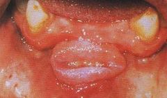
Why stippling is seen in gingiva? The gingiva often possess a textured surface that is referred to as being stippled (engraved points). Stippling is a consequence of the microscopic elevations and depressions of the surface of the gingival tissue due to the connective tissue projections within the tissue.
What is stippling of the gums?
Stippling is seen at the sites of fusion of the epithelial ridges or Rete Pegs and corresponds to the fusion of the valleys created by the connective tissue papillae It is Microscopic elevations and Depressions of the surface of the gingival tissue due to the connective tissue projections within the tissue.
Why is there no stippling in attached gingiva?
Stippling is usually seen in attached gingiva as it is firmly attached to the underlying cementum and alveolar bone with the help of collagen fibers of the connective tissue. Stippling is lost as age progress, in most adult patients above 50 years there is no stippling of Gingiva.
What is the pathophysiology of stippling?
Stippling is a consequence of the microscopic elevations and depressions of the surface of the gingival tissue due to the connective tissue projections within the tissue. The degree of keratinization and the prominence of stippling appear to be related.
What is the attached gingiva?
The attached gingiva extends from the free gingiva coronal to the alveolar mucosa in the apical portion of the tooth. Stippling is usually seen in attached gingiva as it is firmly attached to the underlying cementum and alveolar bone with the help of collagen fibers of the connective tissue.

Is attached gingiva stippled?
Both the attached gingiva and the interdental gingiva are stippled, while the marginal gingiva is not stippled. The attached gingiva is connected to the alveolar bone and not to the freely movable alveolar mucosa.
At what age stippling occurs?
Stippling generally appears at around age 4 or 5 and is absent before this; stippling also begins to disappear in elderly people. Usually, any semblance of stippling that was present in an individual's mouth has disappeared by the age of 50.
What is a stippled?
1 : to engrave by means of dots and flicks. 2a : to make by small short touches (as of paint or ink) that together produce an even or softly graded shadow. b : to apply (something, such as paint) by repeated small touches. 3 : speckle, fleck.
What is a stippling in medical terms?
Medical Definition of stippling : the appearance of spots : a spotted condition (as in basophilic red blood cells, X-rays of the lungs, or bones)
What is stippling in gingiva?
The gingiva often possess a textured surface that is referred to as being stippled (engraved points). Stippling only presents on the attached gingiva bound to underlying alveolar bone, not the freely moveable alveolar mucosaor free gingiva . Stippling used to be thought to indicate health, but it has since been shown that smooth gingiva is not an ...
Why is my gingival smooth?
Stippling used to be thought to indicate health, but it has since been shown that smooth gingiva is not an indication of disease, unless it is smooth due to a loss of previously existing stippling. Stippling is a consequence of the microscopic elevations and depressions of the surface of the gingival tissue due to the connective tissue projections ...
Where does stippling occur?
To be more specific, stippling occurs at sites of fusion of the epithelial ridges (also known as rete pegs -depression of epithelium ) and correspond to the fusion of the valleys created by the connect ive tissue papillae (elevation of connective tissue papilla.An example of stippling could be dots found in basketball or an orange.
What is the attachment of the gingiva?
Stippling is typically seen in the attached gingiva as it is firmly attached to the underlying cementum and alveolar bone with the help of the collagen fibers of the connective tissue. Stippling is often lost as age progresses.
What is the connective tissue of the gingiva?
The connective tissue of the gingiva is covered by the various characteristic gingival epithelia. The cementum, which overlays the tooth’s root, is attached to the adjacent cortical surface of the alveolar bone. It is attached by the alveolar crest, horizontal and oblique fibers of the periodontal ligament.
Which bone is surrounded by the subepithelial connective tissue of the gingiva?
Alveolar bone proper. The tissues found in the periodontium combine together to form an active group of tissues. The alveolar bone is almost completely surrounded by the subepithelial connective tissue of the gingiva. The connective tissue of the gingiva is covered by the various characteristic gingival epithelia.
What is the Marin periodontium?
Marin Contemporary Perio & Implant Concepts. The periodontium is the specialized tissues that provide two functions, surrounding and supporting the teeth. In addition to maintaining them within the maxillary and mandibular bones. The word is derived from the Greek terms peri-, which means "around" and -odont, which means "tooth".
Dr. Zalewsky
Dr. Justin Zalewsky, a graduate of Case Western Reserve University School of Dentistry, then went on to Temple University Kornberg School of Dentistry where he received his Certificate of Periodontology and Oral Implantology.
Dr. Daru
Dr. Antara Daru, a graduate of New York University College of Dentistry, then went on to University of Maryland, Baltimore College of Dentistry where she received her Masters of Science in Oral Biology and Certificate in Periodontology and Oral Implantology.
What is stippling gum?
The gingiva, or the gums, typically contain a textured surface which is referred to as being stippled. This can be thought of as resembling a texture similar to an orange peel. Stippling occurs from the connective tissue projections within the gingival tissue which create microscopic depressions and elevations.
What is attached gingiva?
The attached gingiva is connected to the underlying alveolar bone and not the freely movable alveolar mucosa. Gingiva is comprised of two different categories including the free gingiva and attached gingiva. The free gingiva encompasses the tooth and creates a collar which surrounds the crown.
What age do you not have stippling?
It is common for stippling to be lost completely or at least reduced as a patient ages. Most adult patients over the age of 50 do not have stippling of the gingiva.
What is the free gingiva?
The free gingiva encompasses the tooth and creates a collar which surrounds the crown. This is the portion of the gingiva which extends from the attached gingiva to the surface of the tooth. The attached gingiva extends from the free gingiva coronal all the way to the alveolar mucosa.
What is periodontium in dentistry?
The periodontium includes specialized tissues which serve two different functions including surrounding and supporting the teeth to maintain them in the maxillary and mandibular bones. The word periodontium is derived from the Greek terms peri-, which means "around" and -odont, which means "tooth". When the phrase is taken literally, periodontium translates to "around the tooth". Periodontics is the dental specialty which focuses on the care and maintenance of these tissues. It provides support required to maintain the full function of the teeth. The practice consists of four main areas which includes:
Can gingival stippling be lost?
In cases where gingival inflammation has occurred, gingival stippling can be lost, however, it will be re-established once the gingival health is restored. The presence, configuration, and amount of gingival stippling can vary for each patient based on their age, gender, and the location of the stippling.
Does stippling occur on the alveolar mucosa?
Stippling is only present on the attached gingiva which is connected to alveolar bone. It does not exist on the freely moveable alveolar mucosa. Historically, experts thought that stippling was an indicator of a patient having good oral health.
