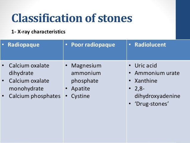
They are radiolucent. The factors that predispose to the development of uric
Uric acid
Uric acid is a heterocyclic compound of carbon, nitrogen, oxygen, and hydrogen with the formula C₅H₄N₄O₃. It forms ions and salts known as urates and acid urates, such as ammonium acid urate. Uric acid is a product of the metabolic breakdown of purine nucleotides, and it is a normal compon…
Full Answer
What is the pathophysiology of uric acid stones?
Uric acid is a purine metabolite and is also found in large quantities within cells. Most patients with uric acid stones have a reduced urinary pH, whereas other less common causes are low urine volume or elevated urinary uric acid levels. Factors associated with uric acid stones are listed in Table 30-3.
What is the difference between kidney stones and uric acid?
Uric acid stones are a type of kidney stone. When you have a high level of uric acid in your blood and urine, small stones can form. These stones can block the passage of urine out of your body, causing pain and other symptoms. Many uric acid stones pass on their own.
What are uric acid stones and how are they treated?
Too much uric acid in the body leads to small stones forming, which can cause pain when you pee and blood in the urine. Small uric acid stones may pass on their own. For larger stones, providers may use minimally invasive or noninvasive treatments such as PCNL and shockwave lithotripsy. What are uric acid stones?
How common are uric acid stones in films?
Uric acid stones are uncommon, and are radiolucent on plain films. They typically have a sheet like / oblong morphology. Christoph Thomas, Martin Heuschmid, David Schilling, Dominik Ketelsen, Ilias Tsiflikas, Arnulf Stenzl, Claus D. Claussen, Heinz-Peter Schlemmer.

Are uric acid stones radiolucent?
Pure uric acid stones are radiolucent but well visualized on renal ultrasound. A 24 h urine collection for stone risk analysis provides essential insight into the pathophysiology of stone formation and may guide therapy.
What type of kidney stones are radiolucent?
Urinary matrix stones are a rare form of urinary calculi that are typically radiolucent [1]. However, the common ureteral stones are easily detected using computed tomography (CT) scanning.
Can you see uric acid stones?
Imaging: You may need a CT scan or ultrasound to find uric acid stones in your urinary tract. These imaging scans can help find even small stones. Usually, providers don't use abdominal X-rays because they may miss smaller stones.
Why are renal stones radiopaque?
They can grow very large and form a cast of the renal pelvis and calyces resulting in so-called staghorn calculi. The struvite accounts for ~70% of these calculi and is usually mixed with calcium phosphate thus rendering them radiopaque.
Which stones are radiolucent and radiopaque?
Cystine calculi are said to be either radiolucent or radiopaque.
Are urate stones radiopaque?
They are correct that urate and cystine are the least radiopaque of the common stones in dogs and cats. However, radiographic appearance of uroliths depends on several factors of which size and mineral type are the most important.
What makes uric acid stones?
Uric acid stones form when the levels of uric acid in the urine are too high, and/or the urine is too acidic on a regular basis. The formation of these types of stones can run in families. Inherited problems in how the body processes uric acid or protein in the diet can increase the acid in urine.
What does a uric acid crystals look like?
Uric acid crystals are characterised by their needle shape and strong double refraction in polarised light, whereas crystals of calcium pyrophosphate dihydrate, which are found in chondrocalcinosis, have a more rhomboid appearance and limited birefringence (2, 3).
What causes uric acid crystals?
Urate crystals can form when you have high levels of uric acid in your blood. Your body produces uric acid when it breaks down purines — substances that are found naturally in your body. Purines are also found in certain foods, including red meat and organ meats, such as liver.
How are radiolucent stones detected?
Computed tomography (CT) can be performed rapidly and can detect radiolucent stones, except for drug-induced stones. It is preferred over intravenous pyelography or urography because it is more sensitive and does not require the use of intravenous contrast medium.
Which kidney stones are not visible on CT?
Many stone types can be visualized using KUB radiography; however cystine and struvite stones often are poorly visible on KUB radiography, and uric acid and matrix stones are not visible at all.
Can radiolucent stones be seen on ultrasound?
Small case series by Edell and Zegel as well as Pollack and colleagues demonstrated the capabilities of grayscale ultrasound by showing how it could detect radiolucent uric acid and matrix stones, respectively, both of which were not appreciated on conventional plain-film urography (Fig. 2).
Is struvite radiolucent?
Struvite stones They can grow very large and form a cast of the renal pelvis and calyces resulting in so-called staghorn calculi. The struvite accounts for ~70% of these calculi and is usually mixed with calcium phosphate thus rendering them radiopaque. Uric acid and cystine are also found as minor components.
Are struvite stones radiopaque?
Like cystine and uric acid stones, struvite stones are radiopaque in nature, but they are not as dense as calcium stones.
How are radiolucent stones diagnosed?
Computed tomography (CT) can be performed rapidly and can detect radiolucent stones, except for drug-induced stones. It is preferred over intravenous pyelography or urography because it is more sensitive and does not require the use of intravenous contrast medium.
Which kidney stones are not seen on ultrasound?
Many stone types can be visualized using KUB radiography; however cystine and struvite stones often are poorly visible on KUB radiography, and uric acid and matrix stones are not visible at all.
What Are Uric Acid Stones?
Uric acid stones are one of four major types of kidney stones, which include calcium stones (calcium oxalate and calcium phosphate), struvite stone...
How Common Are Uric Acid Stones?
It is estimated that one in 10 people in the U.S. will have a kidney stone of one kind or another at some time in their lives. In the late 1970s, a...
What Causes Uric Acid Stones?
Uric acid stones form when the levels of uric acid in the urine is too high, and/or the urine is too acidic (pH level below 5.5) on a regular basis...
What Are The Symptoms of Uric Acid Stones?
All types of kidney stones produce similar symptoms, including one or more of the following: 1. Pain in the lower back, sides, abdomen or groin; th...
What factors predispose to the development of uric acid stones?
They are radiolucent. The factors that predispose to the development of uric acid stones are the urine concentration of uric acid and low urine (pH < 5.5). Low urine volume increases the risk for uric acid stones by the same mechanisms discussed previously for calcium stones.
How to treat uric acid stones?
Treatment of uric acid stones involves increasing urine volume and pH as well as decreasing uric acid excretion. Alkaline urine can prevent uric acid stone formation and may also result in stone dissolution. To raise urine pH, potassium citrate is recommended. Whereas sodium bicarbonate alkalinizes the urine and enhances uric acid solubility, the added sodium increases sodium urate formation, which serves as a nidus for calcium oxalate precipitation. Potassium citrate (40 to 50 mmol/day in divided doses) is given, increasing the dose as necessary to achieve a urine pH of 6.5 to 7. Patients should monitor pH with urine dipsticks at various times of the day and adjust dosing accordingly. If urine pH remains low despite potassium citrate above 100 mmol daily or if that dose results in hyperkalemia, acetazolamide is added. This carbonic anhydrase inhibitor produces an alkaline urine similar to that seen in renal tubular acidosis. Patients should be cautioned not to exceed a urine pH of 7 because this may result in calcium phosphate precipitation.
What is uric acid?
Uric acid is a purine metabolite and is also found in large quantities within cells. Most patients with uric acid stones have a reduced urinary pH, whereas other less common causes are low urine volume or elevated urinary uric acid levels. Factors associated with uric acid stones are listed in Table 30-3.
What are urate stones made of?
All urate stones are composed of salts of uric acid, which means that one or two hydrogen ions are replaced by other cations like NH4+, potassium, sodium, or calcium. In contrast with uric acid, the formation of urate stones mostly starts at physiologic pH levels of greater than 6.5. Nevertheless, no urate stone develops without elevated urinary uric acid excretion. The only urate stone of clinical relevance is the ammonium urate stone, which is known to develop in patients with recurrent urinary tract infections or endemically (see Infection Stones later in this chapter).
How much of a renal stone is uric acid?
Uric acid stones account for 5% to 10% of all renal stones in the United States, but this figure varies in other parts of the globe. The chance of stone formation increases with increasing serum urate levels and urine excretion rates.
What is the risk factor for uric acid stones?
The most important risk factor for uric acid crystallization and stone formation is a low urine pH (below 5.5).
What is the cause of uric acid nephropathy?
A low urine pH is the major cause of uric acid nephropathy. The solubility of uric acid increases sixfold with an increase in urine pH from 5.3 to 6.5 (Fig. 30-7 ). 20,149 Thus, conditions that lower urine pH tend to predispose patients to uric acid lithiasis.
Why do uric acid stones occur?
A decrease in urine output increases highly concentrated urinary solutes and supersaturation that increase susceptibility to crystal and stone formation. This leads to the precipitation of uric acid crystals, which lead to stones . This explains why uric acid stones are more common in tropical and humid climates. [54] [55]
What is the best treatment for uric acid kidney stones?
Management of uric acid kidney stones includes lifestyle changes, medical treatment focusing on decreasing uric acid production and excretion, and urinary alkalinization. [20] Overall, urinary alkalinization is considered the single most effective therapy. The goal is to achieve a urine pH of 6 to 6.5. For uric acid kidney stones, renal ultrasonography can be used for tracking as the calculi will not be visible on a standard KUB.
What is the best way to reduce uric acid?
High intake of fruits and vegetables, low intake of purine-rich diets and animal proteins are necessary to reduce the burden of uric acid production in patients with uric acid nephrolithiasis. Orange juice and lemonade are often recommended but very large amounts are necessary for any meaningful change in urinary pH. [74] A high intake of fluids to maintain adequate urinary output (>2,000 ml daily) is useful to lower supersaturations and help prevent kidney stones. Recommendations to reduce obesity, appropriate management of hypertension, and high blood sugar are also helpful in reducing the burden of uric acid kidney stones.
Does citrate cause urinary stones?
Many components in urine suppress the crystallization of urate and thus inhibit urinary stone formation. The most significant of these is citrate, which lowers urinary pH, but there are also several glycoproteins and glycosaminoglycans present in urine, which specifically suppress precipitation of urate crystals. [59] A decrease in the concentration of urinary glycosaminoglycans has been demonstrated in many patients with uric acid stones. The reason behind the increased formation and development of uric acid stones in the absence of glycosaminoglycans is still not completely understood. [59] [60] [61]
Why is a non-contrast CT scan used?
Non-contrast CT scan studies, often referred to as the "renal colic CT scan", are preferred because the contrast makes the urine "white" on the images. Since stones will also appear white, adding contrast will tend to hide the stones and make them harder to locate and diagnose. It will also interfere with the follow-up KUB which will now only show contrast and not the stone, making it useless for tracking unless it is done prior to the CT scan which is what we recommend in all cases of abdominal pain with any of the following:
Is uric acid uropathy more common in older people?
The prevalence of uric acid uropathy varies with age, gender, and environmental factors. For example, people aged more than 65 were found to suffer from uric acid stones twice as often as younger patients. Males are affected up to three times more than females, but this is changing as more women are developing this disorder. [32] [33] Uric acid stone formers tend to have a somewhat greater risk of recurrences and stone related surgeries than patients with calcium urolithiasis. [34]
Can a uric acid stone be detected on a CT scan?
However, uric acid stones are radiolucent, which will not typically show up on standard plain abdominal x-ray films but can be easily identified by CT scan. [68] The KUB is still very useful since a stone that is visible on the CT but is not seen on the plain abdominal film would suggest the presence of a uric acid stone. If the urine pH is low (5.5 or less), then a presumptive diagnosis of uric acid stone disease can be made.
Is kub normal for gout?
Flank pain and hematuria in a patient with gout. Plain film KUB is normal.
Can uric acid stones be managed?
A: Provided they are not causing obstruction (especially with superimposed infection, in which case they need to be managed as any other stone) uric acid stones usually can be managed with medical and dietetic treatment alone.
Can a gout stone be seen on a KUB?
A: The patient has gout and this sizeable rectangular stone was not visible on a plain film KUB. As such it most likely a uric acid stone, which are radiolucent on KUB films, but can be easily detected on CT, for they are denser than soft tissues or blood clots. The also tend to have a flat, sheet-like form.
Is a urea stone radiolucent?
Uric acid stones are uncommon, and are radiolucent on plain films. They typically have a sheet like / oblong morphology.
Is there a hydroureter in the right renal pelvis?
Within the right renal pelvis a hyperdense focus is present with an oblong form on axial and a more rectangular form on coronal imaged. There is no hydronephrosis or hydroureter although the proximal right ureter is swollen with some surrounding stranding. The left collecting system is unremarkable.
What is the name of the stone that forms in acidic urine?
Cystine stones are also formed in acidic urine and are seen in patients with congenital cystinuria.
How many people have renal stones?
The lifetime incidence of renal stones is high, seen in as many as 5% of women and 12% of males . By far the most common stone is calcium oxalate, however, the exact distribution of stones depends on the population and associated metabolic abnormalities (e.g. struvite stones are more frequently encountered in women, like urinary tract infection as more common) 8.
What is the term for calculi in the urinary tract?
Urolithiasis. Urolithiasis refers to the presence of calculi anywhere along the course of the urinary tracts. For the purpose of the article, the terms urolithiasis , nephrolithiasis, and renal/kidney stones are used interchangeably, although some authors have slightly varying definitions of each.
What is the calcium in renal calculi?
Most renal calculi contain calcium, usually in the form of calcium oxalate (CaC 2 O 4) and often mixed with calcium phosphate (CaPO 4) 1,6. In most instances, no specific cause can be identified, although most patients have idiopathic hypercalciuria without hypercalcemia.
What is the most common stone?
By far the most common stone is calcium oxalate, however, the exact distribution of stones depends on the population and associated metabolic abnormalities (e.g. struvite stones are more frequently encountered in women, like urinary tract infection as more common) 8.
What is brushite stone?
Brushite is a unique form of calcium phosphate stones that tends to recur quickly if patients are not treated aggressively with stone prevention measures and are resistant to treatment with shock wave lithotripsy.
What medications cause calcium oxalate?
Certain medications 14 can predispose to calcium oxalate or calcium phosphate calculi, including: loop diuretics. acetazolamide. topiramate.